2834
Effect of electromagnetic interference on very low-field images1Accessible Magnetic Resonance Laboratory, Biomedical Imaging and Engineering Institute, Department of Diagnostic, Molecular and Interventional Radiology, Icahn School of Medicine at Mount Sinai, New York, NY, United States
Synopsis
Keywords: Low-Field MRI, Brain
Motivation: Very low-field MRI offers several benefits related to accessibility. However, electromagnetic interference (EMI) poses a significant challenge by degrading SNR. Hence, evaluating the effect of EMI on image quality is improvement.
Goal(s): The study aims to investigate the influence of EMI on image quality using 50mT.
Approach: We varied the distance and amplitude of the EMI producing coil from signal generator. We analyzed the effect of EMI on the image quality.
Results: As expected, the SNR decreases with an increase in amplitude. The standard deviation and RMS of the background increase as the increase in amplitude and decrease in distance.
Impact: Highlight the influence of EMI on image quality.The results indicate that as the amplitude of the coil increases or the distance between the transmitter and scanner decreases, the signal-to-noise ratio decreases, the standard deviation and RMS of the background increase.
Introduction
Very low-field (VLF) MRI offers numerous advantages by improving accessibility.1-4 The effect of electromagnetic interference (EMI) on VLF imaging has attracted significant attention in recent research.5-8 With the increasing interest in low-field imaging, understanding the impact of EMI becomes crucial. This study aims to investigate and quantify the effects caused by EMI on VLF images obtained from a phantom and a healthy volunteer.Methods
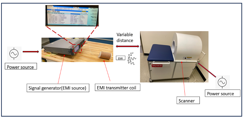
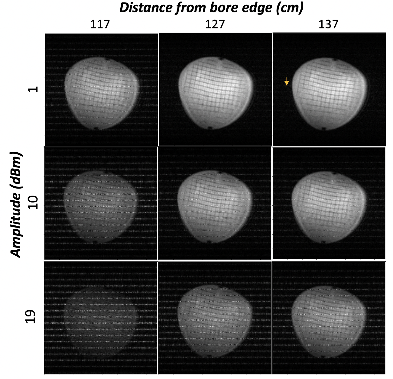
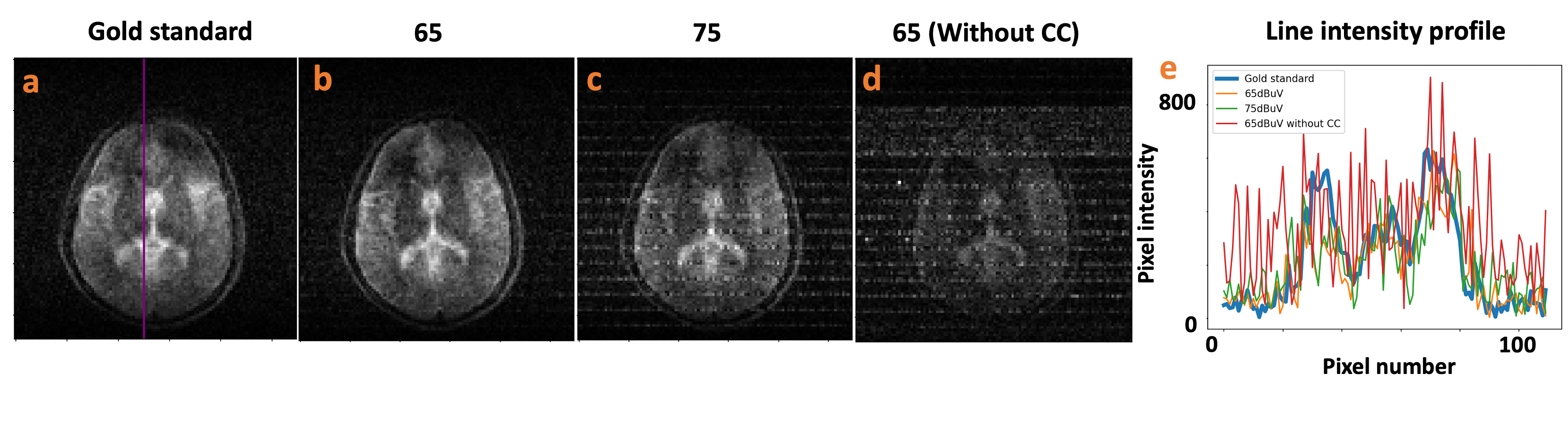
Setup: A Signal Generator was connected to a home-built solenoid (16 turns, 11cm length, and 8.5cm diameter). The solenoid was oriented perpendicular to the bore (see Figure 1). The setup included sweeping signals ranging from 1.98MHz to 2.2MHz, with amplitudes of 10dBuV to 19dBm. The signal amplitude from the transmitter was varied with distance, so altering the distance between the transmitter coil and the scanner resulted in a variation in the signal amplitude received by the RF coil. Acquisition: We acquired data from an in vitro phantom (Pro-MRI by Pro Lab) scanned in a VLF scanner (50mT, Multiwave Technologies SA, France) and a volunteer. We used a 3D TSE sequence. The acquisition parameters for the phantom were: TR/TE-500/20ms, Matrix-155x155x10, FOV-230x230x12mm3, ETL-4, and trajectory-in-out; and the corresponding acquisition parameters for the subject: TR/TE-3000/20ms, Matrix-110x110x8, FOV-220x220x160mm3, ETL-16 and trajectory-linear. In vitro experiment: The solenoid coil was first kept at the edge of the bore with an amplitude of 19bBm. The images were dominated by the artifact suppressing the object. We kept moving the coil away in steps of 10cm. from the scanner until we saw the object along with the artifact EMI interference on the reconstructed image. The optimized distance was 116cm (from the edge of the bore) with the amplitudes 19dBm, 10dBm and 1dBm. We moved the coil to 127cm and 137cm with three amplitudes (9 acquisitions). In vivo experiment: The artifact dominated the brain image when we used the exact distances and amplitudes as in the in vitro experiment. So, we increased the distance to 137cm and the amplitudes to 10dBuV, 65dBuV and 75dBuV. We also acquired an additional scan where we scanned the subject with an amplitude of 65dBuV without the conductive cloth (CC) to see the effect of the CC. Reconstruction and analysis: All in vitro and in vivo data were reconstructed in Python. The preprocessing of the k-space data involved drift correction and squared sine-bell filter to the k-space. The images were reconstructed using a fast Fourier transform. We assessed the image quality and the effect of EMI on the images with four metrics: (i) mean signal intensity of the object (MI); (ii) standard deviation of the background (SD); (iii) signal-to-noise ratio (SNR); (iv) root mean square (RMS) of the background.Results and discussion
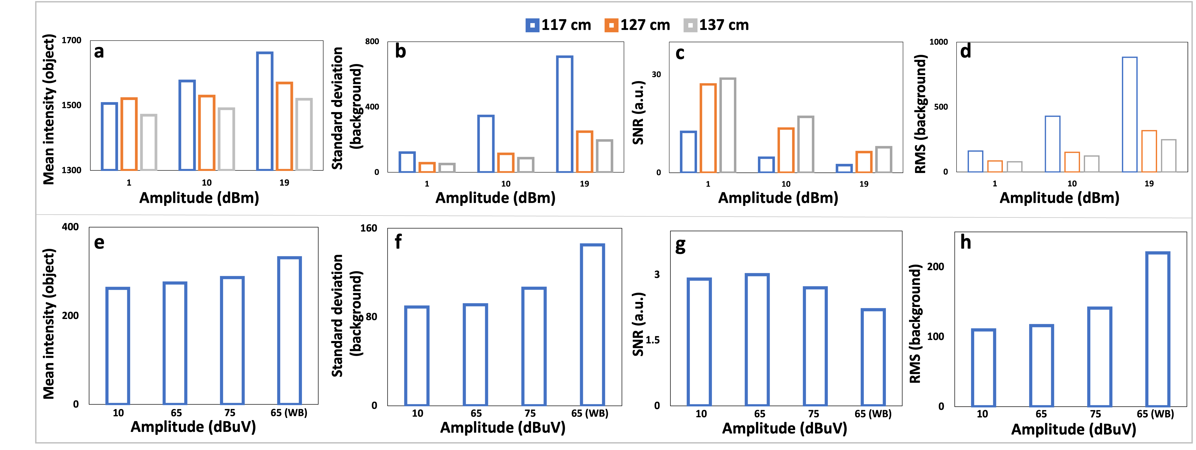
In vitro experiment: Figure 2 shows the effect of artificially generated EMI obtained from a signal generator. The artifact dominated the object for an amplitude of 19dBm and a distance of 117cm. The minimal artifact (see yellow arrow) can be seen for an amplitude of 1dBm and a distance of 137cm. In vivo experiment: Figure 3(a-d) shows the effect of external EMI on brain images. It can be observed that with the amplitude being 10dBuV, there was no effect of EMI on the image and it is prominent from 65dBuV. It can be observed in the line intensity plot (Figure 3(e)) where many peaks can be seen for the image with no CC which shows that the CC significantly blocks the EMI. Image analysis: Figure 4(a-d) shows the plot of MI, SD, SNR and RMS of the phantom for the solenoid coil being at three distances and with three amplitudes respectively. It can be observed that the SNR decreases with an increase in amplitude. Also, the SD and RMS of the background increase as the increase in amplitude and/or decrease in distance (in agreement with the images in Figure 3). Figure 4(e-h) shows the MI, SD, SNR, and RMS plots for the brain. It can be observed that the SD and RMS increase with the increase in amplitude, whereas the SNR decreases with an increase in amplitude. Also, the SNR for an amplitude of 65dBuV with a CC was higher than without a CC, which can be visually compared in Figure 3. The images acquired were low resolution to save acquisition time as the goal was to see the effect of EMI and we have not performed B0 correction.Conclusion
In conclusion, this study investigated and quantified the potential effects of EMI on VLF on vitro phantom and in vivo healthy volunteer images.Acknowledgements
1. Faculty Idea Innovation Prize, Dr. Sairam Geethanath and Dr. Shilpa Taufique, 2022
2. Friedman Brain Institute Research Scholars Fellowship, Dr. Sairam Geethanath and Dr. Shilpa Taufique
3. CEPM-CTSA grant (PI: Dr. Sairam Geethanath)
References
1. Marques, José P., Frank FJ Simonis, and Andrew G. Webb. "Low‐field MRI: An MR physics perspective." Journal of magnetic resonance imaging 49.6 (2019): 1528-154
2. Arnold, Thomas Campbell, et al. "Low‐field MRI: Clinical promise and challenges." Journal of Magnetic Resonance Imaging 57.1 (2023): 25-44
3. Bhat, Seema S., et al. "Low‐field MRI of stroke: challenges and opportunities." Journal of Magnetic Resonance Imaging 54.2 (2021): 372-390.
4. O’Reilly, Thomas, et al. "In vivo 3D brain and extremity MRI at 50 mT using a permanent magnet Halbach array." Magnetic resonance in medicine 85.1 (2021): 495-505.
5. Liu, Yilong, et al. "A low-cost and shielding-free ultra-low-field brain MRI scanner." Nature communications 12.1 (2021): 7238.
6. Yang, Lei, et al. "Active EMI suppression system for a 50 mT unshielded portable MRI scanner." IEEE Transactions on Biomedical Engineering 69.11 (2022): 3415-3426.
7. Zhao, Yujiao, et al. "Electromagnetic Interference (EMI) Elimination via Active Sensing and Deep Learning Prediction for RF Shielding‐free MRI." NMR in Biomedicine (2023): e4956
8. Parsa, Javad, Thomas O'Reilly, and Andrew Webb. "A single-coil-based method for electromagnetic interference reduction in point-of-care low field MRI systems." Journal of Magnetic Resonance 346 (2023): 107355.
Figures