2831
Self-shielded RF coils for imaging in unshielded environments at low-field1Promaxo Inc., Oakland, CA, United States
Synopsis
Keywords: Low-Field MRI, Low-Field MRI
Motivation: Access to MRI worldwide could be improved by reducing cost and increasing portability of the system. This requires imaging to occur in unshielded rooms making them highly susceptible to environmental noise; therefore, low-cost alternatives are needed for noise mitigation
Goal(s): Here we have designed a self-shielded RF coil array, made of resonating coaxial cables, for low-field parallel imaging in unshielded environments.
Approach: The proposed coils were tested against copper loops through phantom and clinical studies in various noise environments.
Results: The results indicate the self-shield coils are robust to external noise and produce images with sufficient signal and contrast to distinguish anatomical features.
Impact: With the increased interest in the community towards developing accessible MR systems, the proposed coil design presents a low-cost and resource efficient method for noise mitigation, thereby allowing MRI to be portable and available to provide point-of-care screening.
Introduction
Portable MRI systems can significantly reduce the cost of and increase access to MR scanners1,2. Such systems require imaging to take place outside of Faraday shielded rooms resulting in increased electromagnetic interference (EMI) and reduced image SNR. Promaxo’s low-field MR system, intended for in-office, image guided prostate biopsies is especially prone to EMI arising from nearby electrical equipment or radiation near the Larmor frequency. Current passive approaches to shielding include applying conductive shielding close to the scanner, or encompassing the body of the patient with a mesh cloth3. However, these techniques limit patient access which is crucial for interventional procedures. A viable alternative is using self-shield resonators or coaxial coils which have been shown to be largely immune to EMI while still providing useful signal information4-6. In this study, we translate self-shielded coils to a low-field environment (60-66mT) for imaging in a single-sided system, and demonstrate the feasibility of using such coils to image in unshielded environments.Methods
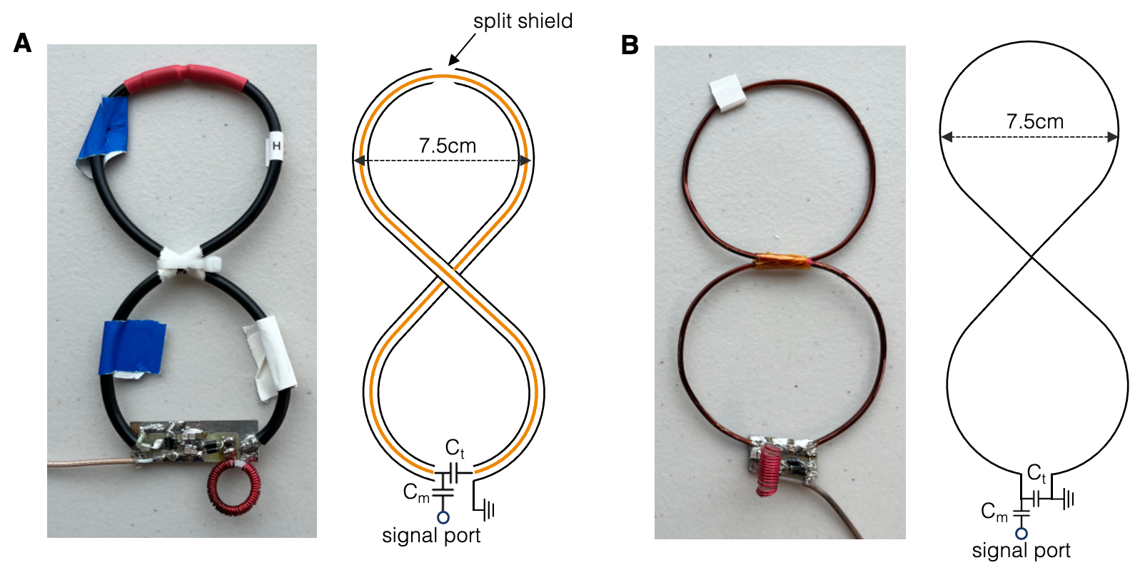
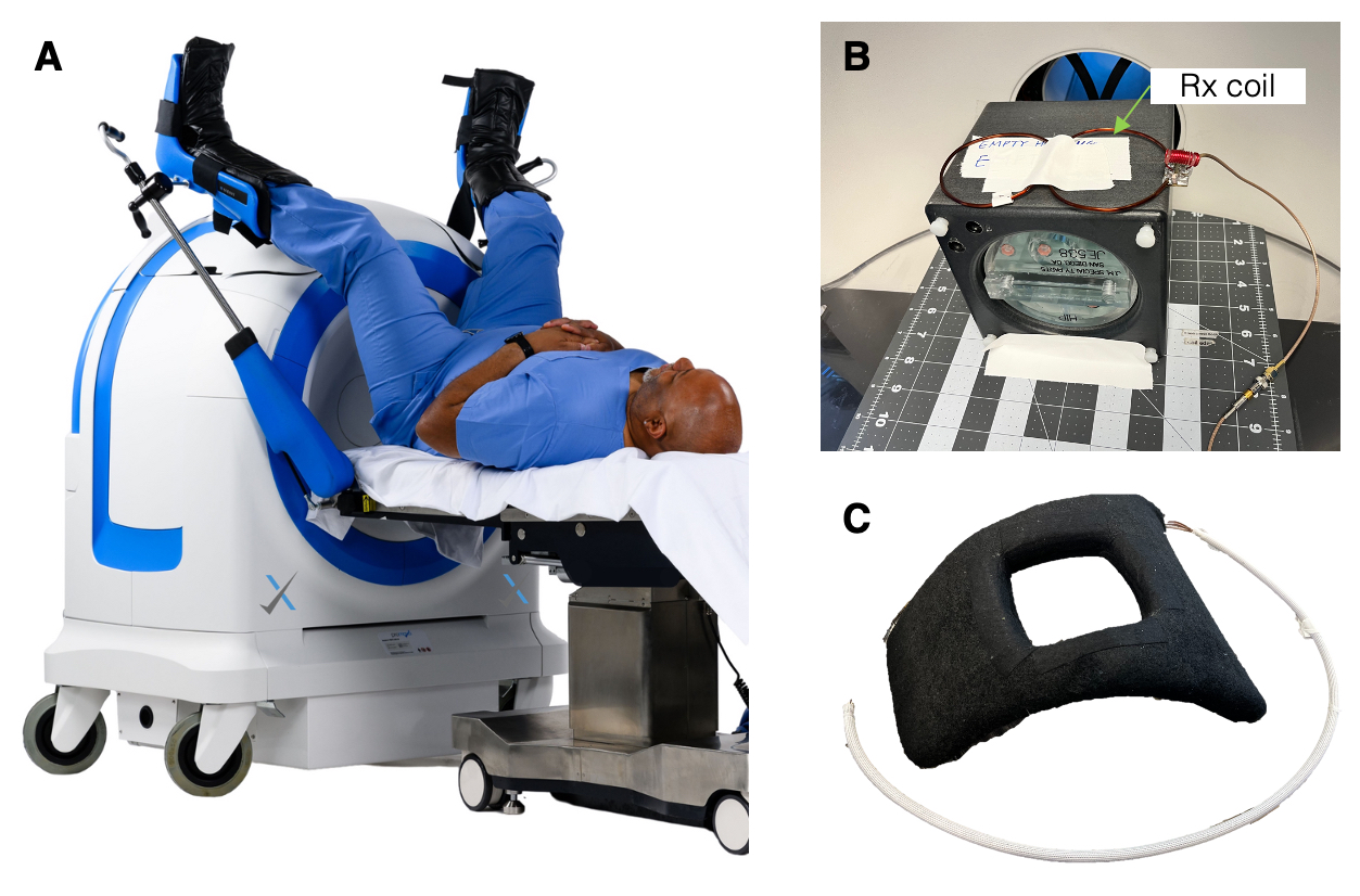
The ability of the self-shielded coil to suppress EMI was evaluated with phantom experiments using the single channel receive (Rx)-only surface coil shown in Figure 1A. The coil consists of two-loops in a figure eight arrangement, where each loop has a diameter of 7.5cm; a small gap was placed away from the tuning and matching circuitry. The coil was tuned to 2.65MHz, with a bandwidth (BW)=20kHz, and Q-factor=50.4. The performance of the self-shielded coil was evaluated against a more conventional copper loop with similar dimensions (Figure 1B). This coil was tuned to 2.65MHz, with BW=20kHz, and Q-factor=152.6. Experiments were conducted on Promaxo’s single-sided low-field MRI system (Figure 2A). The experimental setup and phantom imaged are shown in Figure 2B. Each surface coil was placed on top of an American College of Radiology approved extremity phantom (ACR), positioned in the center of the imaging field of view (FOV). The noise immunity of the coils was tested by introducing external noise signals at frequencies of 2.65MHz, 2.66MHz, and 2.63MHz during signal acquisition. A 3-axis EMI detector probe tuned to 2.65MHz was placed adjacent to the magnet and was used to detect EMI simultaneous to MR signal acquisition. The 5-channel Rx-only coil array shown in Figure 2C was used to test the clinical imaging performance of the self-shielded coils. All images were acquired using a T2-weighted pulse sequence with blipped RARE phase encoding, TE=5.2ms, TR=1.45s, echo train length=12, and FOV=18cmx18cmx10cm.Results
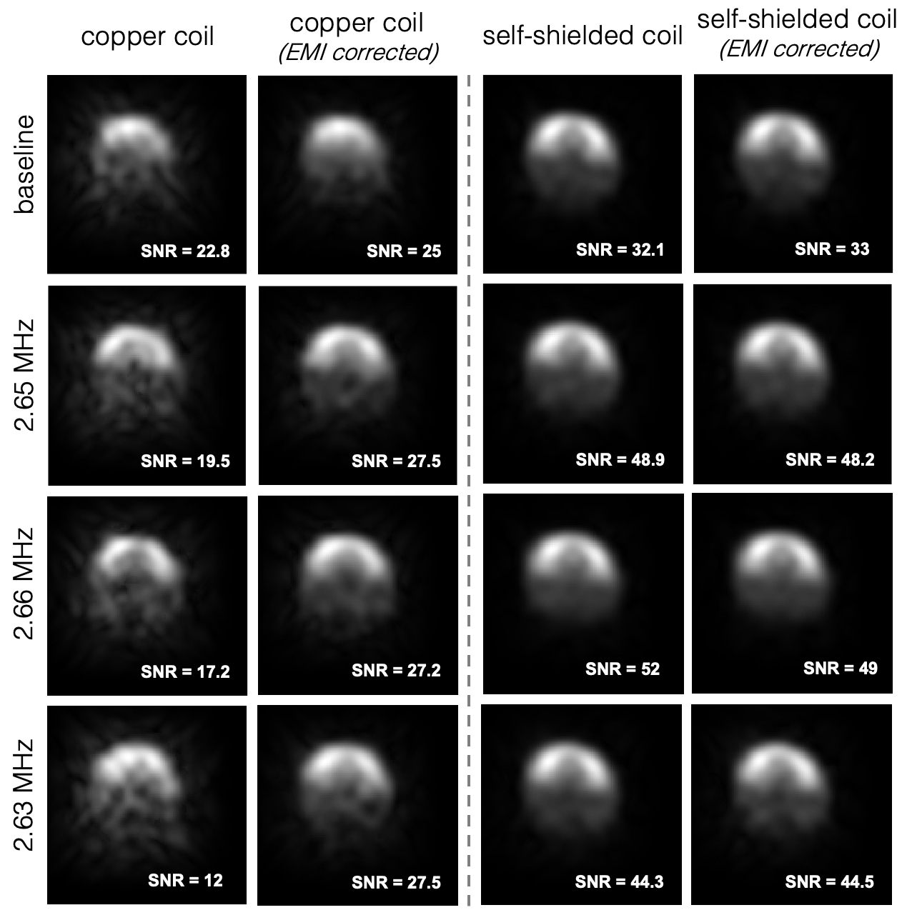
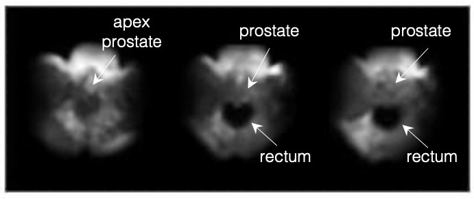
Signal collected from the single channel self-shielded and copper loop coils were reconstructed with and without EMI cancellation for each experimental condition (Figure 3). EMI cancellation was applied retrospectively by assuming a linear convolution model between the EMI in the MR signal and the signal in the noise detectors. The resulting noise estimates were then removed from the primary MR signal. SNR – calculated by taking the mean of the signal amplitude in the phantom region and dividing it by the standard deviation of the noise in the background region – is reported in the bottom right corner of each image.Figure 4 shows prostate images of a human subject obtained using a 5-channel Rx-only, self-shieled RF array coil. Four slices are shown from a 3D volumetric imaging study with an in-plane resolution of 1.5mm x1.5mm. Arrows point to the rectum and prostate anatomy.
Discussion
We have designed and implemented a self-shielded RF coil that allows for low-field MR imaging in unshielded environments. The phantom imaging experiments show that noise suppression of the self-shielded RF coil is much higher than that of a conventional copper loop of similar dimensions. In the case of the copper loop, while EMI cancellation through signal postprocessing was able to remove a significant amount the background noise it is still difficult to delineate phantom features. Moreover, the image quality is not as high as those obtained using the self-shielded loop. Quantitative comparison of image SNR asserts a similar trend. EMI cancellation of signal acquired with the self-shielded coil has little, if any, improvement on image SNR, and the coil appears to be tolerant to external signals are various frequencies.Clinical imaging experiments demonstrate proof-of-concept that RF arrays built from self-shield loops can be used for parallel imaging at low-field and generate images that have sufficient SNR to visualize key anatomical features.
Conclusion
In conclusion, the proposed self-shielded coil design provides SNR robustness to external noise and promising image quality. Such coils can allow for MR imaging to take place in unshielded rooms, increasing the portability of MRI, thereby making it accessible to provide point-of-care screening.Acknowledgements
No acknowledgement found.References
- McDaniel, P.C., et al., The MR Cap: A single-sided MRI system designed for potential point-of-care limited field-of-view brain imaging. Magnetic resonance in medicine, 2019. 82(5): p. 1946-1960.
- Cooley, C.Z., et al., A portable scanner for magnetic resonance imaging of the brain. Nat Biomed Eng, 2021. 5(3): p. 229-239.
- O’Reilly, T., et al., In vivo 3D brain and extremity MRI at 50 mT using a permanent magnet Halbach array. Magnetic Resonance in Medicine, 2021. 85(1): p. 495-505.
- Suits, B.H., J.B. Garroway An Fau - Miller, and J.B. Miller, Noise-immune coil for unshielded magnetic resonance measurements. (1096-0856 (Electronic)).
- Stensgaard, A., Optimized design of the shielded-loop resonator. Journal of Magnetic Resonance, Series A, 1996. 122(2): p. 120-125.
- Harpen, M.D., The theory of shielded loop resonators. Magnetic Resonance in Medicine, 1994. 32(6): p. 785-788.
Figures