2828
A first 3D negative-contrast MR image using a low-Tc SQUID detection: towards portable clinical MRI1Chipiron, Paris, France
Synopsis
Keywords: Low-Field MRI, Low-Field MRI, SQUID
Motivation: At Chipiron we strive to develop the first commercial portable ultra-low field MRI scanner based on SQUID detection.
Goal(s): Here, we aimed to acquire a first 3D image using SQUID-MRI at 1 mT.
Approach: Using our own integrated SQUID-MRI system, we scanned a hand-made water based phantom using a GRE sequence.
Results: We successfully acquired a 3D image of a phantom in 6 h with a resolution of 5x5x40 mm3.
Impact: Aspiring to democratise MRI, we develop a portable ultra-low field scanner that leverages the high sensitivity of SQUID detection. Our first 3D image is just the first step that paves the way to achieving competitive image quality for clinical employment.
Introduction
While low field MRI solutions have been successfully implemented in several academic and commercial groups1, ranging between 50 and 200 mT, field cycling studies have revealed that the most interesting contrast can be obtained in the ultra-low field (ULF) regime (≲ 10 mT)2. However, at such low fields, signal-to-noise ratio (SNR) becomes a problem, especially in the case of inductive detection, where the sensitivity of the antenna scales with B0-¾. The use of superconductive quantum interference devices (SQUID) for signal acquisition has been proposed to tackle this issue3. Here we present our first 3D MR image of a water-based phantom, acquired at 1 mT in 6 h using SQUID detection. Given the superior sensitivity of SQUID sensors over conventional inductive detection at ULF, these results are expected to be significantly improved following a refined integration of the SQUID into the reception line.Methods
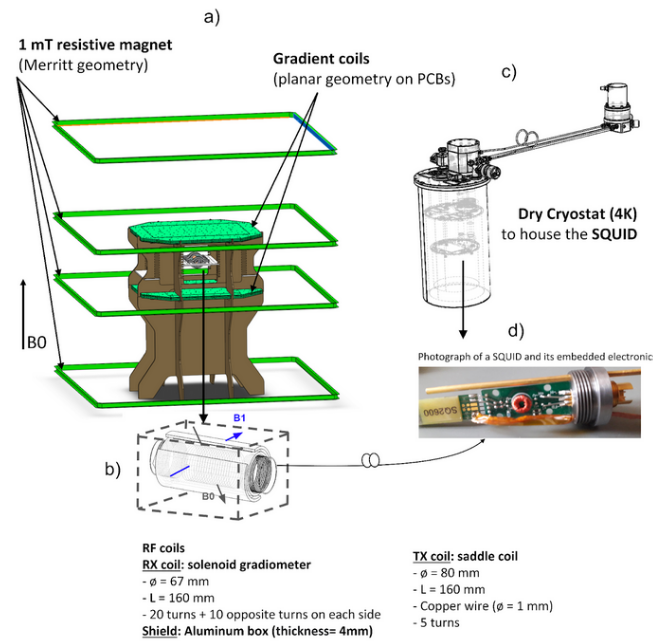
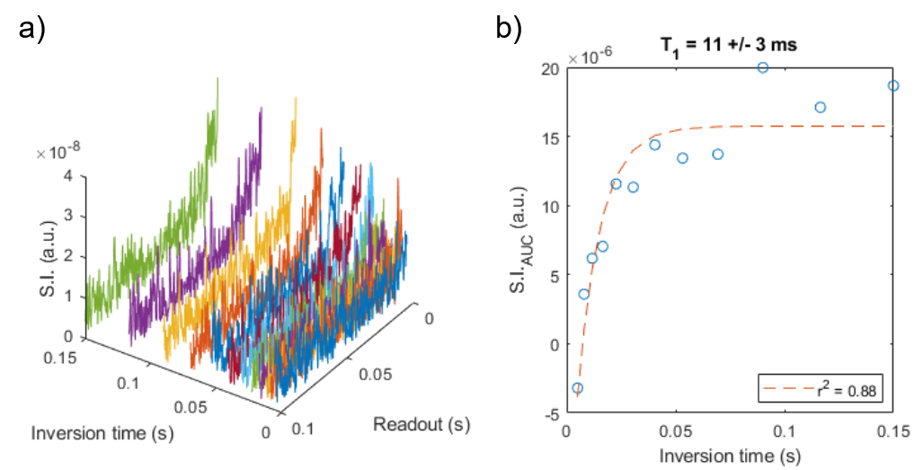
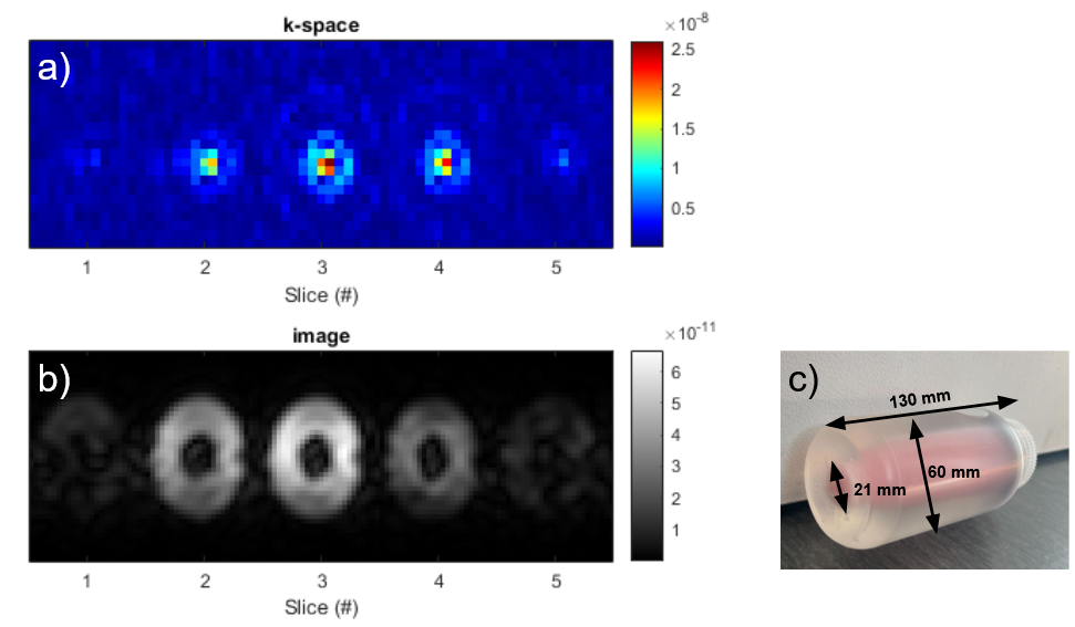
To obtain a negative contrast image at 1 mT, a water-based phantom was built, consisting of a commercial paraffin candle (ø = 21 mm, L = 130 mm) positioned coaxially inside a bottle (ø = 60 mm, L = 130 mm) filled with doped deionised water (1.15 mM of MnCl2). The details of our integrated SQUID-MRI system are as follows4,5: a Merrit coil electromagnet fed by a current source generated the polarisation field. A cylindrical saddle coil and solenoid-based volume gradiometer were used for radiofrequency (RF) field transmission and reception. The signal sensed by the receive coil is then sent to a Niobium-based SQUID detector operating at 4.2 K and acting as a signal amplifier. Both the phantom and the RF coils were positioned inside a 4-mm thick aluminum box, used for electromagnetic shielding. Further details on the SQUID-MRI system are given in Fig. 1.Prior to imaging, a spectroscopic inversion recovery (IR) sequence was employed to assess the T1 of the doped water. Twelve logarithmically spaced inversion times were selected, for a total acquisition time of 16 min. T1 was estimated by calculating the area-under-curve and fitting the processed data to an exponential recovery. A 3D image of the phantom was then acquired using a gradient-recalled echo (GRE) sequence. Based on the magnetic parameters of the doped water, the following sequence parameters were chosen: TR/TE = 20/8.3 ms, flip angle = 70°, matrix size = 23x13x5, spatial resolution ~5x5x40 mm3, bandwidth = 217 Hz/pixel. With 16600 excitations, the whole acquisition lasted 6 h. K-space filtering and in-plane interpolation into a 45x25 matrix were performed in post-processing.
Results
Time traces acquired with IR at the chosen inversion times are plotted in Fig. 2a. A T1 of 11 ± 3 ms was extracted, as shown in Fig. 2b. The acquired 3D k-space and reconstructed image of the phantom are presented in Fig. 3a and 3b. The candle is clearly recognisable as the low-intensity circle surrounded by the high intensity signal given by the doped water. A photograph of the phantom is presented in Fig. 3c, for illustration.Discussion
Thanks to the extreme sensitivity of the SQUID detection, we could obtain a 3D image of a phantom in 6 h. The short T1 of the doped water allowed to maximise the number of excitations per unit time, hence improving SNR. Despite the promising results, further improvements are required to achieve the temporal and spatial resolution required to make this technology competitive in the clinics. Refinement of the transmission line between the RF coils and the SQUID, as well as cooling strategies to reduce Johnson noise are expected to improve SNR between 100 and 1000 times. Furthermore, undersampling strategies and the use of MRI sequences with a higher signal efficiency at ULF, such as bSSFP, would reduce the scan time and improve the spatial resolution. Finally, the use of denoising strategies to reduce electromagnetic interference, such as EDITER6, have proven paramount in SNR-deprived regimes and will be explored.Conclusion
SQUID-MRI is a technology that has the potential to surpass classical inductive detection at ULF. With a preliminary integration of SQUID detection into the MRI signal acquisition chain, we could acquire a first 3D image of a hand-made phantom. This milestone aspires to represent the very first step towards the clinical employment of MRI at such a field regime.Acknowledgements
No acknowledgement found.References
1Sarracanie M. et al., Front. Phys., 8:172, 2020
2Bödenler M. et al., MRM., 86(4):2049–2063, 2021
3Espy M. et al., IEEE Trans. Appl. Supercond, 25(3):1-5, 2014
4Saniour et al., ESMRMB 2023
5Fiorito et al., ISMRM-BIC 2023
6Sri Nivas S.A. et al., MRM, 87(2):614-628, 2022
Figures