2820
Supervised Pretraining and Self-Supervised Finetuning enables Robust Reconstruction of High-Resolution 7T MP2RAGE for Multiple Sclerosis1Advanced Clinical Imaging Technology, Siemens Healthineers International AG, Lausanne, Switzerland, 2Department of Radiology, Lausanne University Hospital, Lausanne, Switzerland, 3LTS5, Ecole Polytechnique Federale de Lausanne, Lausanne, Switzerland, 4Icahn School of Medicine at Mount Sinai, New York, NY, United States, 5CIBM Center for Biomedical Imaging, Geneva, Switzerland, 6Siemens Healthcare GmbH, Erlangen, Germany, 7National Institute of Neurological Disorders and Stroke, National Institutes of Health, Bethesda, MD, United States, 8CIBM Center for Biomedical Imaging, Lausanne, Switzerland
Synopsis
Keywords: Machine Learning/Artificial Intelligence, Machine Learning/Artificial Intelligence, Neuro, Multiple Sclerosis, Reconstruction
Motivation: The infeasibility to collect large datasets of high-resolution, fully-sampled 7T data hinders the training of deep learning reconstructions for accelerated high-resolution 7T scans.
Goal(s): Demonstrate that combining pretraining on fully-sampled scans from 1.5/3T and self-supervised finetuning on a few undersampled 7T scans enables robust reconstruction.
Approach: High-resolution(0.5mm isotropic), 3D, 7T MP2RAGE scans of multiple sclerosis patients are used for finetuning/testing.
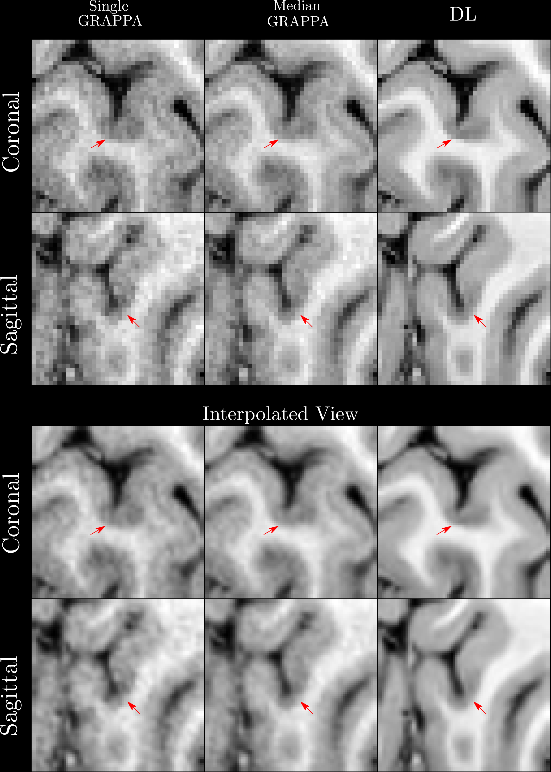
Results: Deep learning reconstructions from one 7T scan have higher apparent SNR than a median of GRAPPA reconstructions from three scans, currently used for assessment, with similar tissue contrast and lesion conspicuity, potentially reducing scan time by a factor of three.
Impact: Very high-resolution 3D scans can require infeasible acquisition time to generate images suitable for clinical use. Our work shows the potential for deep learning reconstructions to reduce scan time by a factor of three, without fully-sampled 7T data.
Introduction
While deep learning$$$\,$$$(DL) methods in MR image reconstruction are state of the art1, they usually require substantial amounts of fully sampled data, the acquisition of which can be infeasible, particularly for high-resolution 3D scans at 7T. We show that high-resolution 3D scans at 7T can be robustly reconstructed through supervised pretraining on a diverse collection of lower-resolution data collected at 1.5T/3T, and self-supervised finetuning on prospectively accelerated, high resolution 7T scans with no access to fully-sampled 7T data. A dataset of ultra-high resolution, 3D, 7T MP2RAGE acquisitions were collected from multiple sclerosis$$$\,$$$(MS) patients who were scanned three times in one session, and the median images were used for assessment of cortical lesions$$$\,$$$(CL), which require high-resolution, high contrast-to-noise-ratio images due to small size and subtle contrast differences from the surrounding cortex2 . We compare the image quality and lesion conspicuity of the DL reconstruction of single scans to the median images.Methods
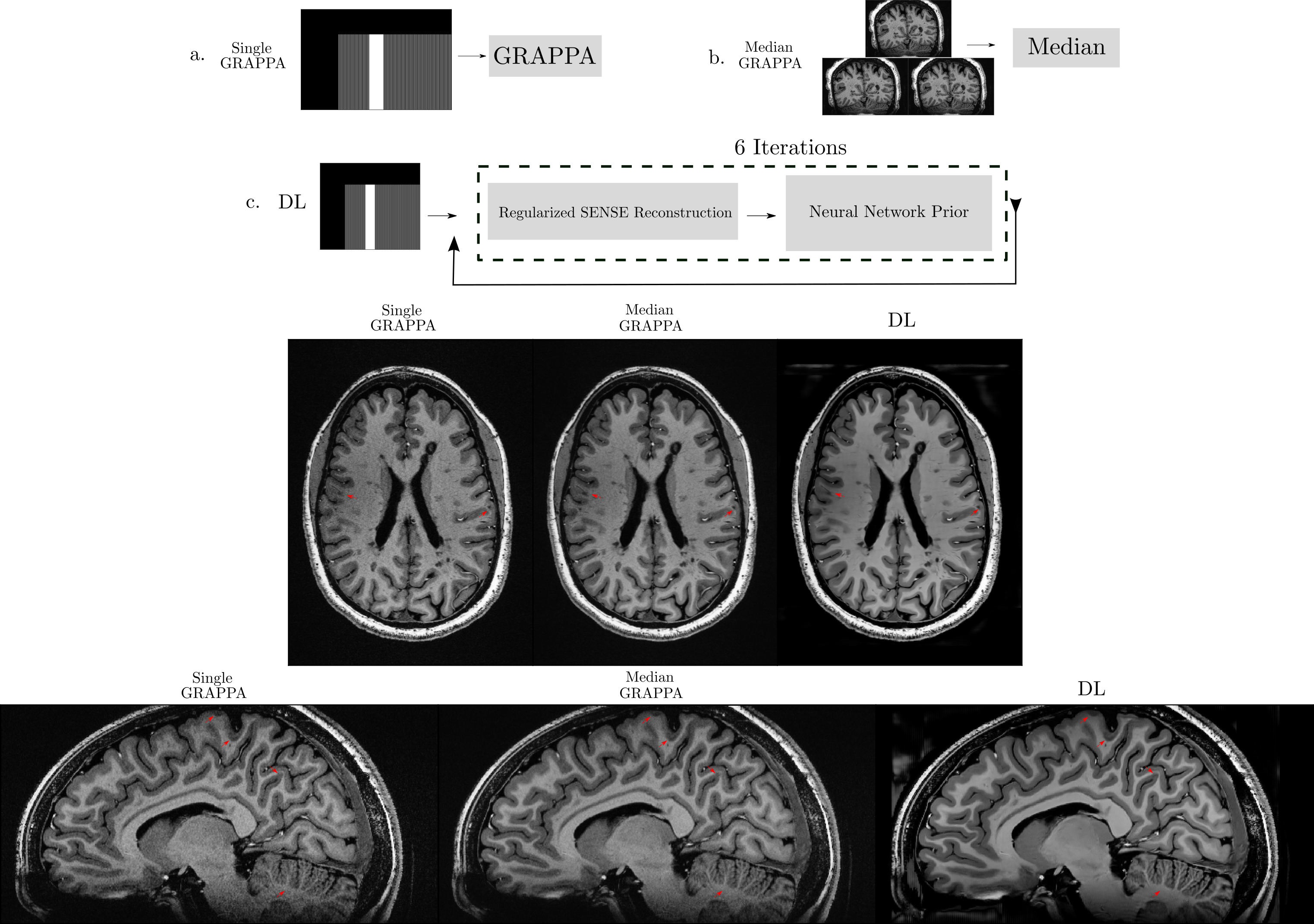
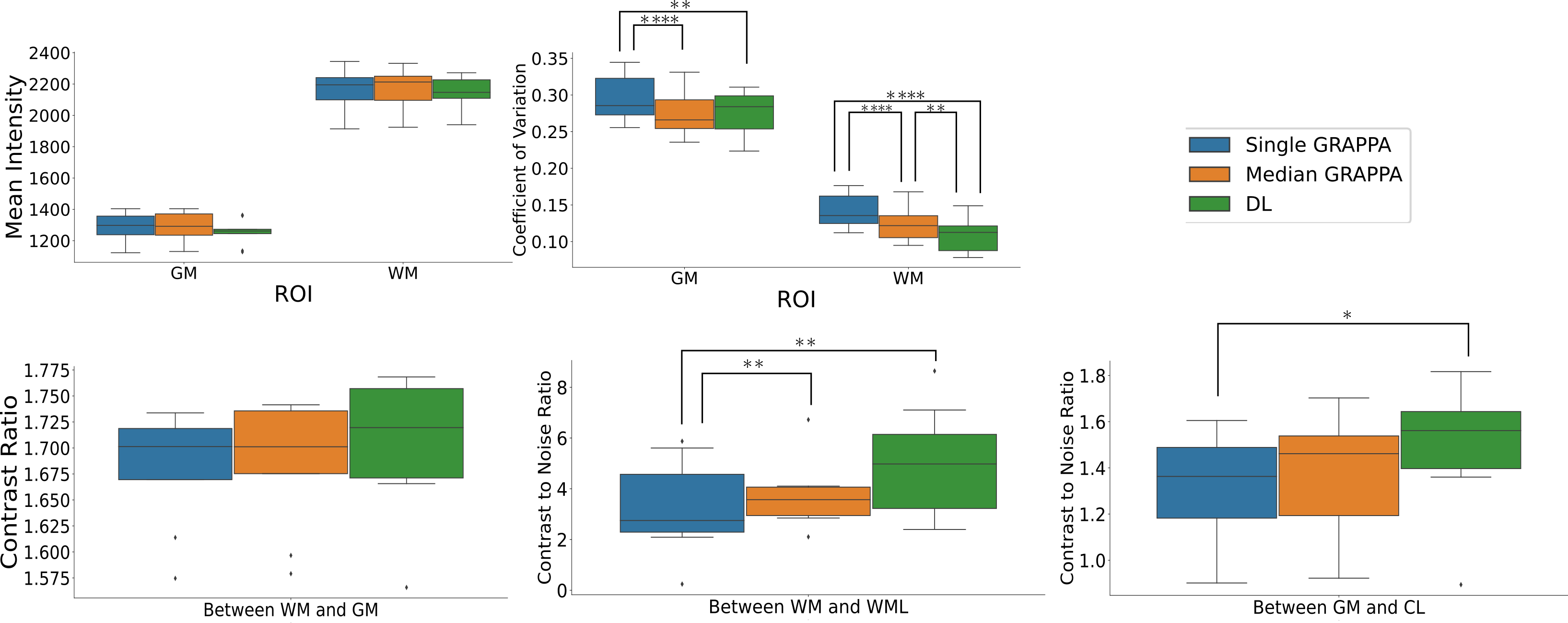
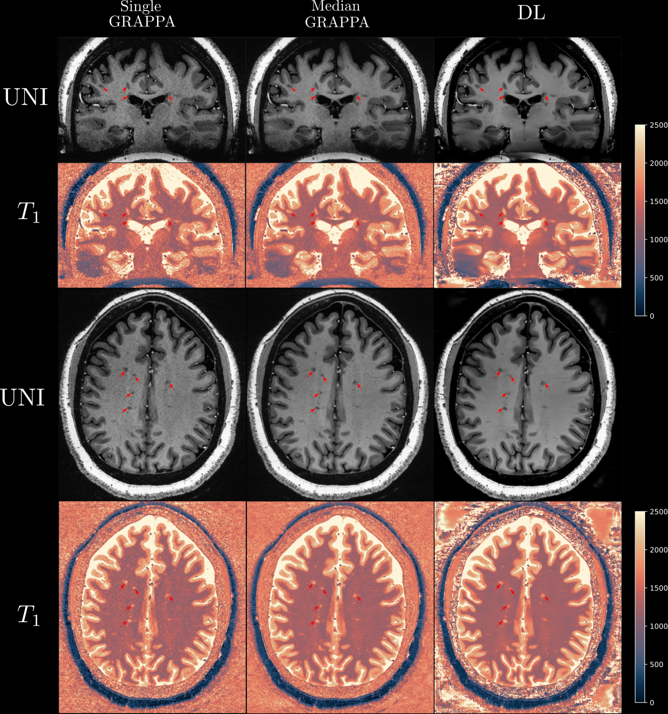
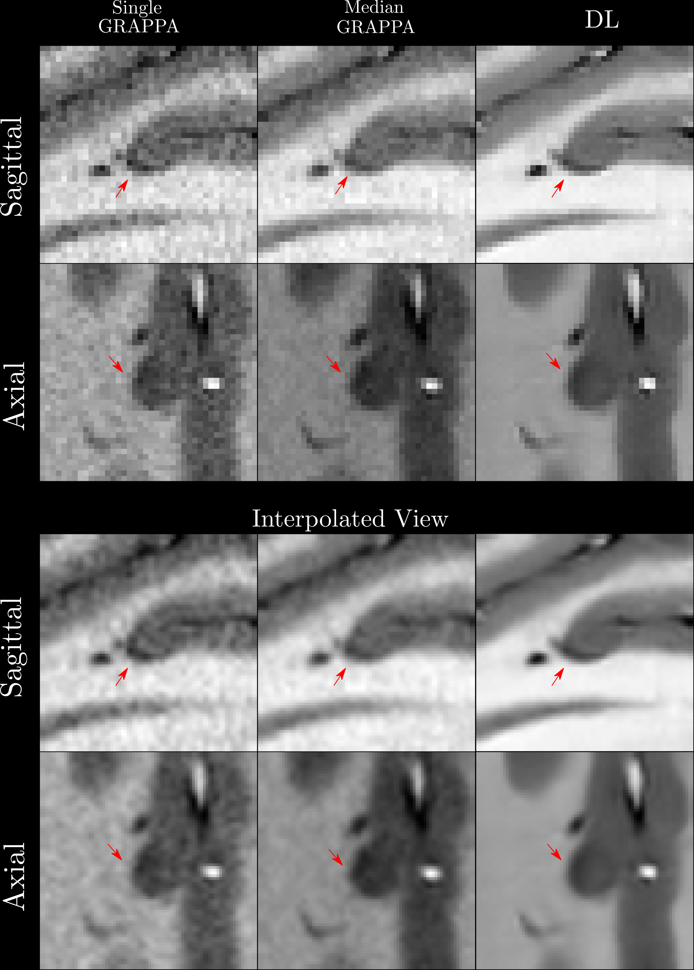
Nine subjects with MS and four healthy volunteers$$$\,$$$(HV) were scanned three times at 7T in a single session using a 3D MP2RAGE research application sequence (TR/TI1/TI2/TE = 6000/800/2700/5 ms, voxel size = 0.5×0.5×0.5 mm3, matrix size=224x336x448, acquisition time=10:30 min) with 6/8 Partial Fourier acceleration in both phase encoding directions and parallel imaging acceleration of 3 along one phase encoding direction. We compare three methods: GRAPPA3 reconstruction of the first scan, the median of GRAPPA reconstructions from three scans$$$\,$$$(co-registered to the first scan), and a DL reconstruction of the first scan. The DL reconstruction uses an unrolled network with 6 iterations of regularized SENSE reconstruction and 3D UNet regularization. The network was pretrained using 5000 training pairs derived from an in-house dataset of 500 fully-sampled 3D scans of healthy volunteers on 1.5 and 3T scanners (MAGNETOM scanners, Siemens Healthcare, Erlangen, Germany) in the head, abdomen, and pelvis. The network is finetuned on the undersampled data from the first scan of two MS patients and 2 HV using a self-supervised approach4,5.The remaining subjects are used for evaluation. Separate models are finetuned for both MP2RAGE inversion contrasts, with image combination after inference to form the MP2RAGE T1-weighted uniform$$$\,$$$(UNI) image without background noise6.For quantitative analysis, an automatic tissue segmentation from an in-house method based on nnUNet7 was used to segment gray matter$$$\,$$$(GM), white matter$$$\,$$$(WM) and WM lesions$$$\,$$$(WML) from the median GRAPPA image and propagated to the other methods. Cortical lesions were segmented manually from the median image and a T2* GRE8 by consensus between a neurologist and a medical doctor. We compared the mean intensity and the coefficient of variation$$$\,$$$(COV) of the intensity in WM and GM, the contrast ratio$$$\,$$$(CR) between WM and GM, and the contrast to noise ratio$$$\,$$$(CNR) between WM/WML and between GM/CL in the UNI images. The CNR is estimated using the standard deviation in WM/GM as the noise. Statistical significance of the mean differences was assessed through a dependent t-test for each pair of methods, with a threshold of 0.0167 after Bonferroni correction. For qualitative comparison, we show slices of MP2RAGE UNI images and T1 maps and closeups of small CL.
Results
Quantitatively$$$\,$$$(Figure 2), we found no significant differences in the intensities in the ROIs or the CRs between WM/GM$$$\,$$$(p=0.29 +- 0.23). DL Recon$$$\,$$$(p=0.0054 and 0.0162) had higher CNRs between WML/WM and CL/GM compared to Single GRAPPA, but no difference to Median GRAPPA. DL Recon also had lower COVs in GM compared to Single GRAPPA$$$\,$$$(p=0.0045), and lower COVs in WM compared to Median and Single GRAPPA$$$\,$$$(p=4.98e-5 and 0.0038). Qualitatively$$$\,$$$(Figures 1/3), DL provides better denoising compared to Median GRAPPA while preserving anatomical detail and contrast between different tissues; furthermore, CL and WML can be clearly delineated. Figures 4-5 show that CL are conspicuous on DL reconstructions.Discussion and Conclusion
We showed that DL reconstructions from a single scan can have the same to higher SNR, same tissue contrast, and same lesion conspicuity compared to the median of three GRAPPA-reconstructed scans, opening the possibility for 0.5mm isotropic acquisitions in clinical research studies. This work improves upon a previous abstract, which used a 2D self-supervised reconstruction9, by adding supervised pretraining and 3D reconstruction, which improved image quality$$$\,$$$(sharpness/denoising), lesion conspicuity, and removed artifacts from slice-wise reconstruction. For future work, we plan to add more subjects for analysis and do a more extensive validation10 by, e.g., comparing prospective CL segmentation over the different methods. In conclusion, the results show the potential for combining supervised pretraining on fully-sampled data from low resolution/low field strengths with self-supervised finetuning on undersampled data from high resolution/high field strengths for robust reconstruction of high resolution, 3D images without access to fully sampled data.Acknowledgements
No acknowledgement found.References
1. Knoll, Florian, Kerstin Hammernik, Chi Zhang, Steen Moeller, Thomas Pock, Daniel K. Sodickson, and Mehmet Akcakaya. "Deep-learning methods for parallel magnetic resonance imaging reconstruction: A survey of the current approaches, trends, and issues." IEEE signal processing magazine 37, no. 1 (2020): 128-140.
2. Mainero, Caterina, Constantina A. Treaba, and Elena Barbuti. "Imaging cortical lesions in multiple sclerosis." Current Opinion in Neurology 36, no. 3 (2023): 222-228.
3. Griswold, Mark A., Peter M. Jakob, Robin M. Heidemann, Mathias Nittka, Vladimir Jellus, Jianmin Wang, Berthold Kiefer, and Axel Haase. "Generalized autocalibrating partially parallel acquisitions (GRAPPA)." Magnetic Resonance in Medicine: An Official Journal of the International Society for Magnetic Resonance in Medicine 47, no. 6 (2002): 1202-1210.
4. Yaman, Burhaneddin, Seyed Amir Hossein Hosseini, Steen Moeller, Jutta Ellermann, Kâmil Uğurbil, and Mehmet Akçakaya. "Self‐supervised learning of physics‐guided reconstruction neural networks without fully sampled reference data." Magnetic resonance in medicine 84, no. 6 (2020): 3172-3191.
5. Batson, Joshua, and Loic Royer. "Noise2self: Blind denoising by self-supervision." In International Conference on Machine Learning, pp. 524-533. PMLR, 2019.
6. O'Brien KR, Kober T, Hagmann P, Maeder P, Marques J, Lazeyras F, Krueger G, Roche A. Robust T1-weighted structural brain imaging and morphometry at 7T using MP2RAGE. PLoS One. 2014 Jun 16;9(6):e99676. doi: 10.1371/journal.pone.0099676. PMID: 24932514; PMCID: PMC4059664.
7. Diekhaus H, Donnay C, Gaitan MI, et al. Pseudo-Label Assisted Nnu-Net (PLAn) Enables Automatic Segmentation of 7T MRI From a Single Acquisition. medRxiv. 2022;2022.12.22.22283866doi: 10.1101/2022.12.22.22283866
8. Liu, Jiaen, Erin S. Beck, Stefano Filippini, Peter Van Gelderen, Jacco A. De Zwart, Gina Norato, Pascal Sati et al. "Navigator-Guided Motion and B0 Correction of T2*-weighted MRI Improves Multiple Sclerosis Cortical Lesion Detection." Investigative radiology 56, no. 7 (2021): 409.
9. Yu et al., Self-Supervised Image Reconstruction of 7T MP2RAGE for Multiple Sclerosis: 0.5mm Isotropic Resolution in 10 Minutes, Abstract at Annual Meeting of ISMRM 2023.
10. Yamamoto, T., C. Lacheret, H. Fukutomi, R. A. Kamraoui, L. Denat, B. Zhang, V. Prevost et al. "Validation of a Denoising Method Using Deep Learning–Based Reconstruction to Quantify Multiple Sclerosis Lesion Load on Fast FLAIR Imaging." American Journal of Neuroradiology 43, no. 8 (2022): 1099-1106.
Figures