2815
Revolutionizing MR Elastography: Deep Learning-Powered Stiffness Map Reconstruction from Sparse Wavefield Data.1Biomedical Engineering, The Ohio State University, Columbus, OH, United States, 2Radiology, The Ohio State University, Columbus, OH, United States
Synopsis
Keywords: Machine Learning/Artificial Intelligence, Elastography, Stiffness Maps
Motivation: AI has proven itself in improving MRI reconstructions, yet its potential in estimating MRE stiffness maps with rapidly acquired data remains unexplored.
Goal(s): Investigating the untapped potential of AI in MR Elastography, promising advancements in diagnostic accuracy and efficiency of this modality.
Approach: 3D FEM was used to create the dataset, and Deep Learning was used to reconstruct the stiffness maps from sparse wavefield data
Results: The Deep Learning model was able to effectively reconstruct the MRE-Stiffness maps at high acceleration rates. The model's performance was reported in terms of SSIM.
Impact: This innovative study leverages deep learning and finite element modeling to reconstruct liver stiffness maps from under-sampled MR Elastography data. The proposed AI approach demonstrates robustness and potential for accelerating stiffness estimation, paving the way for improved tissue stiffness estimation.
Summary of main findings
We propose a method to test the potential of deep learning to generate the stiffness maps from the under-sampled liver MR Elastography data using FEM simulated wavefield.Synopsis
We propose a novel approach to identify the untapped potential of deep learning (DL) in the context of generating MR Elastography (MRE)-derived stiffness estimates. Finite Element Modeling (FEM) was performed in a human liver geometry to create 3D displacement fields to obtain stiffness maps from DL. Subsequently, we evaluated the effectiveness of the DL approach by comparing stiffness maps from under-sampled displacement fields to the stiffness maps computed from fully sampled wavefields. The method was able to reconstruct the stiffness maps with a high acceleration rate effectively.Background
We propose a novel approach to identify the untapped potential of deep learning (DL) in the context of generating MR Elastography (MRE)-derived stiffness estimates. Finite Element Modeling (FEM) was performed in a human liver geometry to create 3D displacement fields to obtain stiffness maps from DL. Subsequently, we evaluated the effectiveness of the DL approach by comparing stiffness maps from under-sampled displacement fields to the stiffness maps computed from fully sampled wavefields. The method was able to reconstruct the stiffness maps with a high acceleration rate effectively.Methodology
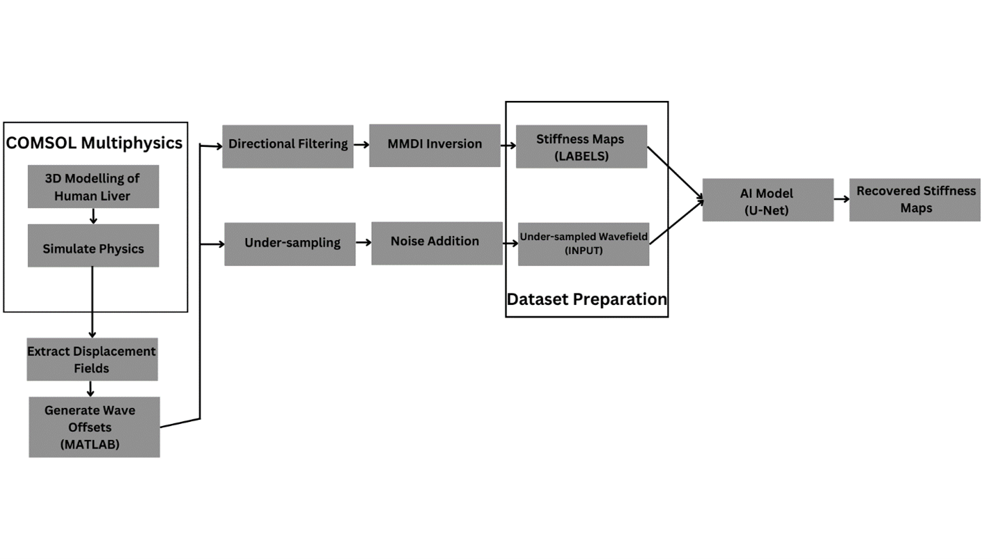
4.1. Deep Learning:An AI model i.e., DL-based U-Net[3] was employed to obtain stiffness maps. The AI model was trained using the stiffness maps generated from multi-modal direct inversion (MMDI) using the displacement fields generated from the FEM simulations as described below. Our dataset consists of under-sampled displacement fields as input and MMDI stiffness maps as ground truth reference, with the model tasked to learn the direct correspondence between the input and the reference data.
4.2. Dataset Preparation:
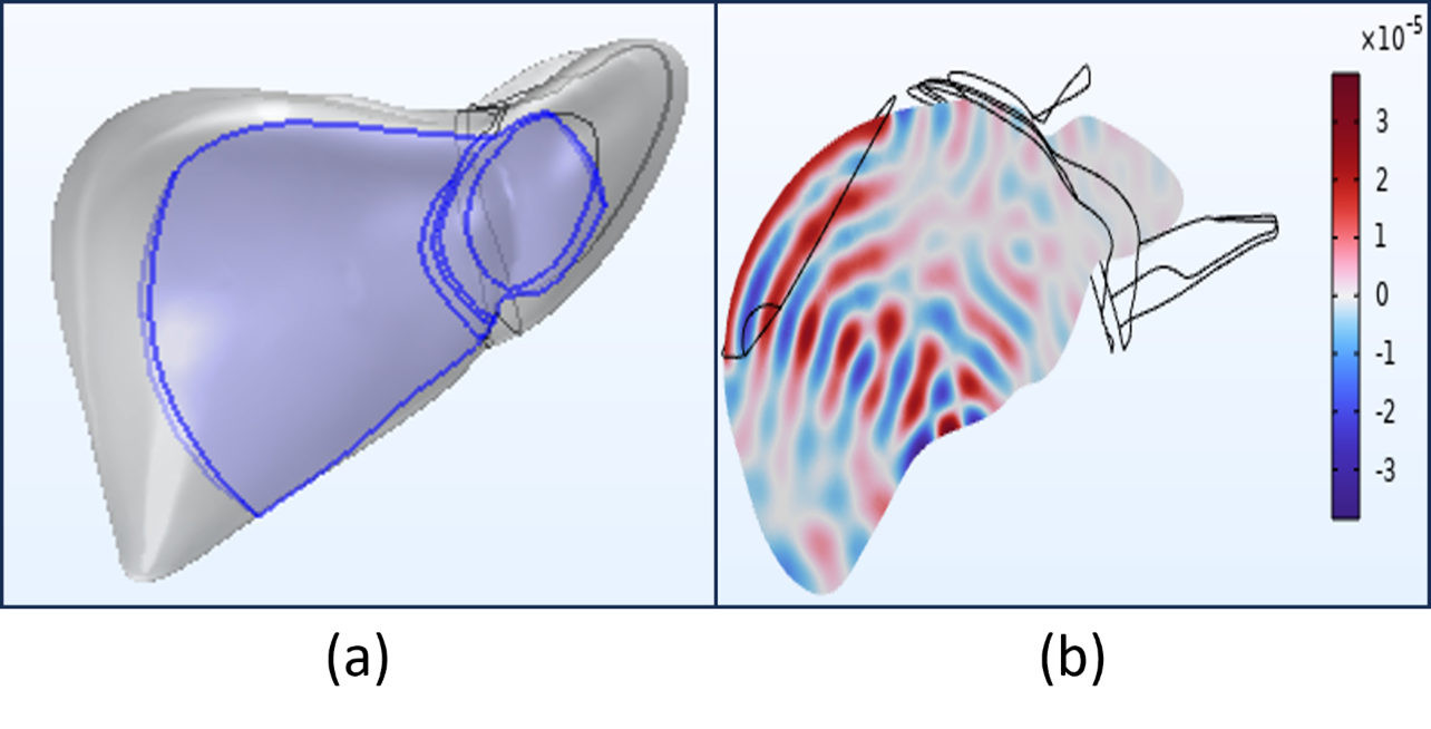
3D FEM was performed in COMSOL with human liver geometry (Fig. 2a), 60 Hz excitation, varied Young's modulus (1-7kPa), Poisson's ratio 0.499 and isotropic loss factor of 0.005. MATLAB processed FEM data for axial slices $$$\{x_{i}\}_{i=1}^{N}$$$ representing 256x256x4 displacement fields with in-plane dimensions and phase offsets. Fig. 1 shows the proposed methodology. The simulation yields complex displacement fields as shown in Fig. 2(b). AI model was trained with fully sampled first harmonic displacement fields and MMDI reference stiffness maps. Testing involved Fourier transforming and undersampling FEM-generated displacements to feed it as input to the AI model for true stiffness map generation, using the undersampling equation. $$k_U = DFS_{i} x_i$$ Utilizing a single coil dataset, coil sensitivity map $$$S_i$$$ was set to one. Undersampled k-space data $$$k_U$$$ was created with downsampling operator $$$D$$$ and Fourier operator $$$F$$$. Undersampled displacement fields were obtained through inverse Fourier transform operation $$$(x_U= Fk_U)$$$. Adding zero-mean Gaussian noise $$$(N(0, \sigma))$$$ to $$$x_U$$$ resulted in noisy input $$$(x_{U}^{'})$$$ for our AI model. $$x'_U = x_U + N(0, \sigma)$$ Ground-truth reference stiffness maps for the corresponding fully sampled displacement fields were generated by applying a 2D Butterworth 4th order bandpass filter in 4 directions with cutoff values 2-128 waves/FOV to remove longitudinal and reflected waves. MMDI in MRELab (Mayo Clinic, Rochester, MN) was performed to obtain the reference stiffness maps $$$\{y_{i}\}_{i=1}^{N}$$$ for comparison against the AI generated stiffness maps from the undersampled displacement fields. The dataset comprises 224 slices of undersampled displacement fields $$$(x_{U}^{'})$$$ and corresponding stiffness maps $$$(y_i)$$$. The AI model aims to learn the one-to-one mapping from $$$(x_{U}^{'})$$$ to $$$(y_i)$$$ , with 156 slices used for training and 67 for validation.
4.3. Model Training:
The model underwent 200 epochs of training with a batch size of 10 slices, utilizing a learning rate of $$$1e^{-4}$$$. The U-Net employed mean-squared error as the loss function, and the Structural Similarity Index (SSIM) was employed for performance evaluation.
Results
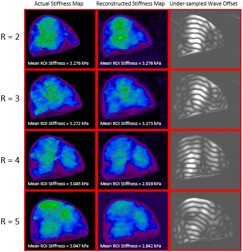
Fig. 3 shows the under-sampled input displacement field at various acceleration rates i.e., R ranging from 2 to 5 and their corresponding stiffness maps generated using AI model and the reference actual stiffness map generated from the MMDI. The red circle highlighted in the figure illustrates the ROI, and the mean stiffness in the ROI for each of the sampling rates is reported. The SSIM values given in figure 3 are close to 1. This shows the fair similarity between AI generated structure, and the reference structure.Discussions
This study demonstrated robust stiffness estimates from a highly accelerated under sampled displacement field using an AI model. However, in this study a homogenous liver was used in generating the displacement fields from FEM simulations. Future studies will involve FEM simulations in liver by incorporating hepatic and portal veins and physiologically varying stiffness measurements to generate required data for AI model.Acknowledgements
This work was funded by NIH R01AR075062References
[1] R. Muthupillai, D. J. Lomas, P. J. Rossman, J. F. Greenleaf, A. Manduca, and R. L. Ehman, “Magnetic Resonance Elastography by Direct Visualization of Propagating Acoustic Strain Waves,” Science, vol. 269, no. 5232, pp. 1854–1857, Sep. 1995, doi: 10.1126/science.7569924.
[2] M. C. Murphy, A. Manduca, J. D. Trzasko, K. J. Glaser, J. Huston, and R. L. Ehman, “Artificial neural networks for stiffness estimation in magnetic resonance elastography: Neural Network Inversion for MRE,” Magn. Reson. Med., vol. 80, no. 1, pp. 351–360, Jul. 2018, doi: 10.1002/mrm.27019.
[3] O. Ronneberger, P. Fischer, and T. Brox, “U-Net: Convolutional Networks for Biomedical Image Segmentation,” 2015, doi: 10.48550/ARXIV.1505.04597.
Figures