2808
DeepSepSTI: Improved Susceptibility Tensor Reconstruction by Anisotropic Susceptibility Source Separation1Department of Biomedical Engineering, Johns Hopkins University, Baltimore, MD, United States, 2Department of Radiology and Radiological Sciences, Johns Hopkins University School of Medicine, Baltimore, MD, United States, 3F.M. Kirby Research Center for Functional Brain Imaging, Kennedy Krieger Institute, Baltimore, MD, United States, 4Department of Neurology, Johns Hopkins University School of Medicine, Baltimore, MD, United States
Synopsis
Keywords: Machine Learning/Artificial Intelligence, Brain, Susceptibility Tensor Imaging, Susceptibility Source Separation
Motivation: Magnetic susceptibility source separation has potential for characterizing pathological tissue changes in disease. However, existing source separation methods assume isotropic susceptibility, ignoring anisotropy in white matter.
Goal(s): To develop a method for anisotropic susceptibility source separation for better susceptibility tensor reconstruction.
Approach: The paramagnetic susceptibility, modeled by an isotropic scalar, and the diamagnetic susceptibility, modeled by an anisotropic tensor, are jointly estimated in each voxel from local frequency and R2’ measurements using a deep learning model, named DeepSepSTI.
Results: DeepSepSTI shows generally improved estimation of susceptibility tensors, anisotropy and PEV than DeepSTI. DeepSepSTI can better describe tissue characteristics in multiple sclerosis lesions.
Impact: The proposed DeepSepSTI approach may help better measure changes in iron, myelin, and susceptibility anisotropy in various neurological diseases such as multiple sclerosis, potentially providing improved biomarkers for better characterization of disease stage and progression.
Introduction
Separation of paramagnetic and diamagnetic susceptibility sources is of immense value for understanding tissue characteristics and disease. However, the existing technique [1] assumes isotropic susceptibility, without considering susceptibility anisotropy in highly ordered tissue, such as white matter myelin [2,3]. On the other hand, a deep learning-based approach, DeepSTI [4], has been recently proposed to estimate the anisotropic susceptibility tensors in human in vivo using a clinically feasible number of measurements, greatly reducing the barrier for practical application of susceptibility tensor imaging. However, DeepSTI is only able to estimate the total susceptibility tensor, without considering contributions of distinct underlying susceptibility sources. In this work, we aim to address these limitations by proposing a novel approach for separating anisotropic susceptibility sources.Methods
We represent the diamagnetic susceptibility component by a tensor image, where the susceptibility at each voxel is modeled by a symmetric second-order tensor. Since the paramagnetic susceptibility component is mainly generated from tissue iron and generally considered isotropic [1], we represent the paramagnetic component by a scalar. A physics model is developed to describe the relation from paramagnetic and diamagnetic susceptibility sources to local frequency and R2’ measurements [6]. First, the local frequency is written as a dipole convolution of the bulk susceptibility tensor [2,3]. Then, the R2’ is modeled as the sum of absolute values of the paramagnetic susceptibility and diamagnetic susceptibility tensor projected onto the respective head orientation, scaled by a constant Dr=114Hz/ppm.Our method, called DeepSepSTI, learns to reconstruct para- and dia-magnetic susceptibility contributions from the observed local frequency and R2’ measurements. The reconstruction problem is formulated as an inverse problem by minimizing the variational objective
$\min_x 1/2 \| y - A x \|_2^2 + R(x)$
where 𝑦 denotes the measurements, 𝑥 denotes the susceptibility maps to be reconstructed, 𝐴 denotes the physics model described above, and 𝑅 is a suitable regularizer. Our deep learning model is constructed by unrolling the proximal gradient descent algorithm for this minimization problem, where the proximal operator for the regularizer is learned by a deep neural network (U-Net [5]). The algorithm is unrolled for four iterations, with the result at the fourth step used as the final reconstruction of susceptibility maps. Training is performed via minimizing the loss between the final output and ground-truth data. To create data for training, realistic brain phantoms with ground-truth paramagnetic and diamagnetic susceptibility maps were generated by combining in vivo DTI data and isotropic chi-separation results. Then, local frequency and R2’ were simulated from the brain phantoms and used for training. The trained model was tested on both simulated and in vivo human brain data for performance evaluation.
For in-vivo data acquisition, 13 normal subjects underwent 3T MRI scans including multiple 3D multi-echo gradient echo (MEGE; resolution=1 mm-isotropic, TR/TE/ΔTE=38/7.7/6.0 ms, 6 echoes) acquired at 6 different head orientations [2], 2D multi-echo spin echo (MESE; resolution=1x1x2 mm3, TR/TE/ ΔTE=7800/15/15 ms, 6 echoes) for reference head orientation, and two diffusion-weighted imaging data (DWI; resolution=2.3 mm-isotropic, max b-value=2000 s/mm2) with opposite phase encoding. For tensor estimation of chi-separation, R2* and local frequency were mapped for each head orientation using MEGE, and R2 map was calculated from MESE, generating multi-orientational R2' (=R2*-R2). DWI was processed using MRtrix [7] to correct noise, signal drift, Gibbs-ringing, motion, and distortion [8,9,10,11] and used to estimate diffusion parameters. Among the 13 subjects, 10 subjects were used for training, 1 for validation, and the remaining 2 for testing. Additionally, another multiple sclerosis (MS) dataset of 34 patients is used for testing.
Results
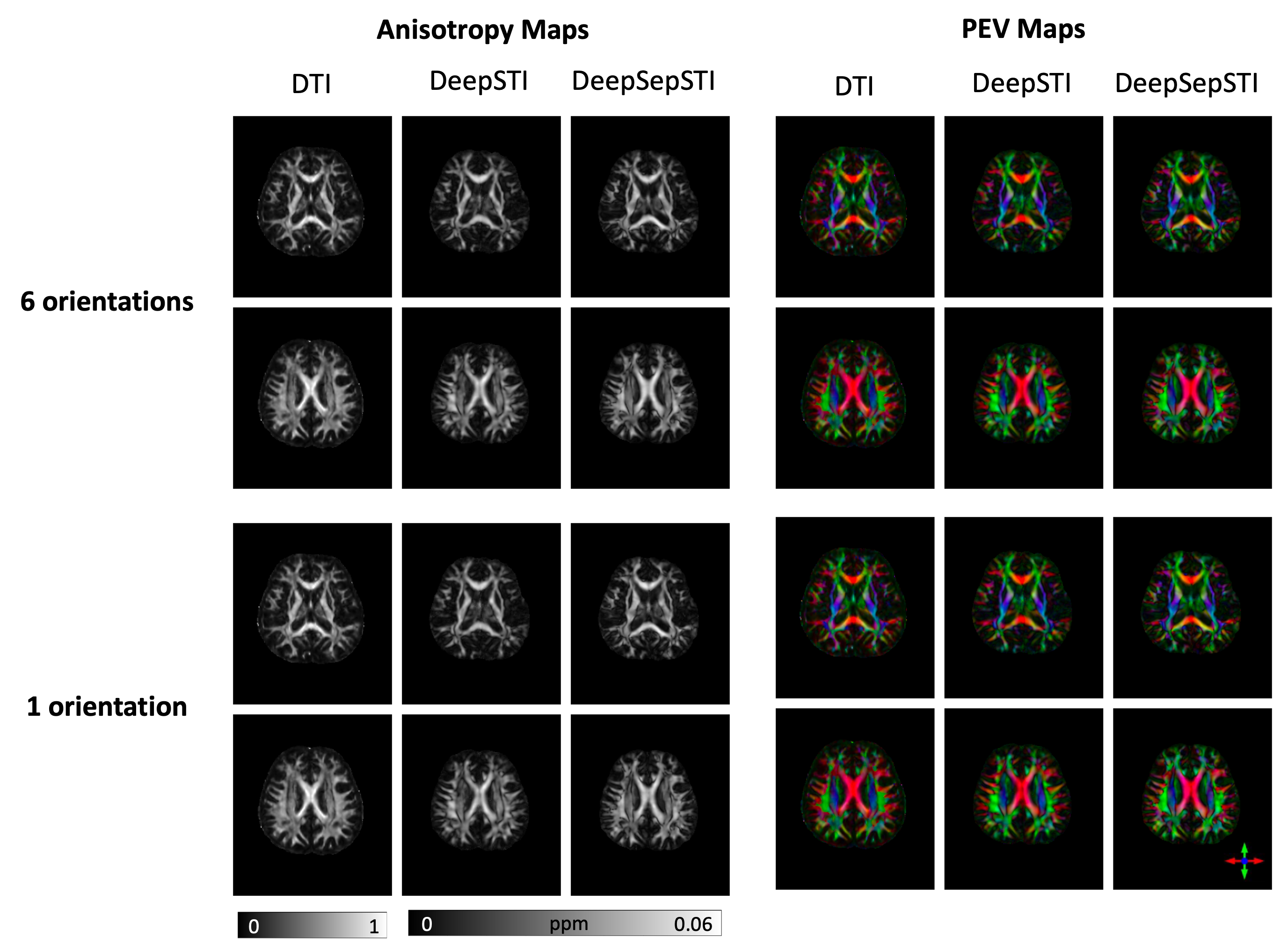
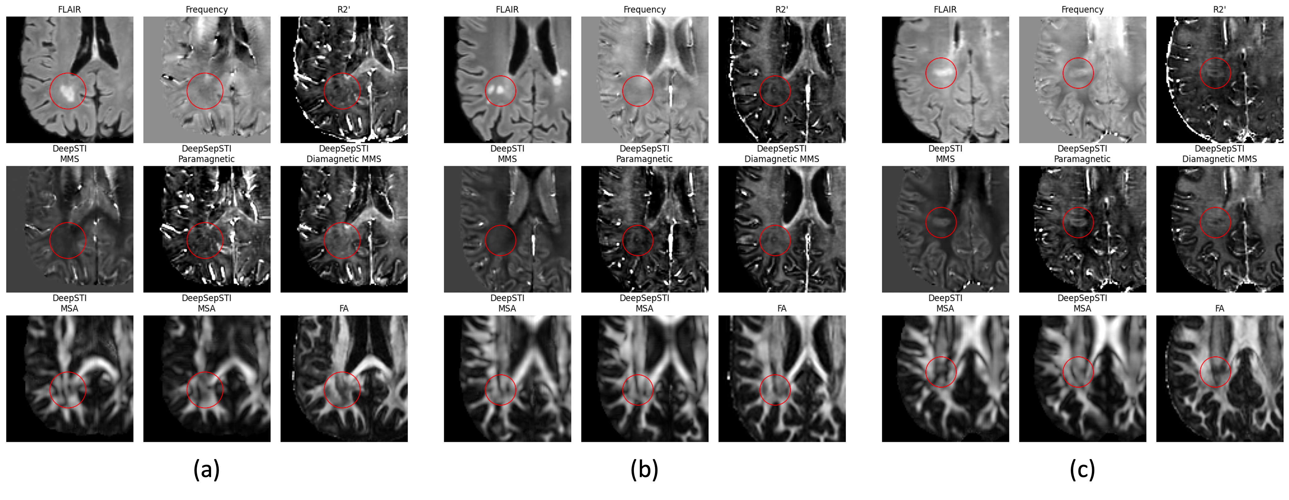
Table 1 shows quantitative metrics for STI estimation on simulation data. By separating paramagnetic and diamagnetic sources, DeepSepSTI outperformed DeepSTI in multiple metrics including SSIM, ECSE and wPSNR. Table 2 shows quantitative metrics for STI PEV estimation on in vivo data, where DeepSepSTI gave lower error than DeepSTI when using 6 and 3 orientations, using DTI PEV as a reference. Table 3 shows the quantitative metrics for magnetic susceptibility anisotropy (MSA) estimation on in vivo data, where DeepSepSTI yielded higher R-squared coefficients and Pearson’s correlation with respect to DTI FA, than DeepSTI. Fig. 4 shows visual results for MSA and PEV maps on in vivo data acquired from a normal subject. Finally, Fig. 5 shows the improved characterization of MS lesions of different types achieved by DeepSepSTI in comparison to DeepSTI.Conclusion
We proposed DeepSepSTI, a method for anisotropic susceptibility source separation. By separating the paramagnetic and diamagnetic sources, DeepSepSTI achieves superior results for estimating susceptibility tensors, anisotropy, and PEV. Promising results from DeepSepSTI in charactering MS lesions were also observed suggesting its potential clinical applications.Acknowledgements
This research has been supported by NIH Grant P41EB031771, as well as by the Toffler Charitable Trust and by the Distinguished Graduate Student Fellows program of the KAVLI Neuroscience Discovery Institute.References
[1] Shin, Hyeong-Geol, Jingu Lee, Young Hyun Yun, Seong Ho Yoo, Jinhee Jang, Se-Hong Oh, Yoonho Nam, et al. 2021. “χ-Separation: Magnetic Susceptibility Source Separation toward Iron and Myelin Mapping in the Brain.” NeuroImage 240 (October): 118371.
[2] Liu, Chunlei. 2010. “Susceptibility Tensor Imaging.” Magnetic Resonance in Medicine: Official Journal of the Society of Magnetic Resonance in Medicine / Society of Magnetic Resonance in Medicine 63 (6): 1471–77.
[3] Li, Wei, Chunlei Liu, Timothy Q. Duong, Peter C. M. van Zijl, and Xu Li. 2017. “Susceptibility Tensor Imaging (STI) of the Brain.” NMR in Biomedicine 30 (4).
[4] Fang, Zhenghan, Kuo-Wei Lai, Peter van Zijl, Xu Li, and Jeremias Sulam. 2023. “DeepSTI: Towards Tensor Reconstruction Using Fewer Orientations in Susceptibility Tensor Imaging.” Medical Image Analysis 87 (July): 102829.
[5] Ronneberger, Olaf, Philipp Fischer, and Thomas Brox. "U-net: Convolutional networks for biomedical image segmentation." Medical Image Computing and Computer-Assisted Intervention–MICCAI 2015: 18th International Conference, Munich, Germany, October 5-9, 2015, Proceedings, Part III 18. Springer International Publishing, 2015.
[6] Fang, Zhenghan, Hyeong-Geol Shin, Peter van Zijl, Xu Li, and Jeremias Sulam. "WaveSep: A Flexible Wavelet-Based Approach for Source Separation in Susceptibility Imaging." In International Workshop on Machine Learning in Clinical Neuroimaging, pp. 56-66. Cham: Springer Nature Switzerland, 2023.
[7] Tournier, J-Donald, Robert Smith, David Raffelt, Rami Tabbara, Thijs Dhollander, Maximilian Pietsch, Daan Christiaens, Ben Jeurissen, Chun-Hung Yeh, and Alan Connelly. "MRtrix3: A fast, flexible and open software framework for medical image processing and visualisation." Neuroimage 202 (2019): 116137.
[8] Veraart, Jelle, Dmitry S. Novikov, Daan Christiaens, Benjamin Ades-Aron, Jan Sijbers, and Els Fieremans. "Denoising of diffusion MRI using random matrix theory." Neuroimage 142 (2016): 394-406.
[9] Veraart, Jelle, Els Fieremans, and Dmitry S. Novikov. "Diffusion MRI noise mapping using random matrix theory." Magnetic resonance in medicine 76, no. 5 (2016): 1582-1593.
[10] Smith, Stephen M., Mark Jenkinson, Mark W. Woolrich, Christian F. Beckmann, Timothy EJ Behrens, Heidi Johansen-Berg, Peter R. Bannister et al. "Advances in functional and structural MR image analysis and implementation as FSL." Neuroimage 23 (2004): S208-S219.
[11] Andersson, Jesper LR, Stefan Skare, and John Ashburner. "How to correct susceptibility distortions in spin-echo echo-planar images: application to diffusion tensor imaging." Neuroimage 20, no. 2 (2003): 870-888.
Figures