2807
Model based rEconstruction by Deep Algorithm unrolLing (MEDAL) for fast 3D whole-heart T2 mapping1MILLENNIUM INSTITUTE FOR INTELLIGENT HEALTHCARE ENGINEERING, Santiago, Chile, 2School of Engineering, Pontificia Universidad Católica de Chile, Santiago, Chile, 3School of Biomedical Engineering, King’s College London, London, United Kingdom, 4Institute for Biological and Medical Engineering, Pontificia Universidad Católica de Chile, Santiago, Chile
Synopsis
Keywords: Machine Learning/Artificial Intelligence, Machine Learning/Artificial Intelligence, model-based
Motivation: T2 mapping provides quantitative myocardial tissue characterization. However, current approaches acquire several 2D contrast images which are then fitted to a model to estimate the T2 values, leading to limited coverage, and long acquisition and reconstruction times.
Goal(s): Here we propose to speed up 3D whole-heart T2 mapping using a model-based deep learning unrolling network (MEDAL) that leverages the power of machine learning and physical knowledge.
Approach: MEDAL reconstructs the T2 maps directly without reconstructing any intermediate contrast weighted images or fitting.
Results: The proposed approach was evaluated in iNAV-based free-breathing 3D T2 mapping 4x accelerated showing promising results.
Impact: A novel method for reconstructing parametric maps using a model-based deep learning unrolling network is presented. The method was demonstrated in a highly accelerated free breathing 3D whole-heart T2 mapping sequence allowing for fast and accurate T2 measurements.
Introduction
Parametric maps are an important tool for diagnosing a number of diseases, including cardiac applications1. Parametric mapping requires acquisition and reconstruction of different contrast images and is typically performed in a two-step process. First, the acquired data is reconstructed to image space using a reconstruction algorithm, such as parallel imaging or compress sensing. Second, the images are fitted to the signal model (typically using a mono exponential function or dictionary matching) to estimate the corresponding relaxometry parameters. The main disadvantage of these approaches is that they require long reconstruction times, and the acceleration is limited by the performance of available reconstruction methods.More recently, model-based reconstruction approaches have been proposed to estimate the parameters directly from the acquired k-space data in a single step2. However, despite promising results, these methods have not gained widespread adoption because of the difficulty of solving the associated non-linear problem efficiently. Here we propose to speed up a free-breathing image-navigator (iNAV)-based 3D whole-heart T2 mapping3 using a Model-based rEconstruction by Deep Algorithm unrolLing (MEDAL) that leverages the power of machine learning and physical knowledge. The proposed approach was evaluated on healthy subjects.
Methods
Model Based ReconstructionModel based reconstruction is achieved by solving the following non-linear optimization:
$$\min_\nu || Eq(\nu) - s ||^2_2$$
Where $$$E$$$ is the sampling operator, $$$q$$$ is the signal equation, $$$\nu$$$ the relaxometry parameters (e.g. $$$T_1, T_2, M_0$$$) and $$$s$$$ the acquired k-space data. To stabilize the solution of this problem, a regularization is typically added4 (e.g. TV or Wavelets).
Proposed algorithm
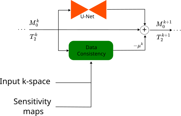
MEDAL leverages the power and inference speed of deep learning with the physical equations of model-based MRI reconstruction (Fig.1). This is achieved by unrolling a regularized gradient descent algorithm similar to variational networks5. The algorithm is given by:
$$\nu^{k+1} = \nu^k - \mu^k \frac{\partial Eq(\nu^k)}{\partial \nu}(Eq(\nu) - s) + f_{\theta^k}(\nu^k)$$
This equates to doing gradient descent with step size $$$\mu^k$$$ on a regularized version of the problem were the network outputs the gradient of the regularizer. The network architecture is a U-net and a different set of weights is used for each iteration. The step size $$$\mu^k$$$ is also learned. A single network with multiple channels is used to regularize all parametric maps simultaneously. Here 5 iterations are used for finding the final solution. MEDAL differs from the method proposed by ref6 in two ways: 1) zero-fill images are used to find the starting maps for optimization while ref6 use a separate network and 2) for regularization ref6 use a different network for each map whereas MEDAL uses a single network for all maps. Coil sensitivities were estimated using ESPIRiT7. The output from the last module are the predicted maps and are compared with the gold standard map using the SSIM loss. As an additional term, we penalize the SSIM difference between the images generated using the predicted maps and the signal model, and the gold standard reconstruction.
Experiments and Results
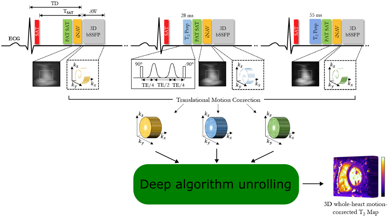
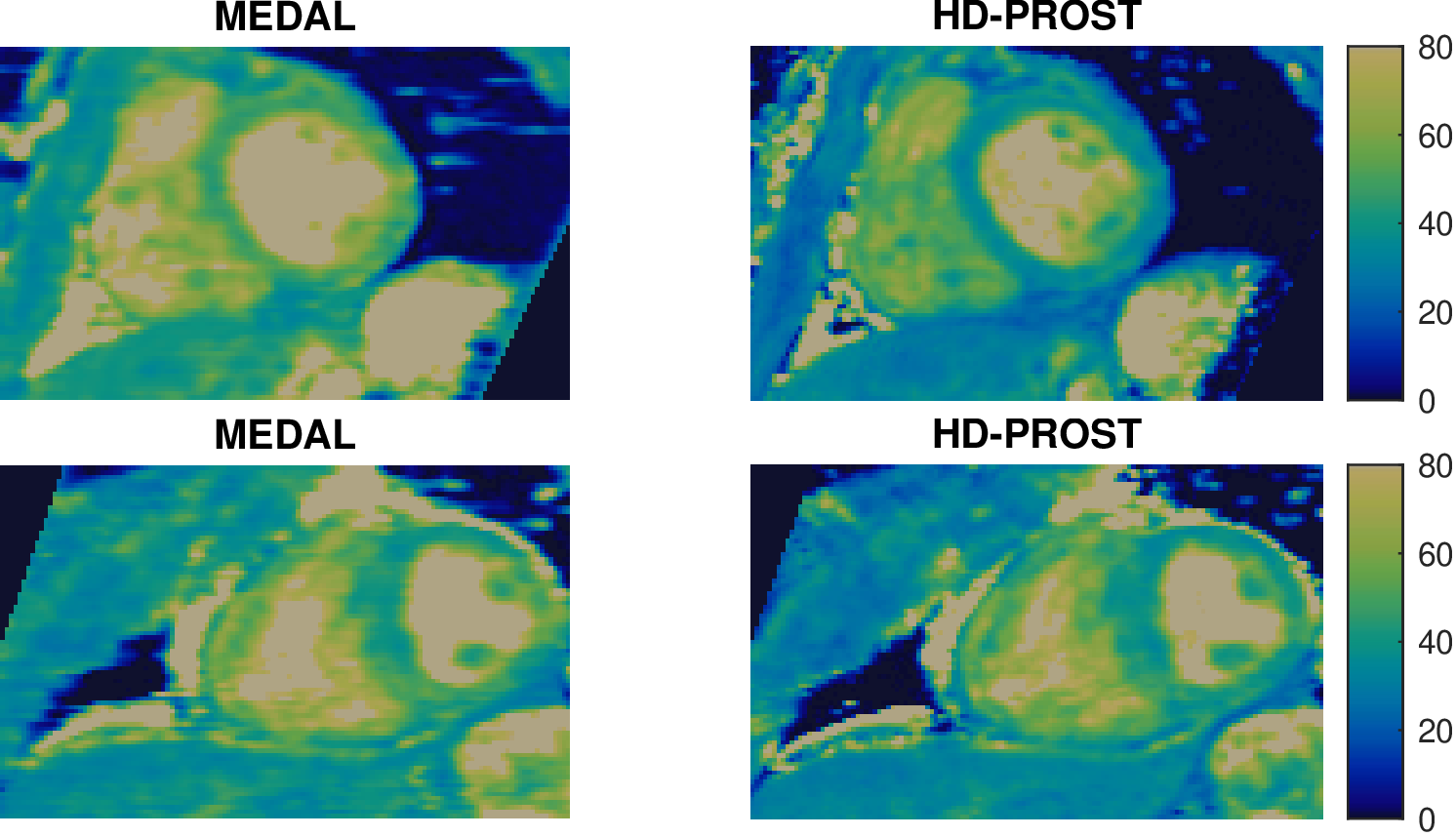
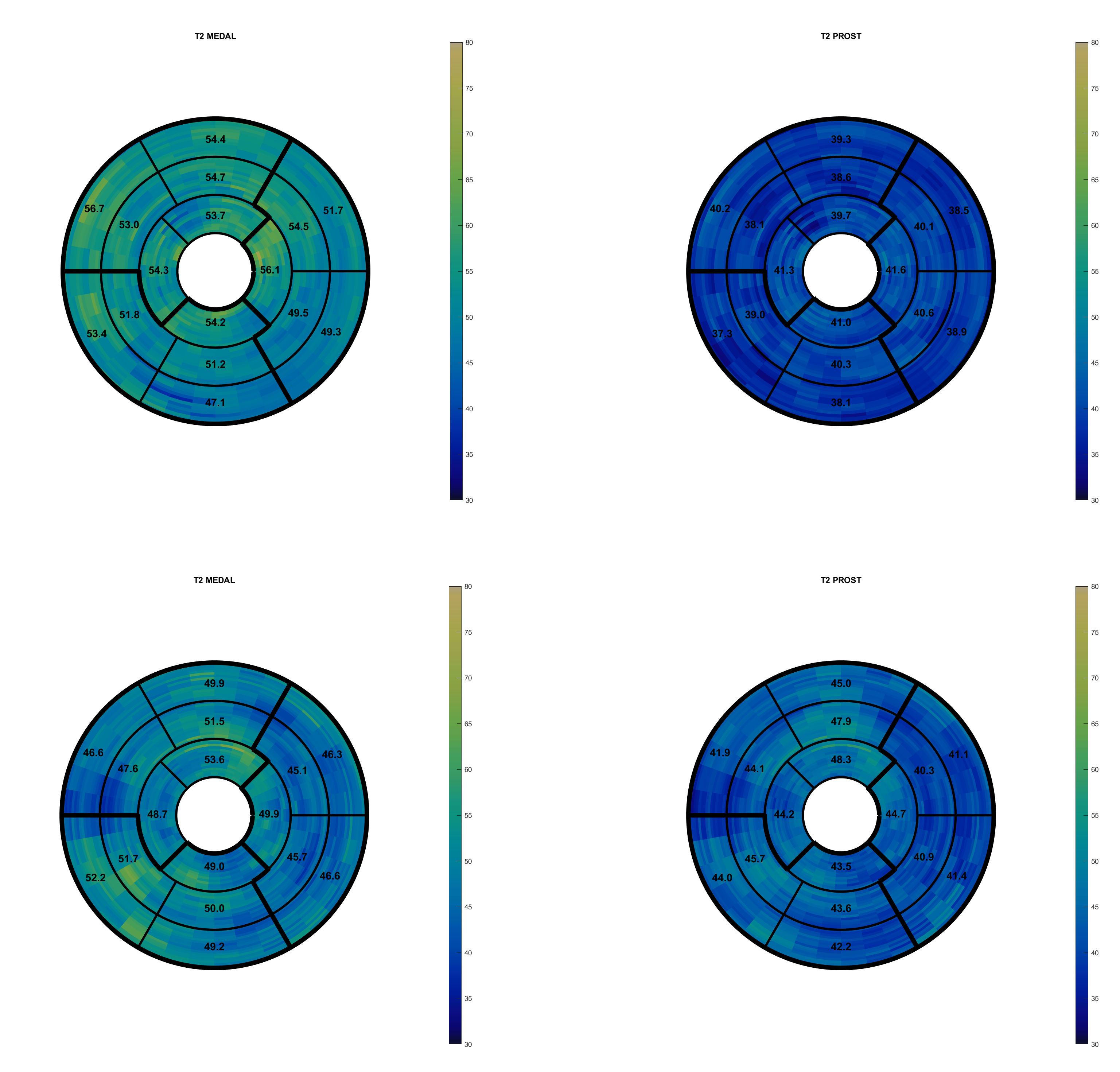
The proposed approach was applied to speed up free-breathing iNAV-based 3D whole-heart T2 mapping. The sequence3 (Fig.2) includes three T2 preparation pulses (T2prep 0, 28, 55ms), a 3D Cartesian variable density with spiral profile order8 with 4x undersampling and iNAVs to correct for beat-to-beat translational motion. To obtaining a gold standard reconstruction, images were reconstructed using HD-PROST9 and an exponential fitting was performed to find $$$T_2$$$ and $$$M_0$$$. The DFT on the read-out direction is performed, and the network is trained on 2D slices. The k-space was zero padded to bring all images to the same matrix size and a Hamming window was applied to reduce ringing artifacts. The proposed approach was trained and evaluated on 23 acquisitions on healthy subjects and patients. Sixteen volumes were used for training, 4 for validation and 3 for testing. To normalize the inputs, the k-space is divided by the maximum of the zero fill for the first T2prep image.Results from 2 subjects (testing set) are shown in Fig. 3 for the proposed MEDAL method in comparison to gold standard HD-PROST. Similar visual map quality is observed between both methods. T2 bullseye plots reconstructed with the proposed method (MEDAL) and the gold standard are shown in Figure 4. Inference reconstruction times were 8s and 5min for MEDAL and HD-PROST.
Conclusions
A novel method for reconstructing parametric 3D whole-heart T2 maps using a model-based deep learning unrolling network is presented. The method enables acquisition in 6min with a fast reconstruction of 8s, achieving similar map quality than the gold standard. Evaluation in a larger cohort of subjects and extension to no-rigid motion correction will be investigated as future work.Acknowledgements
The authors acknowledge financial support from: (1) BHF RG/20/1/34802 (2) EPSRC EP/V044087/1 (3) ANID Millennium Institute iHEALTH, ICN2021_004; Fondecyt 1210637 and 1210638References
1. Ferreira, Vanessa M., and Stefan K. Piechnik. 2020. “CMR Parametric Mapping as a Tool for Myocardial Tissue Characterization.” Korean Circulation Journal 50 (8): 658–76. https://doi.org/10.4070/kcj.2020.0157.
2. Kolbitsch, Christoph, Kirsten Kerkering, and Tobias Schaeffter. 2022. “Chapter 15 - Model-Based Parametric Mapping Reconstruction.” In Advances in Magnetic Resonance Technology and Applications, edited by Mehmet Akçakaya, Mariya Doneva, and Claudia Prieto, 7:419–39. Magnetic Resonance Image Reconstruction. Academic Press. https://doi.org/10.1016/B978-0-12-822726-8.00026-9.
3. Bustin, Aurélien, Giorgia Milotta, Tevfik F. Ismail, Radhouene Neji, René M. Botnar, and Claudia Prieto. 2020. “Accelerated Free-Breathing Whole-Heart 3D T2 Mapping with High Isotropic Resolution.” Magnetic Resonance in Medicine 83 (3): 988–1002. https://doi.org/10.1002/mrm.27989.
4. Wang, Xiaoqing, Volkert Roeloffs, Jakob Klosowski, Zhengguo Tan, Dirk Voit, Martin Uecker, and Jens Frahm. 2018. “Model-Based T1 Mapping with Sparsity Constraints Using Single-Shot Inversion-Recovery Radial FLASH.” Magnetic Resonance in Medicine 79 (2): 730–40. https://doi.org/10.1002/mrm.26726.
5. Hammernik, Kerstin, Teresa Klatzer, Erich Kobler, Michael P. Recht, Daniel K. Sodickson, Thomas Pock, and Florian Knoll. 2018. “Learning a Variational Network for Reconstruction of Accelerated MRI Data.” Magnetic Resonance in Medicine 79 (6): 3055–71. https://doi.org/10.1002/mrm.26977.
6. Jun, Yohan, Hyungseob Shin, Taejoon Eo, Taeseong Kim, and Dosik Hwang. 2021. “Deep Model-Based Magnetic Resonance Parameter Mapping Network (DOPAMINE) for Fast T1 Mapping Using Variable Flip Angle Method.” Medical Image Analysis 70 (May): 102017. https://doi.org/10.1016/j.media.2021.102017.
7. Uecker, Martin, Peng Lai, Mark J. Murphy, Patrick Virtue, Michael Elad, John M. Pauly, Shreyas S. Vasanawala, and Michael Lustig. 2014. “ESPIRiT — An Eigenvalue Approach to Autocalibrating Parallel MRI: Where SENSE Meets GRAPPA.” Magnetic Resonance in Medicine : Official Journal of the Society of Magnetic Resonance in Medicine / Society of Magnetic Resonance in Medicine 71 (3): 990–1001. https://doi.org/10.1002/mrm.24751.
8. Prieto, Claudia, Mariya Doneva, Muhammad Usman, Markus Henningsson, Gerald Greil, Tobias Schaeffter, and Rene M. Botnar. 2015. “Highly Efficient Respiratory Motion Compensated Free-Breathing Coronary Mra Using Golden-Step Cartesian Acquisition.” Journal of Magnetic Resonance Imaging 41 (3): 738–46. https://doi.org/10.1002/jmri.24602.
9. Bustin, Aurélien, Gastão Lima da Cruz, Olivier Jaubert, Karina Lopez, René M. Botnar, and Claudia Prieto. 2019. “High-Dimensionality Undersampled Patch-Based Reconstruction (HD-PROST) for Accelerated Multi-Contrast MRI.” Magnetic Resonance in Medicine 81 (6): 3705–19. https://doi.org/10.1002/mrm.27694.
Figures