2803
High-Resolution Deep Learning Reconstruction (HR-DLR) to Improve Sharpness in Diffusion Weighted Imaging1MRI Systems Development Department, Canon Medical Systems Corporation, Tochigi, Japan
Synopsis
Keywords: Machine Learning/Artificial Intelligence, Machine Learning/Artificial Intelligence, Super Resolution
Motivation: Echo planar diffusion weighted imaging (EPI-DWI) often suffers from Gibbs ringing artifact and/or image blurring, because of limited matrix size. A recently proposed High-Resolution Deep Learning Reconstruction (HR-DLR) may bring a breakthrough to the limitation.
Goal(s): Our goal was to test benefits of HR-DLR when applied to brain EPI-DWI.
Approach: HR-DLR was compared to conventional reconstruction method (zero-filling interpolation[ZIP] and low-pass filtering) with regards to image sharpness and ringing artifact suppression, with a conventional and an accelerated scan conditions.
Results: The advantage of HR-DLR over the conventional method was confirmed by measurements of edge slope width (ESW) and ringing variable mean (RVM).
Impact: A recently proposed High-Resolution Deep Learning Reconstruction successfully improved the sharpness of single shot EPI-DWI while suppressing Gibbs artifacts. The method could help improve clinical confidence by increasing image resolution and gain examination throughput by shortening acquisition time.
Introduction
Single-shot echo planar diffusion weighted imaging sequence (EPI-DWI) is often acquired with small matrix size to shorten the scan time and/or EPI readout duration. The truncated matrix aggravates Gibbs ringing artifact. Typically, low pass filters in k-space and/or image space are used to reduce the ringing artifact, at the expense of image blurring. In a previous study[1, 2, 3], a high-resolution deep learning reconstruction (HR-DLR) was proposed, combining ZIP with two CNN’s: the first one trained to reduce image noise, the second one to reduce Gibbs artifacts. The HR-DLR was evaluated on knee images using 2D Fast Spin Echo sequence[1, 2] and brain with EPI-DWI[3]. Those studies showed better SNR and sharpness with reduced Gibbs artifacts versus standard reconstructions used in routine clinical practice. The goal of this work was to test that HR-DLR method enables high-resolution imaging or shorted acquisition time preserving structural detail and reducing Gibbs artifact on brain EPI-DWI.Method
The reconstruction pipeline of the proposed method is illustrated in Figure 1, same as the previous studies[1, 2, 3]. Although the two neural networks were developed separately, they shared the same ground-truth dataset for training. This ground-truth dataset was collected with higher SNR and resolution than typical clinical images, by averaging repeated scans. The acquisitions included multiple body parts (brain, spine, shoulder, elbow, wrist, hip, knee, and ankle), contrast weightings (DWI, T1w, T2w, FLAIR, T2*w, PDw, etc.), and field strengths (3T and 1.5T). HR-DLR was tested on b0 and isotropic diffusion images(isoDWI) with clinical standard protocol and fast protocol.The brain of a healthy volunteer was scanned at 1.5T (Vantage Orian XGO, Canon Medical Systems Corporation, Tochigi, Japan) with a 16ch head coil. The study was approved by our institutional review board and informed consent was obtained. The relevant parameters of the standard protocol are TR/TE=5700/75[ms], echo train spacing (ETS)=0.9[ms], Bandwidth=1302[Hz], b-value=0 and 1000 in 3-axis, acquisition matrix=160, and number of acquisition (NAQ)=3, while those of the fast protocol are TR/TE=4603/75[ms], ETS=0.7[ms], Bandwidth=1953[Hz], b-value=0 and 1000 in 3-axis, acquisition matrix =128, and NAQ=1. The fast protocol scan time is 42[sec], while the standard protocol is 92[sec].
Three metrics were measured:
1. Edge Slope Width(ESW)[4] in line ROIs: $$(SI_{max}–SI_{min})/Slope,$$ where $$$Slope$$$ is a rate of change of signal intensity near the center of edge, and $$$SI_{max}$$$ and $$$SI_{max}$$$ are the average signal intensity of three neighboring points in the plateaus before and after the edge. Four ROIs were drawn on edge of lateral ventricle. Basically, a smaller ESW means the edge is sharp, i.e., it suddenly rises or falls.
2. Ringing Variable Mean(RVM)[5] in line ROIs which are the mean absolute value of differential of the profile curves: $$\frac{1}{N-1}\sum_{i=1}^{N-1}\left|\frac{SI[i+1]-SI[i]}{\Delta{x}}\right|,$$ where $$$SI[i]$$$ is the signal intensity at the $$$i$$$-th point. Four ROIs were drawn on regions demonstrating Gibbs artifacts in images without filtering (NONE). Generally, a smaller RVM means less ringing artifacts.
3. Signal-to-Noise (SNR) in circle ROIs which are the mean signal intensity divided by the standard deviation:
Two ROIs were drawn in the thalamus.
Test#1. HR-DLR for High Matrix Size:
The three metrics are measured on the standard protocol and compared between ZIP method using an upscaling factor (in both in-plane directions) of 2 without any filter (NONEx2), ZIPx2 with a low pass filter (LPFx2), and HR-DLR with upscaling factor of 3 (HR-DLRx3).
Test#2. HR-DLR for Short Time Acquisition:
The three metrics are measured and compared between LPFx2 of the standard protocol(Standard LPFx2), NONEx2 of the fast protocol(Fast NONEx2) and HR-DLRx3 of the fast protocol(Fast HR-DLRx3).
Results
The resulting images from the retrospective test are shown in Figure 4 and 5. In the Test #1, HR-DLRx3 showed better sharpness(smaller ESW) and SNR with reducing Gibbs artifact(smaller RVM) compared to both conventional methods of LPFx2 and NONEx2.In the Test #2, Fast HR-DLRx3 achieved better or equivalent image quality to Standard LPFx2. Fast HR-DLRx3 reduced Gibbs artifact as well as LPFx2 compared to Fast NONEx2 in terms of the RVM and image appearance. Fast HR-DLR showed similar sharpness with LPFx2 in terms of both ESW and appearance. The Fast HR-DLR achieved better SNR compared to both LPFx2 and Fast NONEx2.Conclusion
This work shows that HR-DLR improves sharpness of EPI-DWI with less or similar Gibbs ringing artifacts compared to conventional ZIP with Low pass filtered images via the metrics of ESW and RVM. It also shows HR-DLR enables shortenerd acquisition times by lowering matrix size and NAQ, while upsampling allows reconstruction of high-resolution images, preserving structural detail and removing the noise.Acknowledgements
No acknowledgement found.References
1. Kutsuna H, et al. High Resolution MR Reconstruction with Functionally Separate Neural Networks. Proc. ISMRM 2023 p.2292
2. Prevost V, et al. Deep learning-based pipeline to improve sharpness in knee imaging at both 1.5T and 3T: a clinical evaluation. Proc. ISMRM 2023 p.4923
3. Matsuo K, et al. Feasibility study of super-resolution deep learning-based reconstruction using k-space data in brain diffusion-weighted images. Neuroradiology 65, 1619–1629 (2023). doi:10.1007/s00234-023-03212-y
4. Michael JMF, et al. Measurement of meningeal blood vessel diameter in vivo with a plug-in for ImageJ, doi:10.1016/j.mvr.2010.04.004
5. Block KT, et al. Suppression of MRI Truncation Artifacts Using Total Variation Constrained Data Extrapolation. International Journal of Biomedical Imaging, Volume 2008, Article ID 184123, 8 pages. doi:10.1155/2008/184123
Figures
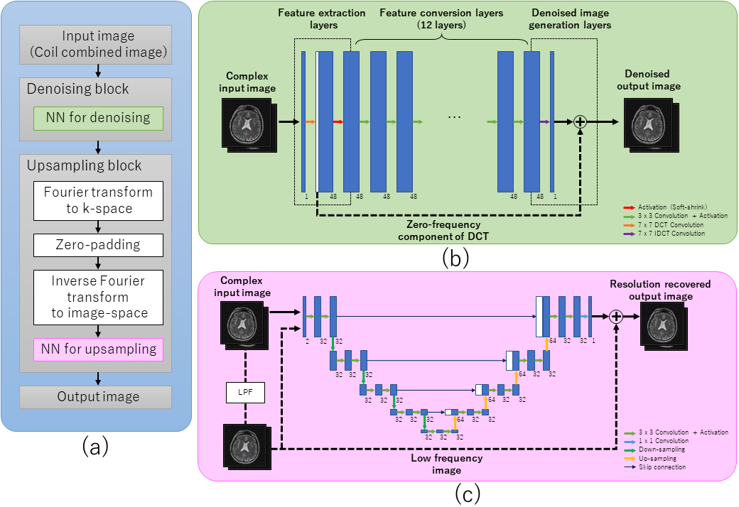
Figure 1 Reconstruction pipeline of HR-DLR
(a) Overview of the reconstruction pipeline
(b) Architecture of the neural network for denoising
(c) Architecture of the neural network for upsampling.
HR-DLR pipeline combined a denoising CNN, a zero padding process compatible with a factor 2 and 3, and a second CNN to reduce Gibbs ringing artifacts.[1]
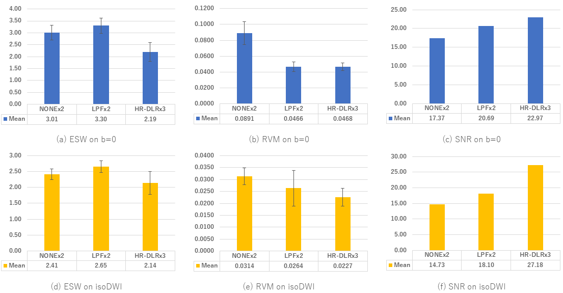
Figure 2 Result metrics of Test #1: HR-DLR utilized for High Matrix Size ( Comparison between NONEx2, LPFx2, and HR-DLRx3 )
All reconstructions are performed on the standard protocol ( acquisition matrix=160). HR-DLRx3 (reconstructed matrix size=480) showed similar sharpness with Standard LPFx2(reconstructed matrix size=320) in term of ESW. HR-DLRx3 could reduce Gibbs artifacts as well as Standard LPFx2 compared to Fast NONEx2(reconstructed matrix size=320) in term of RVM. The HR-DLR achieved better SNR compared to both LPFx2 and NONEx2.
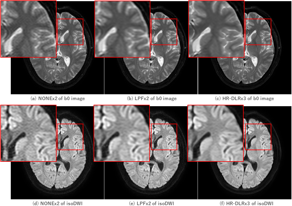
Figure 3 Result images of Test #1: HR-DLR utilized for High Matrix Size ( Comparison between NONEx2, LPFx2, and HR-DLRx3 )
HR-DLRx3 (reconstructed matrix size=480) showed better sharpness and SNR with reducing Gibbs ringing compared to both LPFx2(reconstructed matrix size=320) and NONEx2(reconstructed matrix size=320) on the standard protocol (acquire matrix=160).
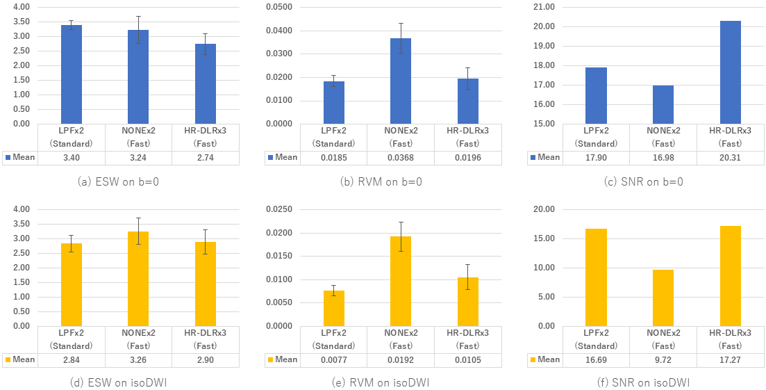
Figure 4 Result metrics of Test #2: HR-DLR utilized for Short Time Acquisition
Fast HR-DLRx3 with the fast protocol (acquisition matrix=128, reconstructed matrix size=384, NAQ=1, scan time=42[s]) could achieve equivalent image quality to LPFx2 with the standard protocol (acquire matrix=160, reconstructed matrix size=320, NAQ=3, scan time=92[s]). Fast HR-DLRx3 could reduce Gibbs ringing as well as LPFx2 compared to Fast NONEx2 in terms of RVM, while showing similar sharpness with LPFx2 in terms of ESW, achieving better SNR compared to both LPFx2 and Fast NONEx2.
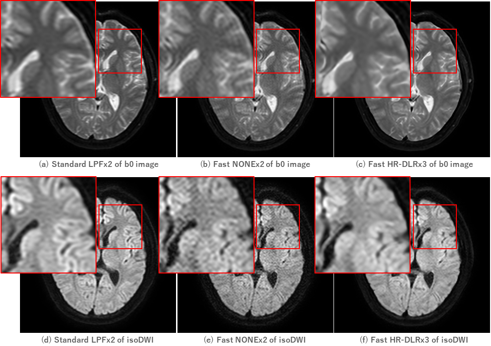
Figure 5. Resulting images of Test #2: HR-DLR for Short Time Acquisition
Fast HR-DLRx3 with the fast protocol (acquisition matrix=128, reconstructed matrix size=384, NAQ=1, scan time=42[s]) could achieve equivalent image quality to Standard LPF with ZIPx2 with the standard protocol (acquire matrix=160, reconstructed matrix size=320, NAQ=3, scan time=92[s]). It could be observed that Fast HR-DLRx3 could reduce Gibbs artifacts and image noise as well as Standard LPFx2 compared to Fast NONEx2 . Fast HR-DLRx3 showed similar sharpness with Standard LPFx2 in image appearance.