2792
Real-time automated assessment of image quality during MRI scanning for optimal image reconstruction1Medicine, UC San Diego, La Jolla, CA, United States, 2Computer Science, UC San Diego, La Jolla, CA, United States, 3Radiology, UC San Diego, La Jolla, CA, United States, 4Bioengineering, UC San Diego, La Jolla, CA, United States, 5Cardiology, Rady Children's Hospital, San Diego, CA, United States
Synopsis
Keywords: Machine Learning/Artificial Intelligence, Machine Learning/Artificial Intelligence
Motivation: MRI scans are often sensitive to subject motion, impacting image quality. One challenge is that it is difficult to detect and mitigate motion-related issues until after the scan has completed.
Goal(s): To create a quantitative method for real-time evaluation of MRI scans, identifying events that may corrupt images and enabling prompt decision-making.
Approach: The study used simulated data from the ACDC dataset, training a ResNET18 neural network to predict image quality using SSIM scores.
Results: Our method can quickly and accurately assess MRI image quality. This could aid motion event detection. However, validation on actual data is needed.
Impact: This study introduces an automated, deep-learning based method for real-time assessment of motion-related image quality for cardiac MRI. This innovation can potentially enhance the reliability and efficiency of MRI scans.
Introduction
Magnetic resonance imaging is often sensitive to subject motion due to the fact that data acquisition time typically exceeds the timescales of many anatomical phenomena. Both voluntary and unvoluntary movements by the subject, such as breathing, heart beating, and even sudden movements such as adjusting positions inside the machine can occur and will impact image quality if not detected and taken into account (1,2). Even if the motion occurs at a brief moment in time during an MRI scan, the change can have a significant impact on the resulting image quality.Therefore, we propose to develop an automated and quantitative method to judge the consistency and quality of acquired k-space data as it is collected. The goal is to assess the progression of the scan and detect any image corrupting events. In this simulation study, we focus on cardiac scans where scans are typically acquired with ECG-gating and obtained data is collected during a breath-hold or via a respiratory navigator. As a result, data for a particular image is collected at a specific point in the cardiac cycle and it is assumed that the heart is in the same physical location.
Dataset and Methods
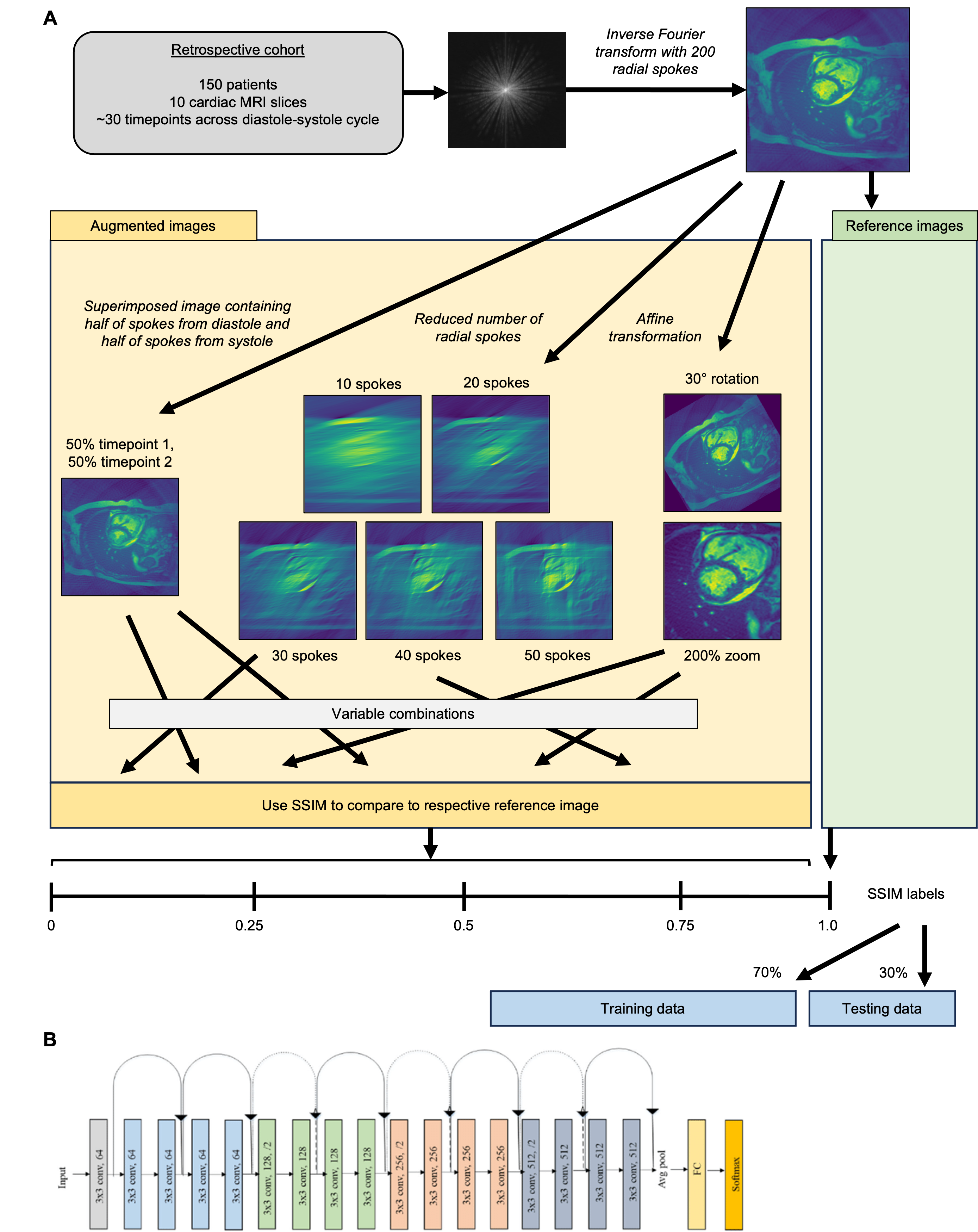
The open-source Automated Cardiac Diagnosis Challenge (ACDC) dataset which consists of cine MR images obtained in 150 patients imaged at approximately ten different slice locations, acquired during breath-holds (3). Golden angle radial k-space spokes were simulated from the cine images. A reference image was generated using 200 radial spokes collected from a diastolic cardiac phase image and inverse Fourier-transformed using the non-uniform FFT (Figure 1A). This reference image then underwent three main alterations to produce a series of images intended to simulate MR imaging phenomena we aim to assess: (1) incorrect ECG gating: in this case, 50% of the k-space lines were collected from the diastolic frame and 50% were collected from the systolic time frame, (2) accumulation of data: images were reconstructed with fewer spoke (10, 20, 30, 40, and 50 spokes), and (3) gross cardiac motion: inability to perform a breath-hold was simulated as one of two affine transformations, a 30° rotation or a 200% zoom.For each image, the specific alteration was computed randomly and then comparison to the reference image (via structural similarity index metric, SSIM) was used to assign a score between 0 and 1. All reference images defaulted with a 1.0 value. 40,000 random images were generated, 70% (28,000) of which were used for training and 30% (12,000) of which were used for testing.
A ResNET18 architecture was trained using PyTorch for 100 epochs with an Adam optimizer and a StepLR scheduler (learning rates = 3e-4, 1e-6). The network was trained as a regression using MSE loss on the predicted SSIM score.
Results
Representative examples of the SSIM created by our manipulations to introduce data-corrupting scenarios are shown in Figure 2A. The predicted SSIM by our neural network are shown below each image.In the testing cohort, the mean MSE was very low (1.533e-5) with low standard deviation (1.051e-5) which suggests strong prediction of SSIM.
To further assess the agreement between SSIM and estimated scores, we assigned each image quality to bins with ranges consisting of 0-0.25, 0.25-0.5, 0.5-0.75, and 0.75-1.0. The resulting confusion matrix is shown in Figure 2B and accuracy per category was very high (>98.0% across all bins, bin 1 98.7%, bin 2: 98.1%, bin 3: 99.4%, and bin 4: 99.9%). A regression analysis determined a linear relationship between SSIM labels and estimates (estimate= 0.9996*SSIM – 0.0005) (Figure 2C).
Discussion and Conclusions
Our neural network-based approach was able to robustly quantify the image quality associated with failures in ECG gating, radial undersampling, and gross cardiac motion. Since it is an automated method and neural networks (once trained) can be calculated quickly, this approach could be used to obtain instantaneous feedback regarding the progress of an MRI acquisition. Specifically, the image quality score over time could be used to detect motion events and decide to abort or re-start the scan.There are several limitations of this study. First, our model was trained on simulated corruptions of the data but needs to be tested further in actual acquisitions and more complex motion simulations. Further, while we hypothesize this could guide decisions during the MRI, our approach has not yet been implemented inline or tested in vivo. Lastly, we trained on images reconstructed using a simple image reconstruction (NUFFT) and need to investigate the effect of different reconstruction methods.
Acknowledgements
Supported by NIH R01HL162671References
- Wang NC, Noll DC, Srinivasan A, Gagnon-Bartsch J, Kim MM, Rao A. Simulated MRI Artifacts: Testing Machine Learning Failure Modes. BME Front. 2022;2022:9807590. doi: 10.34133/2022/9807590.
- Zaitsev M, Maclaren J, Herbst M. Motion artifacts in MRI: A complex problem with many partial solutions. J Magn Reson Imaging. 2015;42(4):887–901. doi: 10.1002/jmri.24850.
- Bernard O, Lalande A, Zotti C, et al. Deep Learning Techniques for Automatic MRI Cardiac Multi-Structures Segmentation and Diagnosis: Is the Problem Solved? IEEE Trans Med Imaging. 2018;37(11):2514–2525. doi: 10.1109/TMI.2018.2837502.
- He K, Zhang X, Ren S, Sun J. Deep Residual Learning for Image Recognition. 2016 IEEE Conference on Computer Vision and Pattern Recognition (CVPR). Las Vegas, NV, USA: IEEE; 2016. p. 770–778. doi: 10.1109/CVPR.2016.90.
Figures
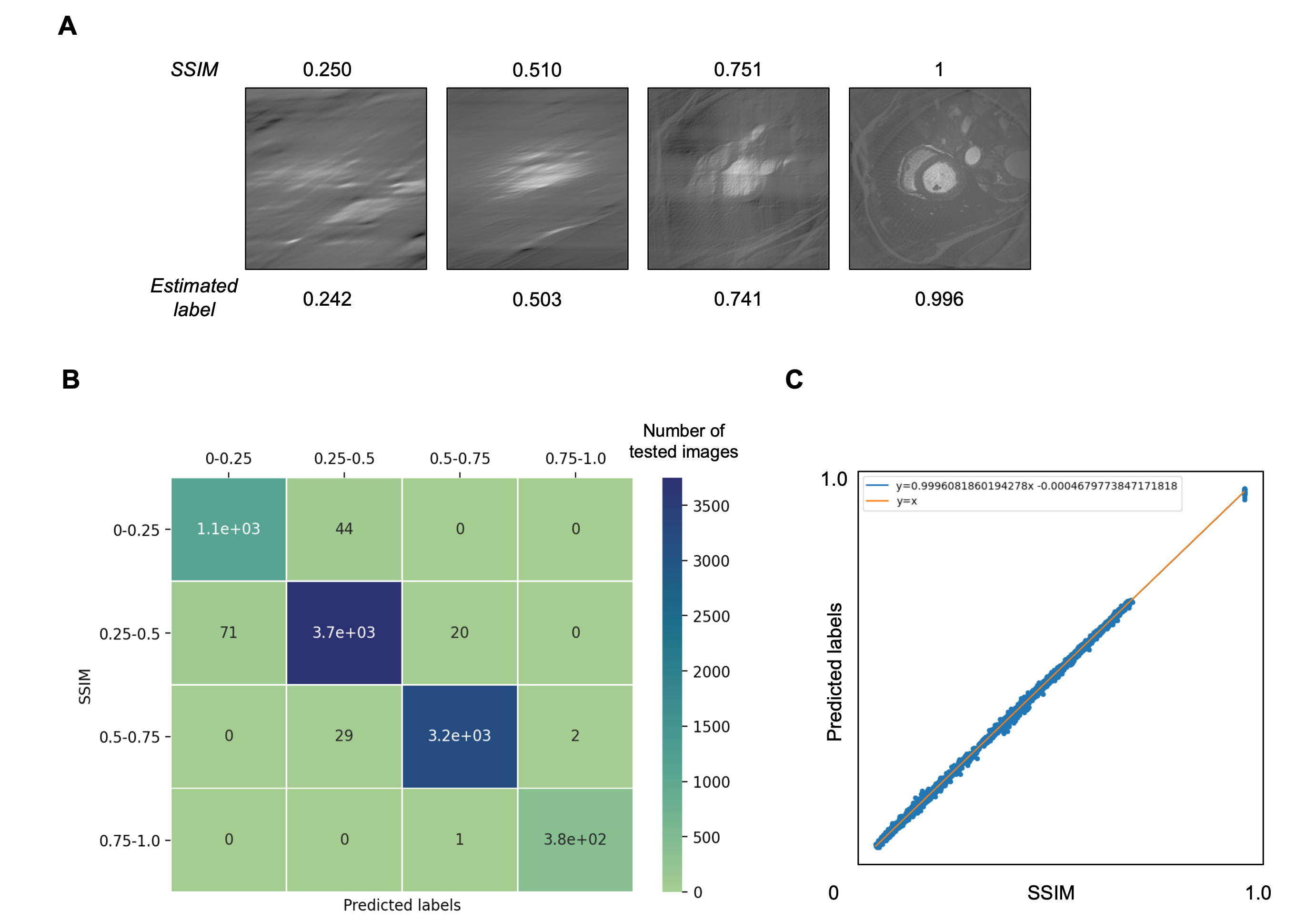
(A) Representative examples of images at different levels of SSIM with the corresponding predictions list below.
(B) Confusion matrix highlighting the strong performance of the ResNET model’s predictions versus the real labels.
(C) Correlation demonstrating strong agreement between true and predicted SSIM