2791
Rapid High-resolution Whole-brain 3D Multi-parametric and Multi-contrast MRI with Deep Learning-based Acquisition & Reconstruction1GE HealthCare, Menlo Park, CA, United States, 2GE HealthCare, Tokyo, Japan
Synopsis
Keywords: Synthetic MR, Neuro, Multi-Contrast, Data Acquisition, Machine Learning, Synthetic MR Neuro
Motivation: As the public demand for MRI grows exponentially, there is an increasing need for a one-click 3D MR exam that can generate multiple image contrasts and parametric maps as an effective way to improve patient throughput.
Goal(s): Our goal was to implement an acquisition and reconstruction method that makes high-resolution whole brain multi contrast examination possible in less than 3 minutes.
Approach: We implemented a deep learning-guided vast undersampled MR acquisition and a time efficient recon algorithm that uses a densely connected unrolled neural network.
Results: Our proposed method preserved image quality and quantitative accuracy of the multicontrast and multiparametric images.
Impact: This study demonstrates that with highly undersampled 3D QALAS acquisition combined with the DL recon algorithm, a 3-minute one-click exam is feasible that generates whole-brain high-resolution brain volumes of multiple contrasts and quantitative maps, which can enhance patient workflow in a busy clinical practice.
INTRODUCTION
As the public demand for MRI grows exponentially, there is an increasing need for a one-click 3D MR exam that can generate multiple image contrasts and parametric maps as an effective way to improve patient throughput.The QALAS sequence1,2 is one such technique that employs a T2 prep module, an inversion pulse, and a series of fast 3D SPGR readouts to capture a high-resolution image of the whole brain with 5 weightings over the inversion recovery, which then can be combined to synthesize multiple image contrasts and quantitative maps relevant for clinical diagnosis.
In this study, we implemented a highly accelerated acquisition of this sequence and coupled it with DL-based image reconstruction (DL Speed)3,4 to greatly reduce its acquisition time while retaining image quality and quantitative accuracy of the synthetic images.
METHODS
A healthy male volunteer was scanned on a Discovery MR750 3.0T scanner (GE HealthCare, Waukesha WI) with the 32-channel Nova coil (Nova Medical, Wilmington, MA). The axial images were acquired with 3D QALAS sequence using the following parameters: FOV = 24×24 x15cm3, matrix = 208x208, slice thickness = 1.2mm, TR/TE = 6.5ms/2.6ms, NEX = 1. The sequence was first run with parallel imaging acceleration (2x1) as the baseline (scan time = 8:42 min) followed by 6 additional scans with variable density sampling scheme of increased acceleration factors (see Table 1 for acceleration factors and corresponding scan times).K-space data acquired with variable density undersampling scheme were reconstructed off-line using a densely connected unrolled neural network3,4. Each unroll block includes a data consistency term and a CNN based regularizer, and the output of previous unrolls are concatenated and input to the next unroll block. We refer to this technique as DL Speed (DLS).
The reconstructed DICOMS from all scans were post processed using SyMRI® software (v23Q2, SyntheticMR, Linkoping, Sweden) to generate T1, and T2 parameter maps, as well as synthetic T1w, T2 STIR, T2 FLAIR, and PSIR images. The software was also used to extract segmented gray matter and white matter brain volumes, as well as tissue specific T1 and T2 values (mean ± STD). These metrics were compared relative to the baseline values acquired with 2x1 parallel imaging.
RESULTS
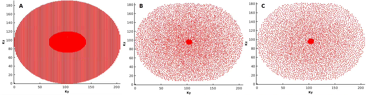
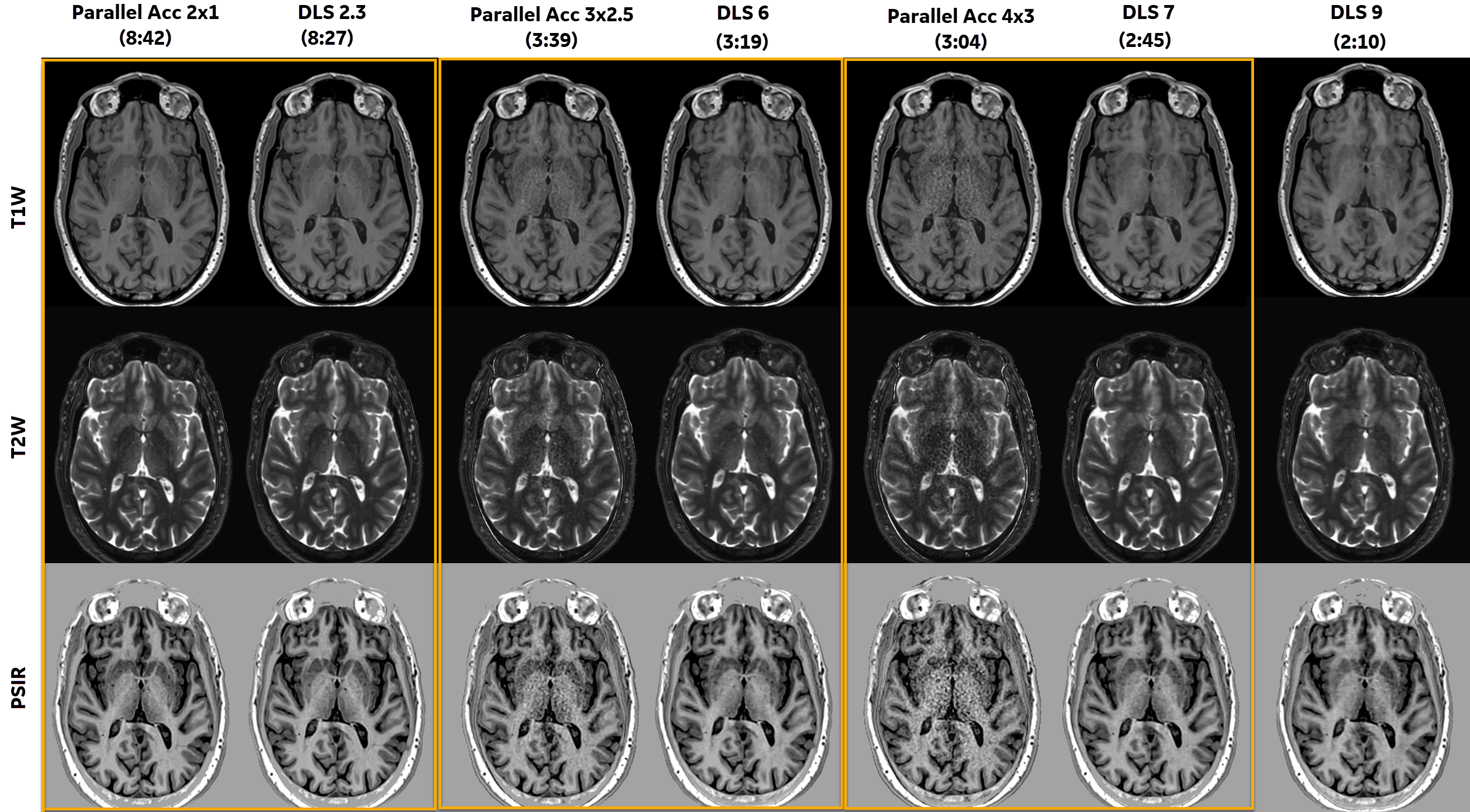
The ky-kz plane sampling patterns corresponding to 2x1 parallel imaging, and two DLS acquisitions are shown in Figure 1.Synthetic T1, T2, and PSIR images generated from the DLS acquisitions showed higher image quality (IQ) relative to their parallel imaging accelerated counterparts (yellow boxes in Fig. 2), especially in the deep brain region. The IQ difference became more apparent as the acceleration factor increased and acquisition time was reduced.
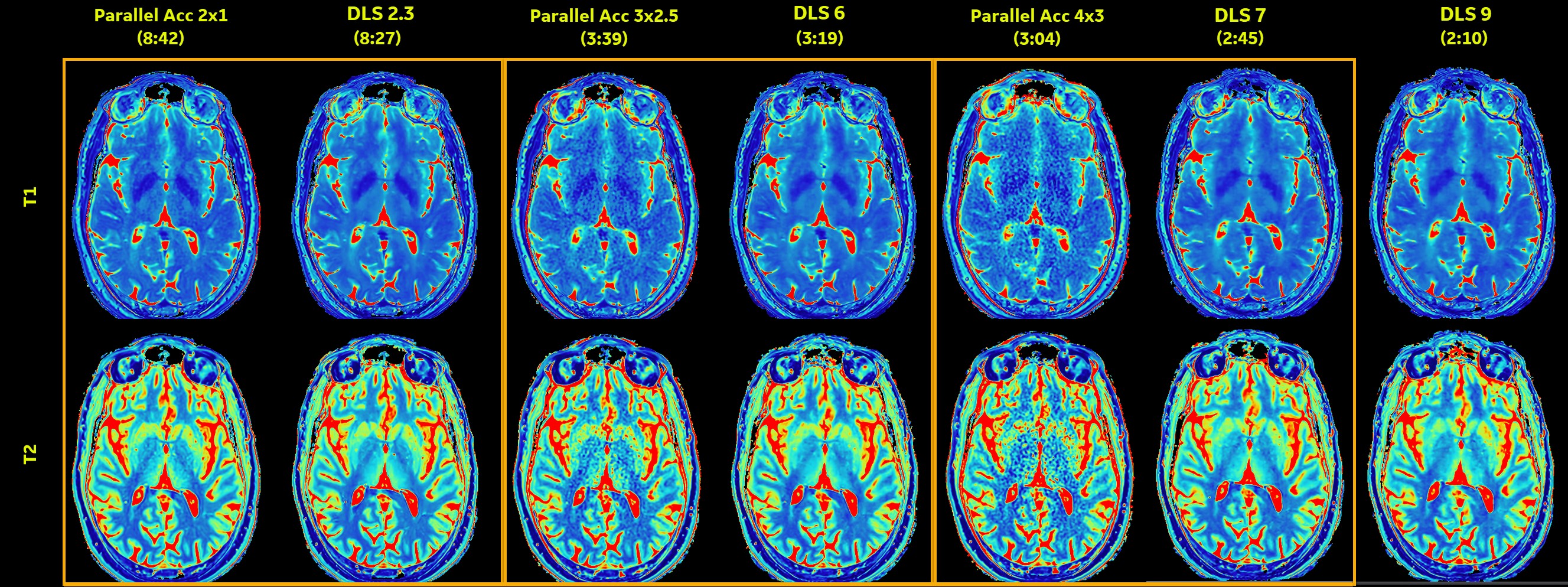
T1 and T2 parametric maps with increased acceleration factors and corresponding scan times are shown in Figure 3.
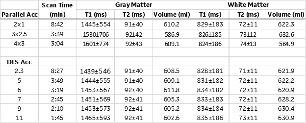
Table 1 lists the tissue specific T1 and T1 values and brain tissue volumes from all acquisitions.
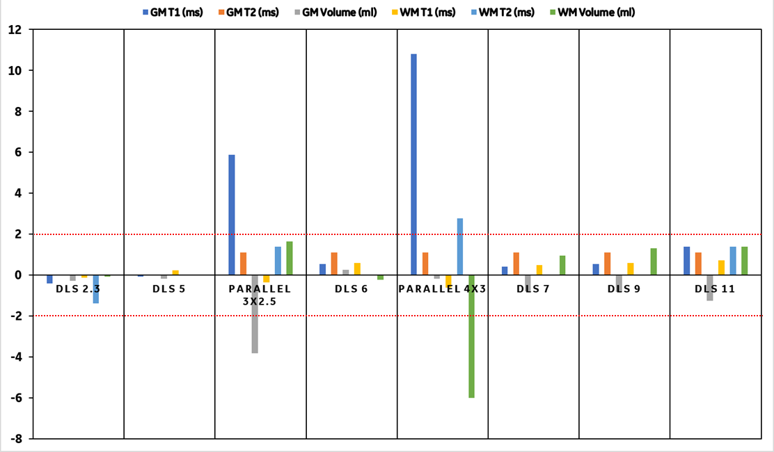
Using the T1/T2 and brain tissue estimates from the parallel imaging acceleration 2x1 as the baseline, Figure 4 shows the percent difference in these values. Eight different accelerated acquisitions are positioned in the order of scan times on the x-axis (left to right = longest to shortest). T1/T2 values, and tissue volume estimates are all within ±2% of the baseline (see red dotted lines) for all DLS accelerations. However, more significant deviations are observed for parallel imaging accelerated acquisitions, particularly the gray matter T1 and both gray matter and white matter volumes, likely due to amplified noise seen in Fig. 2 and Fig. 3.
CONCLUSION
This study demonstrates that with highly undersampled 3D QALAS acquisition combined with DL Speed, a 3-minute one-click exam is feasible that generates whole-brain high-resolution brain volumes of multiple contrasts and quantitative maps, which may enhance patient workflow in a busy clinical practice.Acknowledgements
No acknowledgement found.References
- Kvernby S, Warntjes MJ, Haraldsson H, et al. Simultaneous three-dimensional myocardial T1 and T2 mapping in one breath hold with 3D-QALAS. J Cardiovasc Magn Reson. 2014;16(1):102.
- Fujita S, Hagiwara A, Takei N, et al. Accelerated Isotropic Multiparametric Imaging by High Spatial Resolution 3D-QALAS With Compressed Sensing: A Phantom, Volunteer, and Patient Study. Invest. Radiol. 2021; 56:292–300.
- Ahn et al. Deep learning-based reconstruction of highly accelerated 3D MRI. arXiv: 2203.04674, 2022.
- Ahn, Sangtae, et al. “Task-based evaluation of deep learning-based reconstruction for highly-accelerated 3D T1-weighted brain MRI scans” Proceedings of the International Society of Magnetic Resonance in Medicine (ISMRM). 2023.
Figures