2790
Data-driven Image Reconstruction for Ultra-low-field Knee and Spine MRI at 0.05T1Laboratory of Biomedical Imaging and Signal Processing, The University of Hong Kong, Hong Kong SAR, China, 2Department of Electrical and Electronic Engineering, The University of Hong Kong, Hong Kong SAR, China
Synopsis
Keywords: Machine Learning/Artificial Intelligence, Image Reconstruction, knee, c-spine
Motivation: Deep learning (DL) is a powerful tool for MR image formation tasks and MR data at ultra-low-field (ULF) strength has significantly lower SNR than high-field.
Goal(s): Enhancing the image quality of ULF knee and c-spine data at 0.05T via DL reconstruction.
Approach: We extend our recently developed 3D DL partial Fourier reconstruction and superresolution (PF-SR) method on PF-sampled low-resolution noisy brain data to knee and c-spine data.
Results: The preliminary results demonstrate PF-SR, trained on synthetic ULF data simulated from high-field data, can reduce noise and artifacts, and enhance spatial resolution in experimental ULF knee and c-spine data, acquired from 0.05T MRI platform.
Impact: Through leveraging the homogeneous human knee and spine anatomy available in high-field data to enhance the image quality of ultra-low-field knee and spine MRI at 0.05T via deep learning reconstruction in a low-cost and shielding-free 0.05T MRI platform.
Introduction
Deep learning (DL) is a powerful tool for various MR image formation tasks, such as reconstruction from undersampled k-space, artifact suppression, and denoising1-4. This stems from its capability to extract features from large-scale MR database. Meanwhile, MRI at ultra-low-field (ULF) strength has limited clinical adoption due to its significantly lower signal-to-noise ratio in contrast to high-field5-8, which is challenging for traditional image reconstruction methods. Recent studies have demonstrated the effective reduction of noise and artifacts, and enhancement of spatial resolution in 3D brain MR data at ULF via DL9,10. Recently, we developed 3D DL partial Fourier reconstruction and superresolution (PF-SR) method to reconstruct 3mm isotropic noisy ULF brain MR data with 2D PF sampling of a fraction of 0.7 to 1.5mm isotropic data11. In this study, we extend PF-SR to anisotropic knee and c-spine data and demonstrate their preliminary results on a 0.05 Tesla MRI platform similar to the system used in previous research5.Method
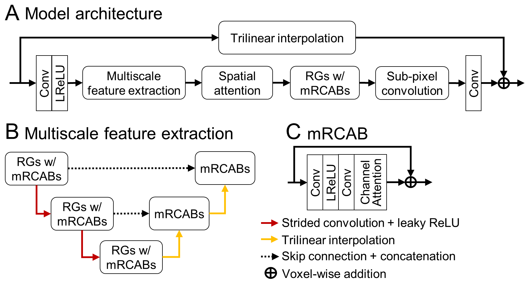
Figure 1 shows the overall architecture of 3D PF-SR model. It contains residual group with modified residual channel attention block, multiscale feature extraction, and spatial attention. Multiscale feature extraction allows the extraction of local and semiglobal features through downsampling and upsampling of features12-14. Spatial attention exploits the interspatial relationships among the extracted features15. Sub-pixel convolution layer projects the low-resolution features to the high-resolution feature space16. The global residual connection between high-resolution model output and trilinearly upsampled model input enforces the model to learn the image residue17.For knee model training data, a publicly available 3T knee OAI dataset18 was used, which included 2D SE sagittal magnitude knee data with echo times of 10 and 70, and 0.31x0.31x3.00mm3 resolution. For c-spine model training data, private HKU and publicly available Spine Generic dataset19 were used. The HKU dataset contained 1.5T 2D FSE sagittal magnitude data with 0.43x0.43x3.00mm3 resolution while the Spine Generic dataset included 3T 3D SE magnitude data with 0.8x0.8x0.8mm3 resolution. To simulate the PF-sampled low-resolution noisy 3D ULF data, image downsampling was performed via local mean to downsample the data to approximately the resolution of the training target. It was further downsampled through symmetric k-space truncation, undersampled by 2D PF sampling of a fraction of 0.8 along two PE directions, and degraded by addition of Rician noise, to generate the PF-sampled low-resolution noisy training input.
All 3D convolution layers had 3x3x3 kernel size and 64 channels, except for channel-attention and the last convolution layer, which had 8 and 1 channel, respectively. Random patch extraction was performed during training. L1 loss and AdamW optimizer with initial learning rate of 10-4, β1 = 0.9, β2 = 0.999, and weight decay of 0.1 were used. 1899 OAI knee, 137 HKU, and 214 Spine Generic c-spine data were used for training of the corresponding model. All models were trained for 350 epochs and took ~3.5 hours and <2 hours on four A100 GPUs for knee and c-spine models, respectively.
The experimental ULF knee and c-spine data were acquired from a 0.05T MRI platform, which is free from magnetic and radiofrequency (RF) shielding. The sagittal knee data was acquired using a 3D FSE sequence with TR/TE = 420ms/45ms and 1500ms/106ms, and 1.9x2.0x7.0mm3 resolution. The sagittal c-spine data was acquired using 3D FSE with TR/TE = 210ms/76ms and 2300ms/136ms, and ~2.0x2.0x8.0mm3 resolution. Each data was acquired within 8 minutes.
Results
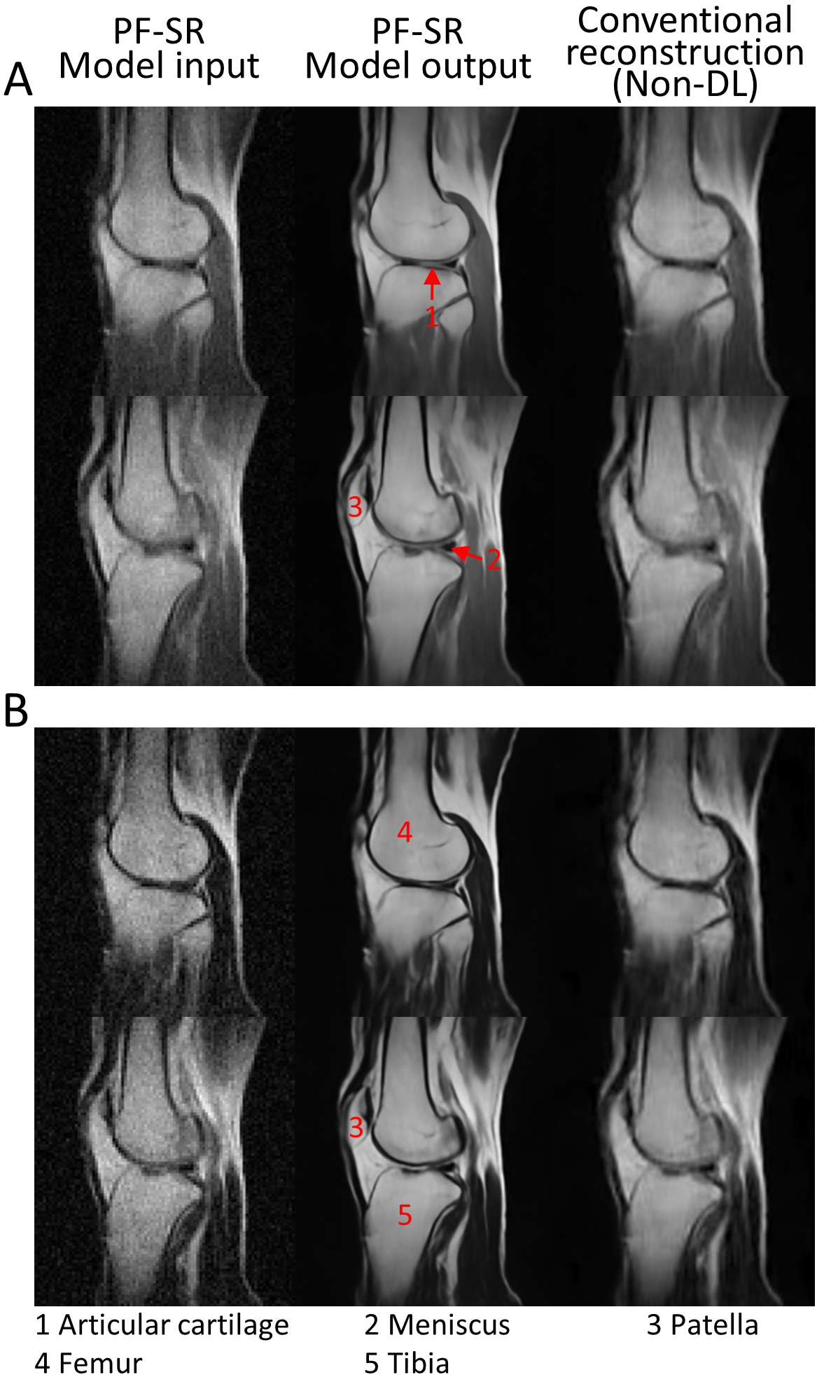
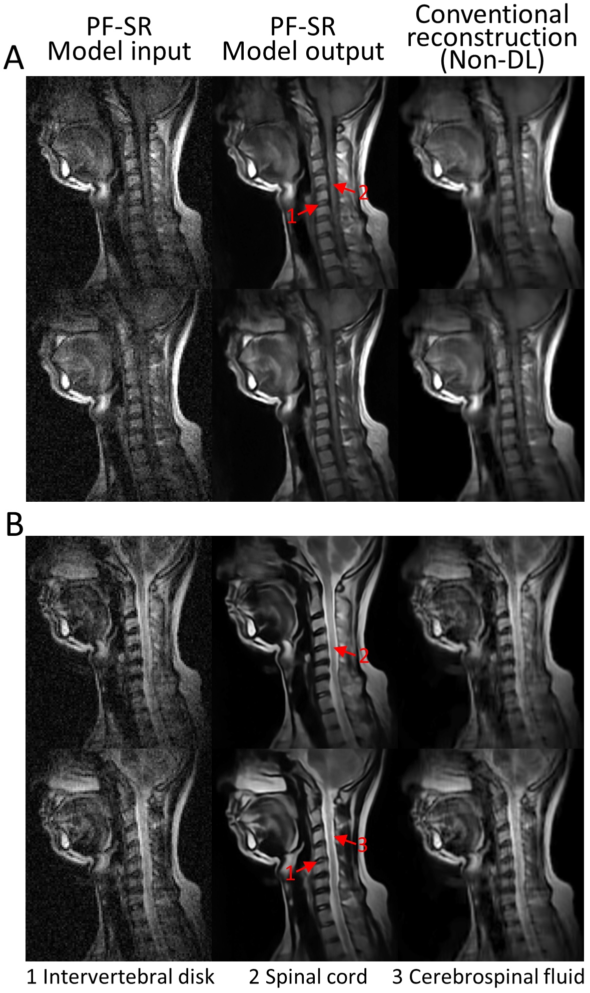
Figure 2 shows the results of experimental ULF knee data, which is acquired from a magnetic and RF shielding-free 0.05T MRI platform, using conventional non-DL method and PF-SR. Non-DL method consists of 2D POCS20,21 for PF reconstruction, BM4D22 denoising, and tricubic interpolation. Reduction of noise, and ringing and blurring artifacts, together with the enhancement of spatial resolution, can be observed using PF-SR. Structures, such as articular cartilage, meniscus, and patella, can also be delineated.The results of experimental ULF c-spine data from the same platform using conventional non-DL method and PF-SR (Figure 3). Noise and artifacts can be reduced using PF-SR. Through the increase in spatial resolution, the intervertebral disk, spinal cord, and cerebrospinal fluid inside spinal cord can be observed.
Discussion and Conclusion
In this study, we extend PF-SR to experimental anisotropic knee and c-spine data at 0.05T. Through leveraging the homogeneous human knee and c-spine anatomy in high-field data, the preliminary results demonstrated the model can reduce noise and artifacts, and enhance spatial resolution on experimental ULF knee and c-spine data. In future work, they should be compared to 3T to evaluate the structural fidelity and a larger cohort of subjects should be tested.Acknowledgements
This work was supported in part by Hong Kong Research Grant Council (R7003-19F, HKU17112120, HKU17127121, HKU17127022 and HKU17127523 to E.X.W).References
[1] Zhu B, Liu JZ, Cauley SF, Rosen BR, Rosen MS. Image reconstruction by domain-transform manifold learning. Nature 2018;555(7697):487-492.
[2] Hammernik K, Klatzer T, Kobler E, Recht MP, Sodickson DK, Pock T, Knoll F. Learning a variational network for reconstruction of accelerated MRI data. Magn Reson Med 2018;79(6):3055-3071.
[3] Xiao L, Liu Y, Yi Z, Zhao Y, Xie L, Cao P, Leong ATL, Wu EX. Partial Fourier reconstruction of complex MR images using complex-valued convolutional neural networks. Magn Reson Med 2022;87(2):999-1014.
[4] Muckley MJ, Ades-Aron B, Papaioannou A, Lemberskiy G, Solomon E, Lui YW, Sodickson DK, Fieremans E, Novikov DS, Knoll F. Training a neural network for Gibbs and noise removal in diffusion MRI. Magn Reson Med 2021;85(1):413-428.
[5] Liu Y, Leong ATL, Zhao Y, Xiao L, Mak HKF, Tsang ACO, Lau GKK, Leung GKK, Wu EX. A low-cost and shielding-free ultra-low-field brain MRI scanner. Nat Commun 2021;12(1):7238.
[6] Sarracanie M, Salameh N. Low-Field MRI: How Low Can We Go? A Fresh View on an Old Debate. Frontiers in Physics 2020;8.
[7] Coffey AM, Truong ML, Chekmenev EY. Low-field MRI can be more sensitive than high-field MRI. Journal of Magnetic Resonance 2013;237:169-174.
[8] Hoult DI, Richards R. The signal-to-noise ratio of the nuclear magnetic resonance experiment. Journal of Magnetic Resonance (1969) 1976;24(1):71-85.
[9] Lau V, Xiao L, Zhao Y, Su S, Ding Y, Man C, Wang X, Tsang A, Cao P, Lau GKK, Leung GKK, Leong ATL, Wu EX. Pushing the limits of low-cost ultra-low-field MRI by dual-acquisition deep learning 3D superresolution. Magn Reson Med 2023;90(2):400-416.
[10] Iglesias JE, Schleicher R, Laguna S, Billot B, Schaefer P, McKaig B, Goldstein JN, Sheth KN, Rosen MS, Kimberly WT. Quantitative Brain Morphometry of Portable Low-Field-Strength MRI Using Super-Resolution Machine Learning. Radiology 2023;306(3):e220522.
[11] Man C, Lau V, Su S, Zhao Y, Xiao L, Ding Y, Leung GKK, Leong ATL, Wu EX. Deep learning enabled fast 3D brain MRI at 0.055 tesla. Science Advances 2023;9(38):eadi9327.
[12] Lin T-Y, Dollár P, Girshick R, He K, Hariharan B, Belongie S. Feature pyramid networks for object detection. Proceedings of the IEEE conference on computer vision and pattern recognition 2017:2117-2125.
[13] Çiçek Ö, Abdulkadir A, Lienkamp SS, Brox T, Ronneberger O. 3D U-Net: learning dense volumetric segmentation from sparse annotation. Medical Image Computing and Computer-Assisted Intervention–MICCAI 19th International Conference 2016:424-432.
[14] Ronneberger O, Fischer P, Brox T. U-net: Convolutional networks for biomedical image segmentation. Medical Image Computing and Computer-Assisted Intervention–MICCAI 18th International Conference 2015:234-241.
[15] Woo S, Park J, Lee J-Y, Kweon IS. CBAM: Convolutional Block Attention Module. Computer Vision – ECCV 2018:3-19.
[16] Shi W, Caballero J, Huszár F, Totz J, Aitken AP, Bishop R, Rueckert D, Wang Z. Real-time single image and video super-resolution using an efficient sub-pixel convolutional neural network. Proceedings of the IEEE conference on computer vision and pattern recognition 2016:1874-1883.
[17] He K, Zhang X, Ren S, Sun J. Deep residual learning for image recognition. Proceedings of the IEEE conference on computer vision and pattern recognition 2016:770-778.
[18] Nevitt M, Felson D, Lester G. The osteoarthritis initiative. Protocol for the cohort study 2006;1.
[19] Cohen-Adad J, Alonso-Ortiz E, Abramovic M, Arneitz C, Atcheson N, Barlow L, Barry RL, Barth M, Battiston M, Büchel C. Open-access quantitative MRI data of the spinal cord and reproducibility across participants, sites and manufacturers. Scientific data 2021;8(1):219.
[20] McGibney G, Smith MR, Nichols ST, Crawley A. Quantitative evaluation of several partial Fourier reconstruction algorithms used in MRI. Magn Reson Med 1993;30(1):51-59.
[21] Haacke EM, Lindskogj ED, Lin W. A fast, iterative, partial-fourier technique capable of local phase recovery. Journal of Magnetic Resonance (1969) 1991;92(1):126-145.
[22] Maggioni M, Katkovnik V, Egiazarian K, Foi A. Nonlocal transform-domain filter for volumetric data denoising and reconstruction. IEEE Trans Image Process 2013;22(1):119-133.
Figures