2788
The use of deep learning based reconstruction in accelerating rectal cancer imaging1Radiology department, West China Hospital of Sichuan University, Chengdu, China, 2West China Clinical Medical College, Sichuan University, Chengdu, China, 3GE Healthcare, MR Research, Beijing, China., Chengdu, China
Synopsis
Keywords: Machine Learning/Artificial Intelligence, Machine Learning/Artificial Intelligence
Motivation: Clinical routine rectal cancer imaging requires high resolution and hence increased number of excitations to achieve sufficient signal to noise ratio (SNR).
Goal(s): The deep learning method is applied to improve the image quality and imaging speed of rectal magnetic resonance imaging.
Approach: We investigate the use of deep learning based reconstruction in shortening the scan time of the T2-weighted TSE imaging (T2DL) sequence in rectal cancer imaging.
Results: The results show that the DL reconstruction improves the SNR and CNR of the images. Also, the image acquisition time can be reduced by reconstructing images with reduced number of excitations by deep learning.
Impact: Deep learning reconstruction may lead to unprecedented improvements in SNR and CNR compared to conventional reconstruction algorithms, which may be used to obtain higher quality images. In addition, deep learning methods can indirectly shorten image acquisition time.
Objective
Clinical routine rectal cancer imaging requires high resolution and hence increased number of excitations to achieve sufficient signal to noise ratio (SNR)1. In this work, we investigate the use of deep learning based reconstruction in shortening the scan time of the T2-weighted TSE imaging (T2DL) sequence in rectal cancer imaging. The impact on images were assessed on overall image quality diagnostic confidence, and tumor boundaries as compared to standard high resolution T2-TSE (HRT2) imaging2.Methods
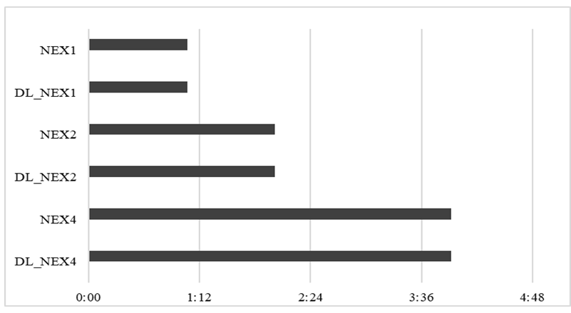
This study included 18 patients. Standard HRT2 sequences with different excitation numbers (NEX=4, 2 and 1) and T2DL sequences with different excitation numbers (NEX=4, 2 and 1) were obtained by scanning. Figure 1 shows the axial acquisition times were 3min55s, 2min1s, and 1min4s min for three excitation numbers (NEX=4, 2 and 1), respectively. Two experienced radiologists independently assessed the image quality using an ordered 5-point Likert scale 3,4. The images were evaluated qualitatively by measuring the mean signal (SI) and the standard deviation (SD) of the background of the muscle and tumor in the images, and calculating the signal-to-noise ratio (SNR) and the contrast-to-noise ratio (CNR) of the muscle and tumor using the following equations.SNRtumor = SItumor/SDbackground (1)
SNRmuscle = SImuscle/SDbackground (2)
CNR = |SNRtumor - SNRmuscle| (3)
The diagnostic value of extra-mural vascular invasion and fascial rectus status was assessed using TN staging5. The Friedman and Bonferroni tests were used for comparative analysis.
Results
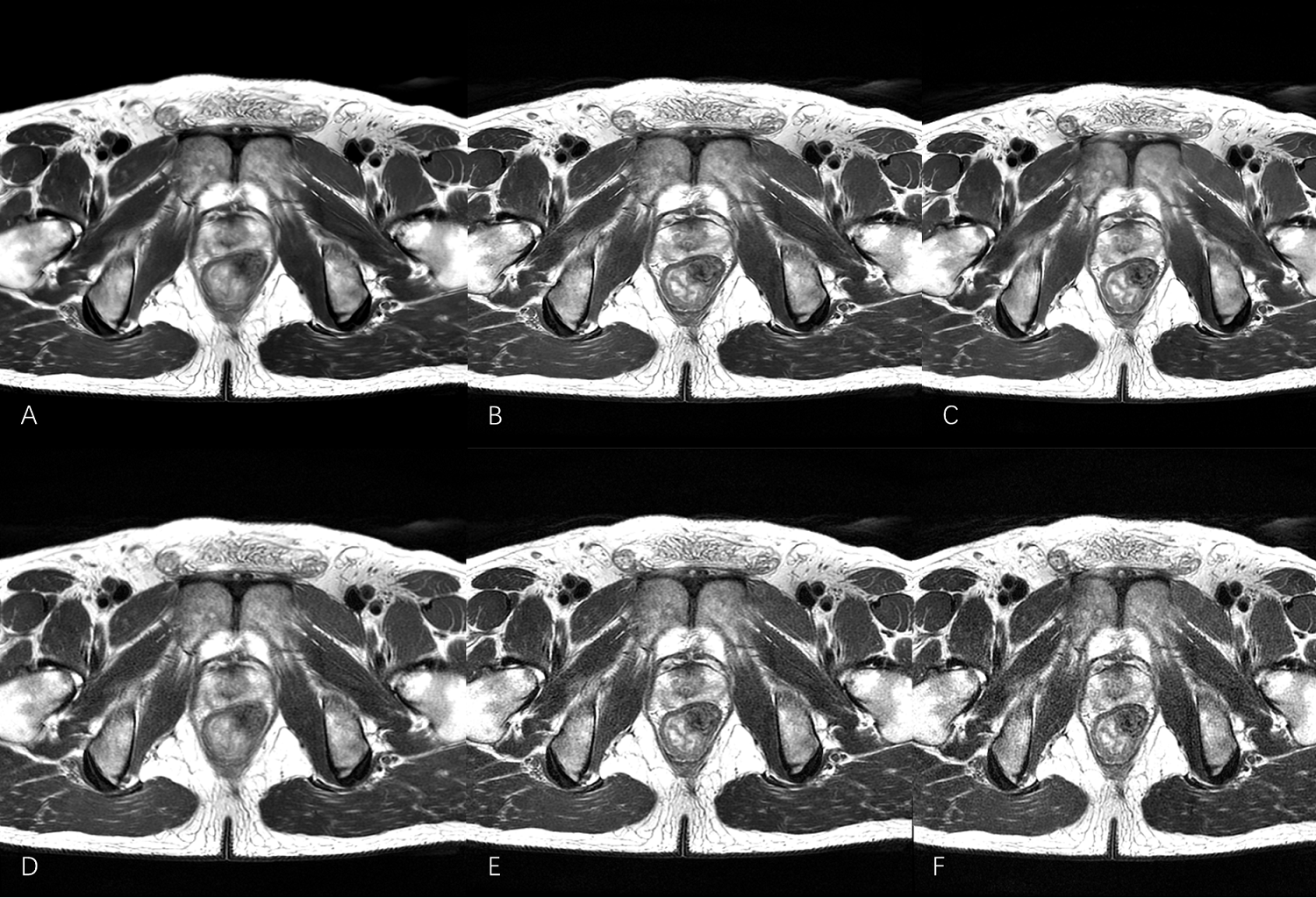
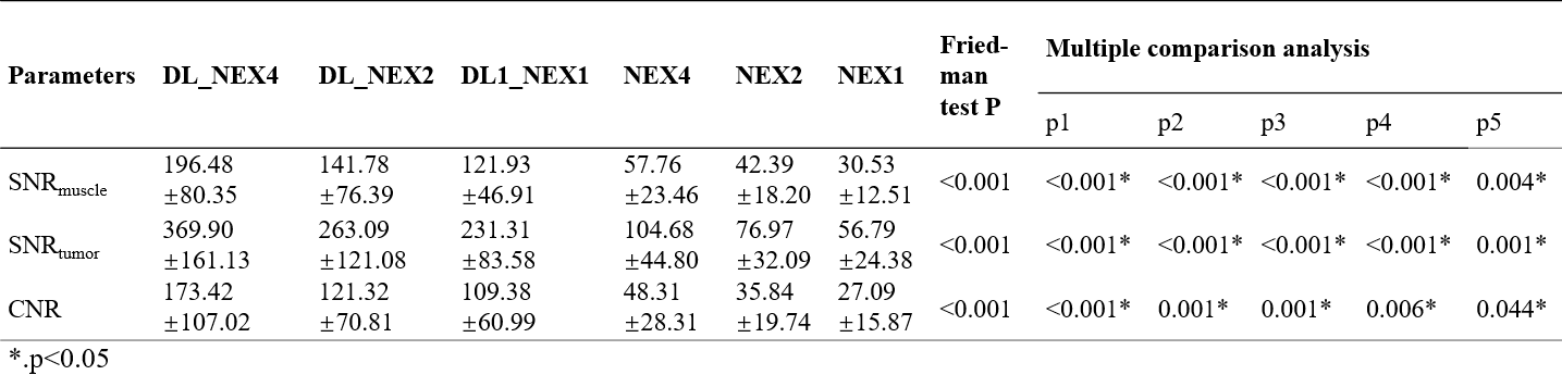
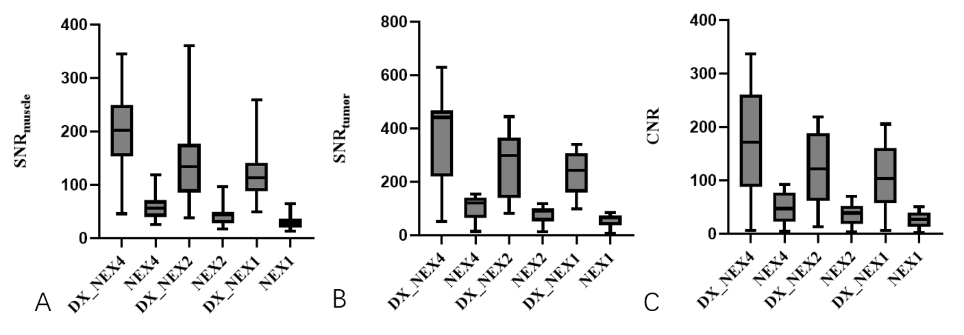
Figure 2 shows a typical set of DL reconstructed and conventional reconstructed images with different NEXs, and the improved signal-to-noise ratio of the DL reconstruction can be clearly seen. The overall image scores of different images are summarized in Table 1. Firstly, for images with the same number of excitations, the signal-to-noise ratio of images reconstructed by deep learning is significantly better than that of images reconstructed by conventional methods (p<0.05). Secondly, the scores of DL reconstructed images with NEX =2 are higher than those of conventionally reconstructed images with NEX =4, while the scores of DL reconstructed images with NEX =1 are similar to those of conventionally reconstructed images with NEX =4. The summary of SNR and CNR of muscle and tumor for deep learning reconstructed and conventional reconstructed images are shown in Table 2. The results show that the DL reconstruction improves the SNR and CNR of the images. Also, the images with reduced number of excitations (NEX=1 and 2) reconstructed by deep learning are of similar or even better quality than the images of standard sequences (NEX=4), so the image acquisition time can be reduced by reconstructing images with reduced number of excitations by deep learning.Conclusion And Discussion
First, deep learning reconstruction may lead to unprecedented improvements in SNR and CNR compared to conventional reconstruction algorithms, which may be used to obtain higher quality images. In particular, when NEX at a lower level, deep learning reconstruction can yield higher quality images with the same NEX, which can greatly help to improve the accuracy of diagnosis. Second, in this work, studies using DL accelerated T2 rectal cancer imaging used a standard rectal imaging prototype as a benchmark. We observed that deep learning reconstructions of rectal T2w axial TSE imaging reduced acquisition time by more than 50% while maintaining image quality level and diagnostic value. This means that the same or even higher quality images can be obtained in less time, which is a great help to acquire stable quality images more quickly. We believe that deep learning reconstruction has some application value in routine imaging of rectal cancer.Acknowledgements
No acknowledgement found.References
1.Zhao, L., et al. A preliminary study of synthetic magnetic resonance imaging in rectal cancer: imaging quality and preoperative assessment. Insights Imaging 12, 120 (2021).
2.Gormly, K. Rectal MRI: the importance of high resolution T2 technique. Abdom Radiol (NY) 46, 4090-4095 (2021).
3.Gong, X., et al. Comparison of compressed sensing-sensitivity encoding (CS-SENSE) accelerated 3D T2W TSE sequence versus conventional 3D and 2D T2W TSE sequences in rectal cancer: a prospective study. Abdom Radiol (NY) 47, 3660-3670 (2022).
4.Xia, C.C., et al. Readout-segmented echo-planar imaging improves the image quality of diffusion-weighted MR imaging in rectal cancer: Comparison with single-shot echo-planar diffusion-weighted sequences. Eur J Radiol 85, 1818-1823 (2016).
5.Peng, Y., et al. Comparison of reduced field-of-view diffusion-weighted imaging (DWI) and conventional DWI techniques in the assessment of rectal carcinoma at 3.0T: Image quality and histological T staging. J Magn Reson Imaging 47, 967-975 (2018).
Figures