2786
Hankel-based data preparation method for radial MRI artifact removal from undersampled zero-filled images1Cognitive Sciences, Institute for Research in Fundamental Sciences (IPM), Tehran, Iran (Islamic Republic of)
Synopsis
Keywords: Image Reconstruction, Cardiovascular, Deep learning, Radial MRI
Motivation: Hankel-based reconstruction distorts the image's center less than its periphery. This prompts us to examine Hankel-based reconstruction for neural network training data preparation.
Goal(s): To train the model-agnostic neural network on Hankel-based reconstruction data to improve image center reconstruction.
Approach: A neural network trained on Hankel-based reconstruction data was compared to an equivalent network trained on NUFFT-based reconstruction data.
Results: In the context of radial dynamic imaging, where the ROI can be placed in the center of the image, our approach achieved better results than when using NUFFT-based data preparation for reconstruction of undersampled radial data.
Impact: This study might influence dynamic radial-MRI reconstruction. Our data preparation for training and testing the network improved cardiac-MRI qualitative outcomes, especially in the heart region. The radial-MRI society may find the proposed solution appealing when paired with DL-based approaches.
Purpose
To propose an efficient method of data preparation for radial MRI reconstruction using the Hankel-based reconstruction method.Introduction
Unlike the NUFFT, the Hankel-based radial reconstruction, originally proposed for CT reconstruction1 and later adapted for MRI applications2,3, does not require frequency interpolation. No previous research has used fast MRI methods like DL in conjunction with Hankel-based reconstruction. We speculate that, compared to the NUFFT, the Hankel-based reconstruction for DL-based radial-MRI reconstruction may provide some benefits. In this research, we used Hankel-reconstructed data to train a model-agnostic GAN. Due to the no frequency interpolation during data preparation, it is expected that the proposed network will outperform an identical network trained on the same data but prepared using NUFFT.Method
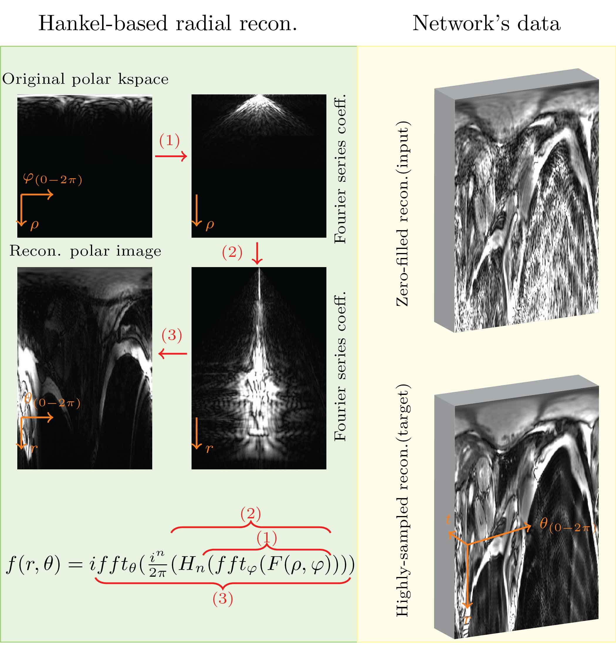
Hankel-based radial reconstruction. The Hankel-based radial reconstruction, which directly reconstructs the polar kspace without frequency interpolation, produces an image in the polar spatial space using the following equation:$$f(r,\theta)=ifft_{\theta}(\frac{i^{n}}{2\pi}(H_{n}(fft_{\varphi}(F(\rho,\varphi))))(1)$$
Where $$$ifft(.)$$$ and $$$fft(.)$$$ represent the one-dimensional inverse-$$$fft$$$ and the $$$fft$$$ along the specified dimension. $$$H_{n}(.)$$$ is the $$$n^{th}$$$-order Hankel transform.
Dataset and data-preparation. We used Harvard public data4. The dataset includes Radial MRI scans of 108 people—101 patients and 7 healthy volunteers. The scans were taken using a 3T MRI scanner and acquired mid-ventricular slices using Breath-holding bSSFP cine sequences. Each scan consists of 25 cardiac frames and 196 radial spokes ($$$196\times2$$$ of half-spokes). The dataset was randomly split into 28 training and 80 testing cases. This study uses a model-agnostic GAN that requires input (reconstructed from undersampled polar k-space) and target data. We retrospectively undersampled the highly-sampled polar k-space using the uniform distribution over the radial spokes at 5x, 10x, and 15x acceleration factors to prepare the input. The Hankel-based reconstruction method in Equation 1 was used to reconstruct the undersampled data. Figure 1 illustrates input and target at 10x acceleration factor. The Hankel-based reconstruction is entirely polar, as shown in Figure 1, therefore the input and target are in polar space. We stacked real and imaginary parts in the channel dimension to handle complex data.
Implementation. Aliasing artifacts from under sampled data were reduced using 3D-GAN (generator = 4-staged 3D-UNet, discriminator = classifier with 6 average-pooling). Alongside the adversarial loss, L1 and SSIM losses were incorporated into the generator with weights determined through experimentation. To ensure stable training, the network used the progressive growing method described in our previous publication5. All experiments were performed on a computer with an Intel Core i7 processor, 64 GB of RAM, and an NVIDIA -Titan RTX GPU (24 Gb) using PyTorch. For evaluation, we will present qualitative results and SSIM for 5x, 10x, and 15x acceleration factors. The suggested method will be qualitatively compared to the same network trained with adjoint NUFFT for input and target preparation.
Results
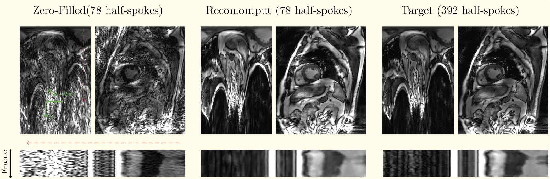
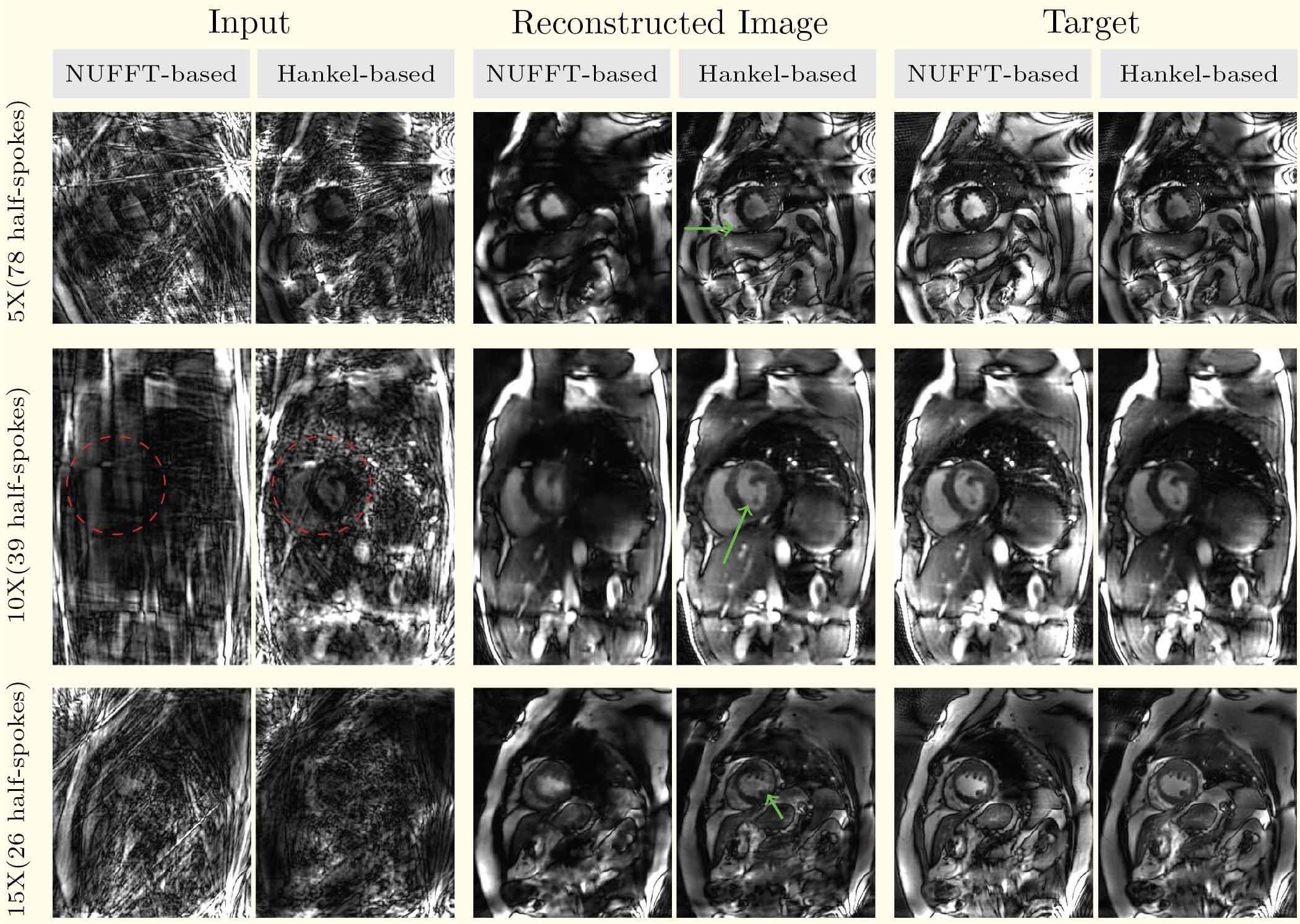
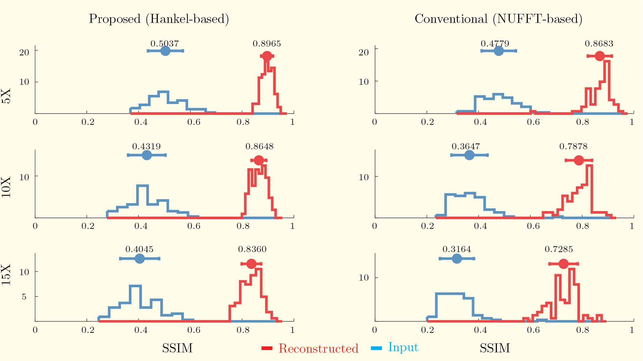
The proposed method removes aliasing in the reconstructed image, as illustrated in Figure 2. The intensity profile demonstrates that the temporal sharpness closely resembles the target. Figure 3 contrasts the NUFFT-based and Hankel-based methods at various acceleration rates. Both methods eliminate aliasing artifacts in input images. However, Hankel-based reconstruction generates sharper images, particularly at higher acceleration rates. NUFFT and Hankel-based SSIM measurements are shown in Figure 4. SSIM values in reconstructed images are always higher than input. Our proposed method shows slower decline in SSIM with increasing acceleration factor compared to NUFFT-based method.Discussion
A unique and effective data preparation strategy for training DL-based radial MRI was proposed in this study. We demonstrated that the proposed method can preserve sharpness and remove artifacts from undersampled radial kspace input images. Importantly, the recommended technique produced sharper and higher-quality images at 5x, 10x, and 15x acceleration factors than the adjoint-NUFFT-based data preparation. Figure 3 shows that the suggested method's cardiac input images exhibit less artifact than NUFFT-based input. Our technique worked well since the network started with good inputs. We must assess the results radiologically to establish a definite conclusion, and this is the next step in our research. In addition, we intend to include functional analysis, such as EF analysis, to determine whether or not the results are functionally compatible with the target images.Acknowledgements
No acknowledgement found.References
1. Higgins WE, Munson DC. A Hankel transform approach to tomographic image reconstruction. IEEE Transactions on Medical Imaging, vol. 7, no. 1, pp. 59-72, March 1988, doi: 10.1109/42.3929.
2. Guo H, Song AW. MRI image reconstruction by polar Fourier trans-form. In: Proceedings of the 12th Annual Meeting of ISMRM, Kyoto, Japan, 2004. Abstract 350.
3. Mohammadi, E, Nasiraei-Moghaddam, A, Uecker, M. Real-time radial tagging for quantification of left ventricular torsion. Magn Reson Med. 2022; 87: 2741–2756. doi:10.1002/mrm.29169
4. El-Rewaidy, H, Fahmy, AS, Pashakhanloo, F, et al. Multi-domain convolutional neural network (MD-CNN) for radial reconstruction of dynamic cardiac MRI. Magn Reson Med. 2020; 85: 1195–1208. https://doi.org/10.1002/mrm.28485
5. Ghodrati, V, Bydder, M, Bedayat, A, et al. Temporally aware volumetric generative adversarial network-based MR image reconstruction with simultaneous respiratory motion compensation: Initial feasibility in 3D dynamic cine cardiac MRI. Magn Reson Med. 2021; 86: 2666–2683. https://doi.org/10.1002/mrm.28912
Figures