2783
DeepEMC-T2 Mapping: Deep Learning-Enabled Echo Modulation Curve T2 Mapping1Bernard and Irene Schwartz Center for Biomedical Imaging, Department of Radiology, New York University Grossman School of Medicine, New York City, NY, United States, 2Center for Advanced Imaging Innovation and Research (CAI2R), Department of Radiology, New York University Grossman School of Medicine, New York City, NY, United States, 3Department of Electrical and Computer Engineering, NYU Tandon School of Engineering, New York City, NY, United States, 4Department of Radiation Oncology, New York University Grossman School of Medicine, New York City, NY, United States, 5Department of Biomedical Engineering, Tel Aviv University, Tel Aviv, Israel, 6Sagol School of Neuroscience, Tel Aviv University, Tel-Aviv, Israel
Synopsis
Keywords: Machine Learning/Artificial Intelligence, Machine Learning/Artificial Intelligence
Motivation: Echo Modulation Curve T2 Mapping (EMC-T2) mapping can generate highly accurate, precise, and reproducible T2 quantification. However, the standard EMC-T2 framework requires ~10 echoes and a cumbersome post-processing step for pixel-wise dictionary matching.
Goal(s): This work proposes a deep learning version of EMC-T2 mapping, called DeepEMC-T2, to enable efficient and accurate estimation of T2 maps from fewer echoes without requiring a dictionary.
Approach: DeepEMC-T2 was developed using a spatiotemporal convolutional neural network, which estimates both T2 and PD maps directly from multi-echo spin-echo images.
Results: DeepEMC-T2 enables efficient and accurate T2 mapping and requires only smaller number of echoes compared to standard EMC-T2.
Impact: Standard EMC-T2 enables accurate T2 quantification but previously required a complicated post-processing step that made clinical translation challenging. DeepEMC-T2 enables efficient and accurate T2 quantification with fewer echoes. This could facilitate more widespread translation of this technique into clinical practice.
Introduction
Quantitative T2 relaxation has great potential to provide valuable clinical information1. An echo modulation curve-based T2 mapping (EMC-T2) technique can generate rapid accurate T2 mapping based on the Bloch simulation to model the exact signal evolution in multi-echo spin-echo (ME-SE) sequences2. However, the EMC-T2 method requires a cumbersome pixel-wise dictionary searching step with sufficient echoes (e.g., 10 echoes) to ensure quantification accuracy. This may limit volumetric coverage and result in an increased specific absorption rate (SAR). In this study, we propose a deep learning version of EMC-T2 mapping, referred to as DeepEMC-T2, to enable efficient and accurate T2 mapping directly from a flexible number of echoes without requiring a dictionary.Methods
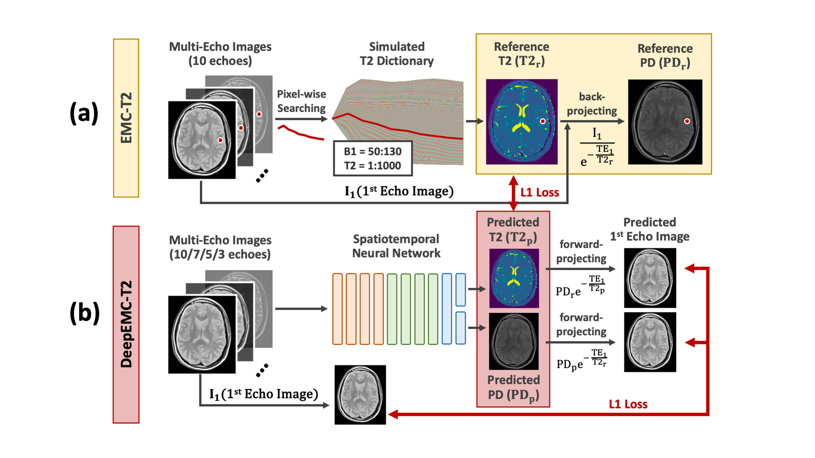
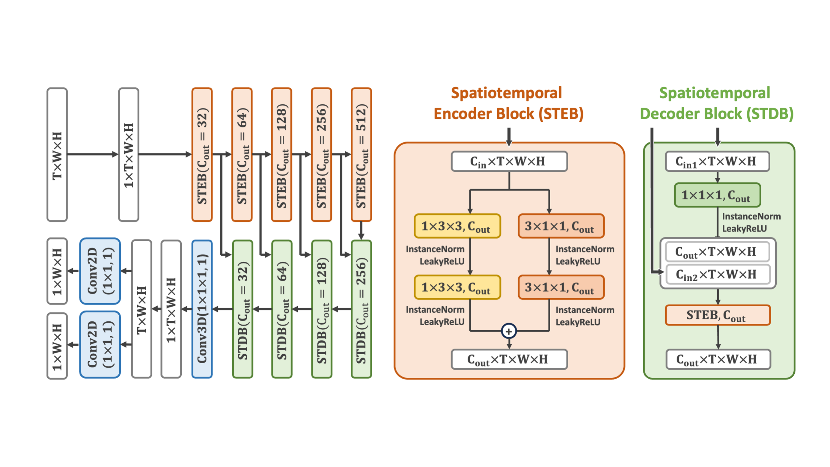
Standard EMC-T2 mapping uses a Bloch simulation-based dictionary for T2 estimation (Figure 1a). Specifically, a T2 map is generated by matching the acquired ME-SE images with the pre-generated dictionary at each pixel location. Based on the estimated T2 map, a proton density (PD) map can then be computed by back-projecting the first echo image.DeepEMC-T2 implements a modified version of U-net3 to directly estimate both T2 and PD maps from the acquired ME-SE images, as shown in Figure 1(b). The modified U-net employs spatiotemporal convolutions and removes the standard downsampling and unsampling layers, which are two features to enable more accurate pixel-wise parameter generation. The network is optimized by enforcing an L1 loss between the predicted T2 and PD maps and the reference T2 and PD maps, which are calculated using the standard EMC-T2 algorithm from ME-SE images with 10 echoes. In addition, DeepEMC-T2 also incorporates a data-consistency loss by minimizing the difference between the reference first-echo image (from the reference ME-SE images) and predicted first-echo images computed through forward-projecting using the predicted T2 and reference PD, as well as predicted PD and reference T2 separately. The DeepEMC-T2 network structure is shown in Figure 2.
A total of 68 datasets previously acquired on 3T MRI scanners (TimTrio/Skyra, Siemens Healthineers) using a vendor-provided ME-SE sequence were used in this study. Each dataset has 26 slices with 10 echoes each. Other imaging parameters included: FOV=220x206mm2, matrix size=128x120, slice thickness=3mm, TR=4.1s, TE/DTE=15ms/15ms, GRAPPA factor=2. 46 datasets were used for training (33 healthy controls and 13 patients with multiple sclerosis [MS]). 7 datasets were used for validation (5 healthy controls and 2 MS patients). 15 datasets (all MS patients) were used for evaluation.
The T2 and PD maps generated from DeepEMC-T2 were compared with those from standard EMC-T2 using different numbers of echoes. T2 and PD maps also were generated using a standard U-Net with only spatial convolution and with the original structure (3) for additional comparison. For each testing dataset, the error of T2 estimation was assessed in different T2 ranges as follows: $$Error\left(\%\right)=\frac{\left|Pred-Ref\right|}{Ref}\times100\%$$
Results
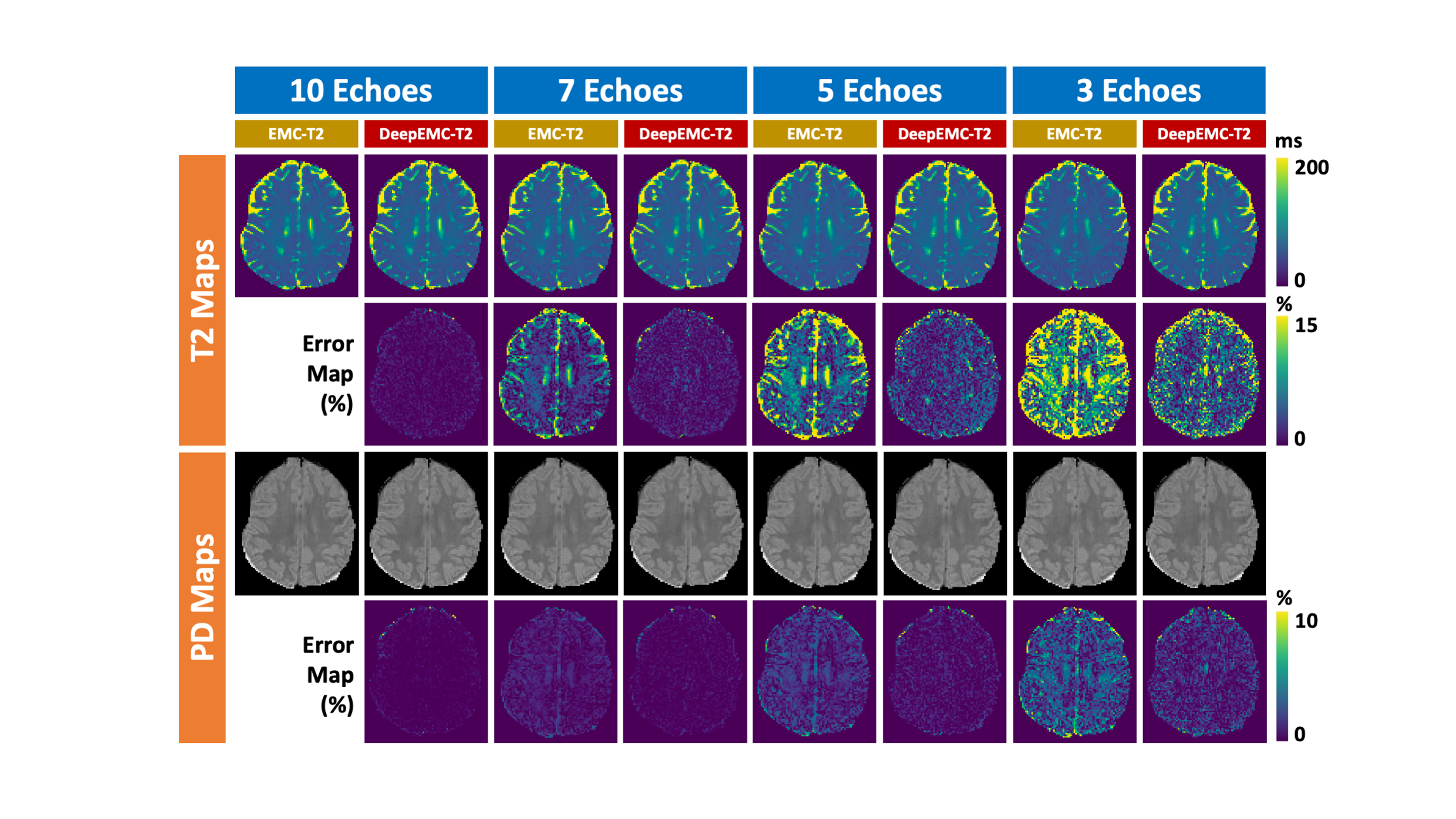
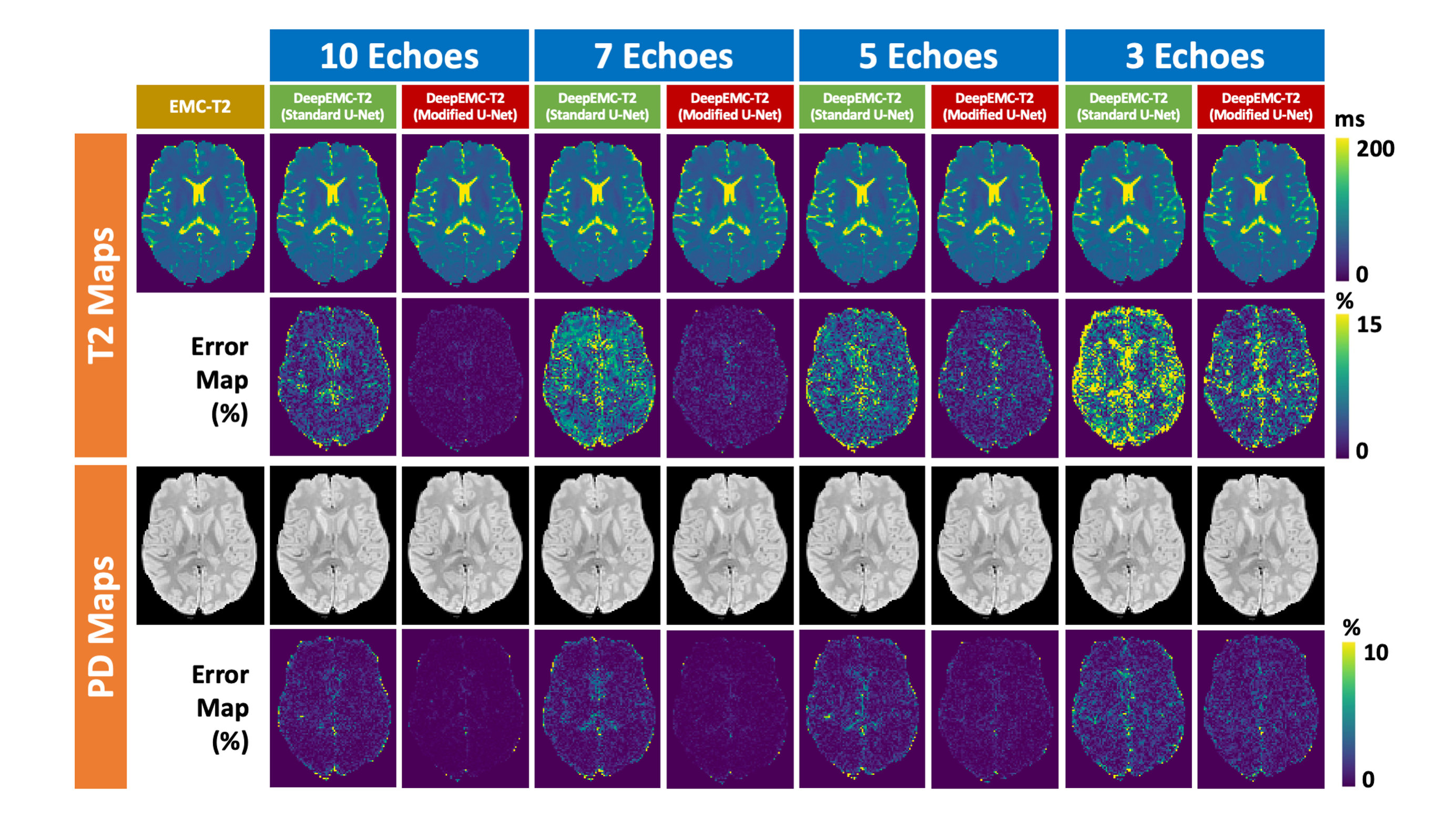
Figure 3 shows a representative case comparing T2 and PD maps estimated using EMC-T2 and DeepEMC-T2 from ME-SE images with different numbers of echoes. When the number of echoes reduces (e.g., from 10 echoes to 7/5/3 echoes), DeepEMC-T2 enables more accurate parameter estimation than EMC-T2. Also, during the inference step, DeepEMC-T2 does not require a dictionary and enables much more efficient quantification.Figure 4 shows another case comparing the T2 and PD maps estimated using DeepEMC-T2 based on modified U-net and standard U-Net with different numbers of spin echoes. The error maps suggest that a modified U-net can improve the accuracy of T2 quantification.
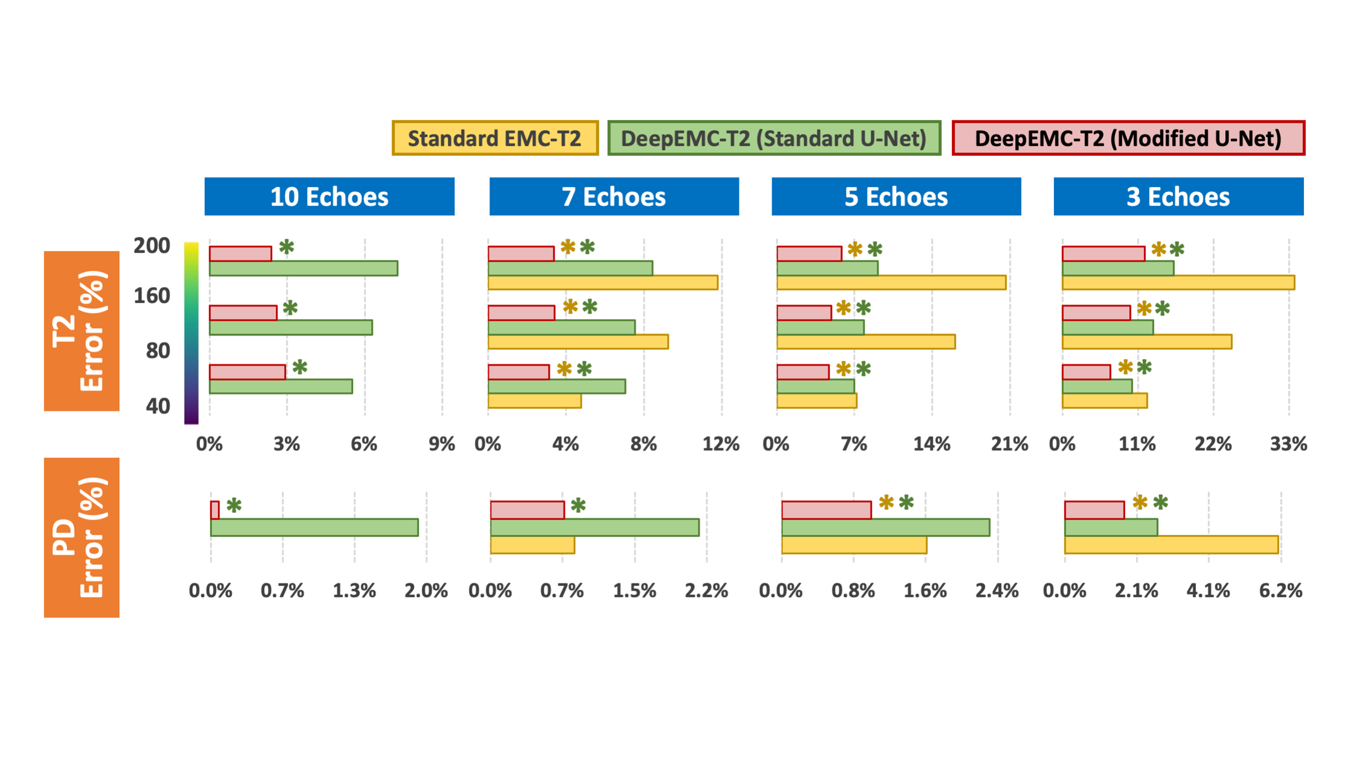
Figure 5 summarizes the quantitative comparison of EMC-T2 with DeepEMC-T2 using both modified and standard U-Net in all 15 testing datasets with different numbers of echoes based on averaged pixel-wise errors in different T2 ranges. The results indicate that DeepEMC-T2 enables accurate PD and T2 estimations across different T2 ranges, and the use of the modified U-Net yields higher accuracy compared to standard U-Net. The yellow and green stars indicate improvements that reach statistical significance for different comparisons.
Conclusion
This work proposed a novel deep learning framework to implement EMC-T2 mapping. It addresses major challenges presented in standard EMC-T2: the need for a complicated dictionary matching step and sufficient echoes. DeepEMC-T2 enables simple, efficient, and accurate T2 quantification directly from acquired ME-SE images with a fewer number of echoes. This allows for increased volumetric coverage and/or reduced SAR by reducing the number of 180o refocusing pulses. Moreover, the DeepEMC-T2 framework is directly implemented using DICOM images acquired from a standard ME-SE sequence. It is expected that DeepEMC-T2 also can be performed on accelerated ME-SE images, enabling reduced total scan times and/or improved spatial resolution.Acknowledgements
This work was supported by the NIH (R01EB030549, R21EB032917, and P41EB017183) and was performed under the rubric of the Center for Advanced Imaging Innovation and Research (CAI2R), an NIBIB National Center for Biomedical Imaging and Bioengineering.References
1. Poon, Colin S., and R. Mark Henkelman. "Practical T2 quantitation for clinical applications." Journal of Magnetic Resonance Imaging 2.5 (1992): 541-553.
2. Ben‐Eliezer, Noam, Daniel K. Sodickson, and Kai Tobias Block. "Rapid and accurate T2 mapping from multi–spin‐echo data using Bloch‐simulation‐based reconstruction." Magnetic resonance in medicine 73.2 (2015): 809-817.
3. Ronneberger, Olaf, Philipp Fischer, and Thomas Brox. "U-net: Convolutional networks for biomedical image segmentation." Medical Image Computing and Computer-Assisted Intervention–MICCAI 2015: 18th International Conference, Munich, Germany, October 5-9, 2015, Proceedings, Part III 18. Springer International Publishing, 2015.
Figures