2782
Ten-fold accelerated multi-echo spiral fMRI using self-supervised physics-driven DL reconstruction1Department of Medicine, University of Hawaii, Honolulu, HI, United States, 2Center for Magnetic Resonance Research, University of Minnesota, Minneapolis, MN, United States, 3Electrical and Computer Engineering, University of Minnesota, Minneapolis, MN, United States
Synopsis
Keywords: Machine Learning/Artificial Intelligence, fMRI
Motivation: Multi-echo fMRI holds the promise of more potential applications, however it suffers from long readout lengths.
Goal(s): Explore the possibility of using deep learning(DL) reconstruction for highly under-sampled spiral multi-echo fMRI acquisition.
Approach: Multi-echo data from four subjects were collected for DL training. Multi-echo fMRI data from another subject was used for testing the DL model. The DL model has been designed and modified to enable the reconstruction of ten-fold under-sampled fMRI images for BOLD analysis.
Results: The DL model has not only reconstructed the multi-echo spiral fMRI with good image quality, but also preserved its BOLD sensitivity with the highly under-sampled data.
Impact: With the help of DL, multi-echo fMRI may become more versatile for clinical use and future studies.
Introduction
Multi-echo fMRI has the potential to improve the accuracy of fMRI measurements and extract multi-quantitative maps by utilizing the extra echo information1. Nevertheless, the relatively long readout poses a challenge in integrating multi-echo acquisition into fMRI due to its time constraints. One potential solution to circumvent this challenge involves employing parallel imaging2. However, conventional parallel imaging methods, such as compressed sensing3-6, may struggle to maintain optimal image quality when the under-sampling rate is excessively high.In recent years, deep learning (DL) has emerged as a promising approach for image reconstruction7-9, particularly at high accelerations. In this study, we implemented a spiral multi-echo sequence along with a self-supervised physics-driven DL reconstruction to get multi-echo fMRI data with an under-sampling rate of ten, and investigated its utility for BOLD analysis.
Methods
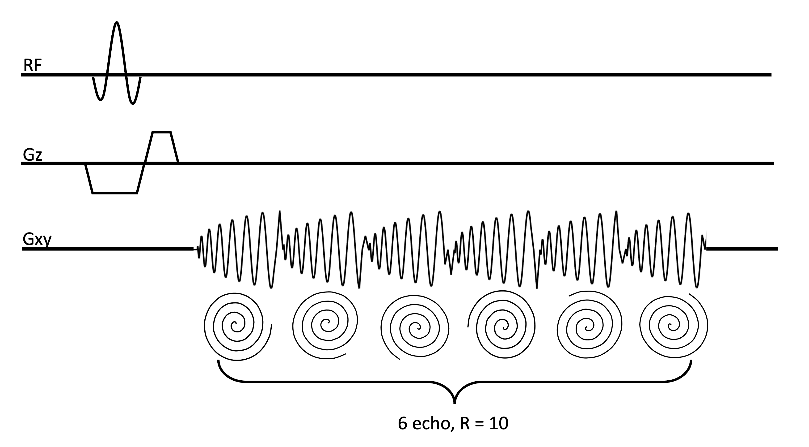
Image Acquisition: A prototype multi-echo 2D GRE sequence was used (Fig.1). The experiments were comprised of two acquisitions. First, training data for the DL algorithm was collected from four healthy subjects. The multi-echo dataset for training was acquired with flip angle 20°, TR=4464ms. TEs=[3.4,9.5,15.6,21.8,27.9,34.1]ms. 72 slices were collected with in-plane FOV=240$$$\times$$$240mm2, isotropic resolution2$$$\times$$$2$$$\times$$$2mm3. A corresponding fully sampled dataset with the same parameters was acquired to generate the coil maps for training. This data did not include a time-series.Subsequently, in-vivo fMRI experiments were performed on another subject using the 2D multi-echo sequence and a standard single-echo spiral sequence for comparison. The protocol for the multi-echo sequence was set similar to the training dataset with same flip angle, TEs and in-plane FOV, but different TR was used by acquiring 24 slices within 1488ms to fulfill the fine time resolution for fMRI while also covering the visual cortex. The single-echo spiral sequence was using flip angle 20°, TE/TR=27.91/1920ms, in-plane FOV=240$$$\times$$$240mm2, resolution3$$$\times$$$3$$$\times$$$2mm3. The longer TR value is necessary to accommodate a lengthier spiral readout.
The fMRI tasks consisted of six blocks of 20sec flickering checkerboard for visual stimulation, separated by 20sec resting periods leading to a paradigm duration of 4:20 min. BOLD analyses were carried out using a general linear model with a canonical hemodynamic response function after removal of first and second order temporal trends in the data. We used weighted echo summation10 based on the local T2* (the T2* map was extracted from the fully sampled multi-echo data) to prepare the multi-echo data for BOLD analyses. All scans were acquired on a 3T Siemens Prisma scanner.
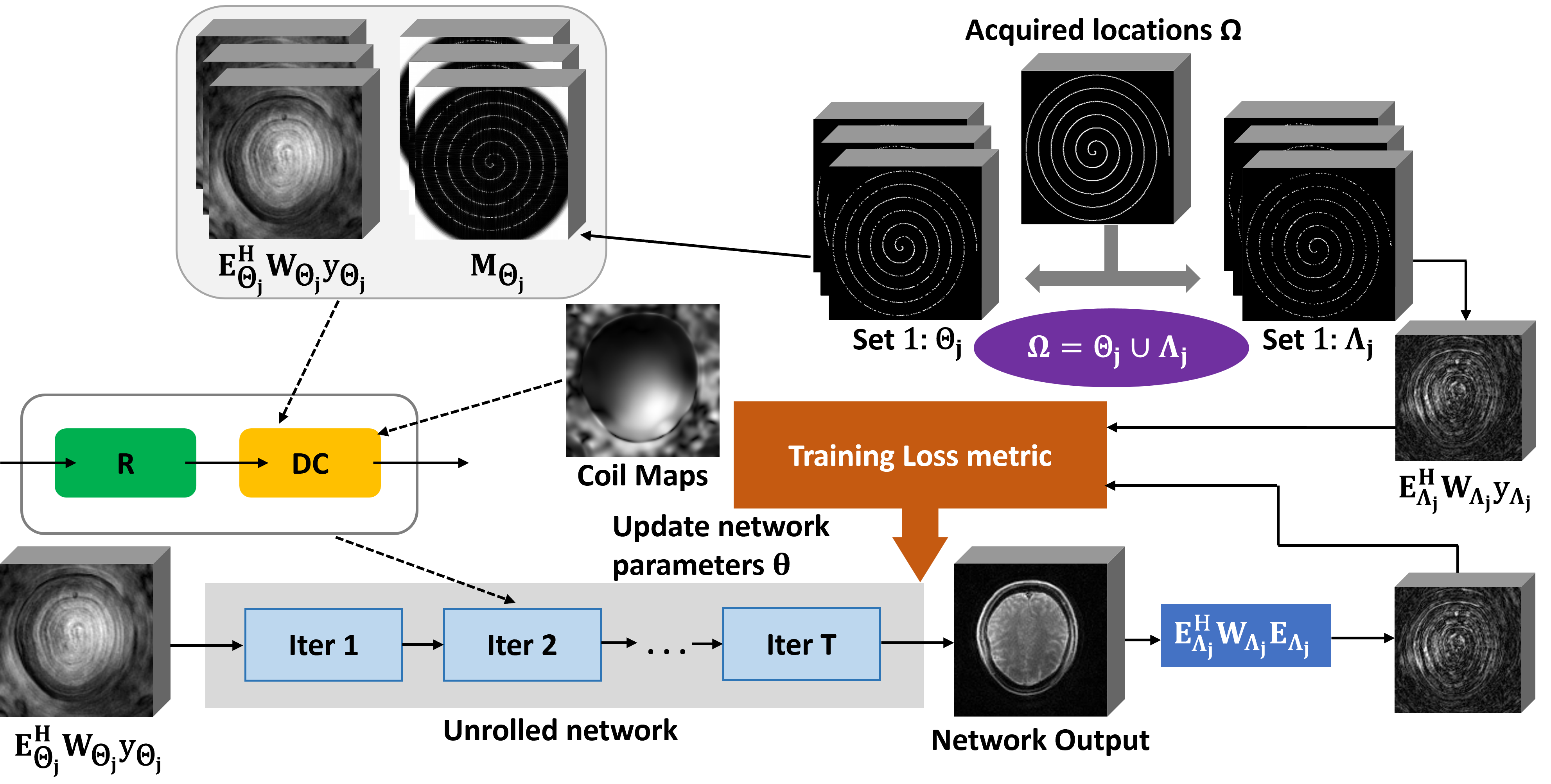
Image Reconstruction: We extended multi-mask self-supervision via data undersampling(SSDU)11 method for non-Cartesian MRI(Fig.2). Specifically, physics-driven DL solves:$$arg\min_{\bf{x}}{||\sqrt{\bf{W}_{\Omega}}(\bf{{E_\Omega}x-y_{\Omega}})||}^2_2+{\mathcal{R({\bf{x}})}}$$where $$$\bf{x}$$$ is the image to be reconstructed, $$$\bf{y_{\Omega}}$$$ is the acquired non-Cartesian k-space samples,$$$\bf{E_\Omega}$$$is the forward operator that incorporates non-uniform fast Fourier transform12(NUFFT) and coil sensitivities.$$$\bf{W_\Omega}$$$refers to density compensation13 in $$${\Omega}$$$, and$$${\mathcal{R({\cdot})}}$$$is a regularizer. Using variable splitting with quadratic penalty (VSQP)7,8,14, the problem is solved via algorithm unrolling that alternate between DC and a proximal operator. In DL the proximal operator is implicitly solved via neural networks.
Multi-mask SSDU11 allows training the network with only subsampled points by splitting $$${\Omega}$$$ into multiple pairs of disjoint sets $$$\bf{\Theta_{\it{j}}}$$$and$$$\bf{\Lambda_{\it{j}}}$$$. Here we modify the training loss of multi-mask SSDU into a form amenable to the use of the Toeplitz decomposition6,15 in non-Cartesian MRI:$$\min_{\bf{\theta}}\sum_{\it{n}}\sum_{\it{j}}\mathcal{L}(\bf{E_{\Lambda_{\it{j}}}^{\it{H}}W_{\Lambda_{\it{j}}}E_{\Lambda_{\it{j}}} {\it{f_{\bf{\theta}}}}(z_{\Theta_{\it{j}},{\it{n}}}),r_{\Lambda_{\it{j}},{\it{n}}}})$$where$$${\mathcal{L}({\cdot},{\cdot})}$$$is the loss function, $$${f_{\bf{\theta}}}$$$is the unrolled network with parameters $$${\bf{\theta}}$$$, $$$\bf{z_{\Theta_{\it{j}},{\it{n}}}}={E_{\Theta_{\it{j}},{\it{n}}}^{\it{H}}}{W_{\Theta_{\it{j}},{\it{n}}}}{y_{\Theta_{\it{j}},{\it{n}}}}$$$ is zero-filled image with acquisition $$$\bf{\Theta_{\it{j}}}$$$for nth slice, $$$\bf{r_{\Lambda_{\it{j}},{\it{n}}}}={E_{\Lambda_{\it{j}},{\it{n}}}^{\it{H}}}{W_{\Lambda_{\it{j}},{\it{n}}}}{y_{\Lambda_{\it{j}},{\it{n}}}}$$$ is zero-filled image with acquisition $$$\bf{\Lambda_{\it{j}}}$$$for nth slice. Note loss is calculated in image domain instead of k-space to avoid using NUFFT that slows down the training. Instead$$$\bf{E_{\Lambda_{\it{j}}}^{\it{H}}}{W_{\Lambda_{\it{j}}}}{E_{\Lambda_{\it{j}}}}$$$are implemented using Toeplitz decomposition. The testing is then performed using all available$$${\Omega}$$$.
Results
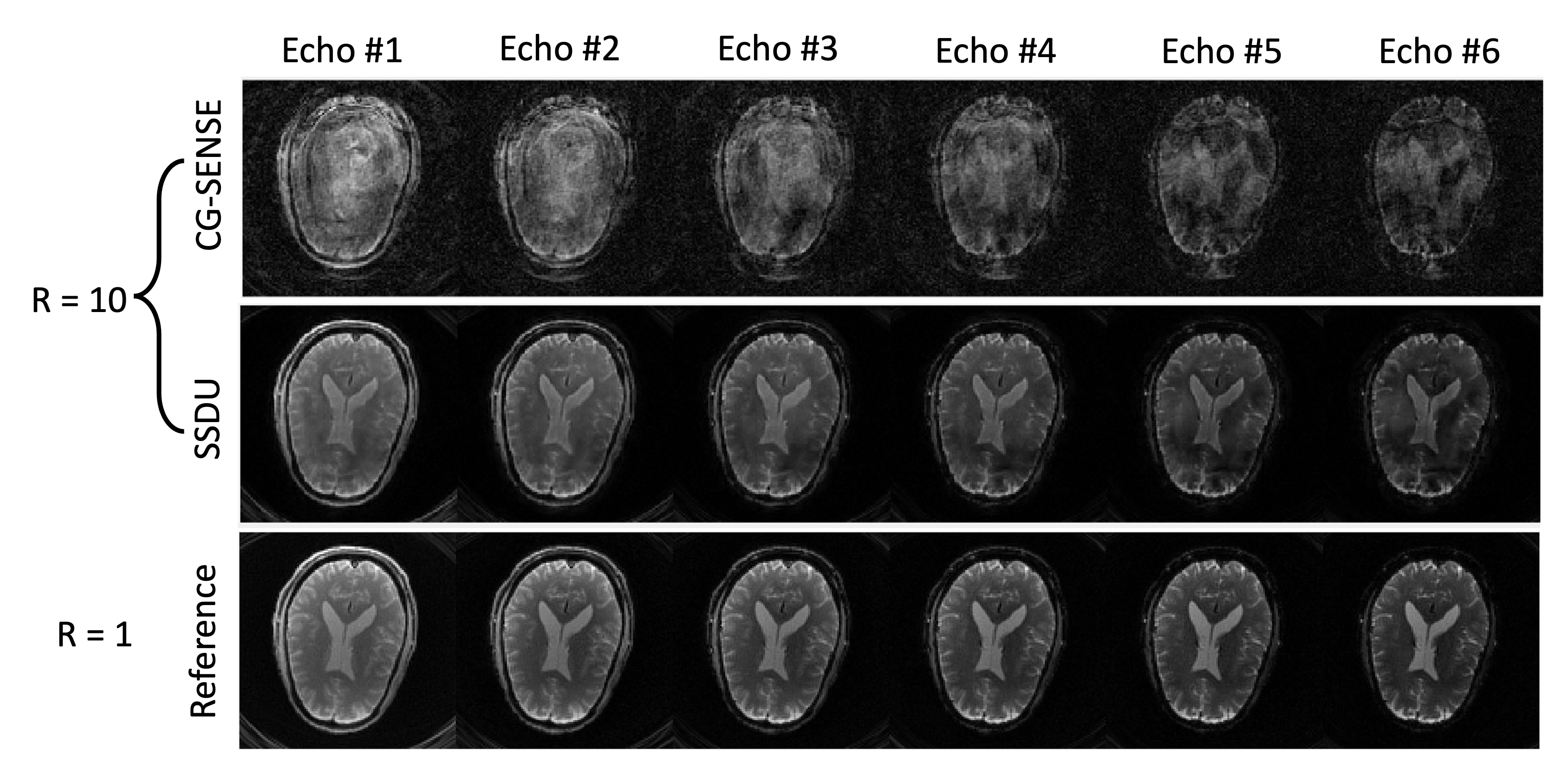
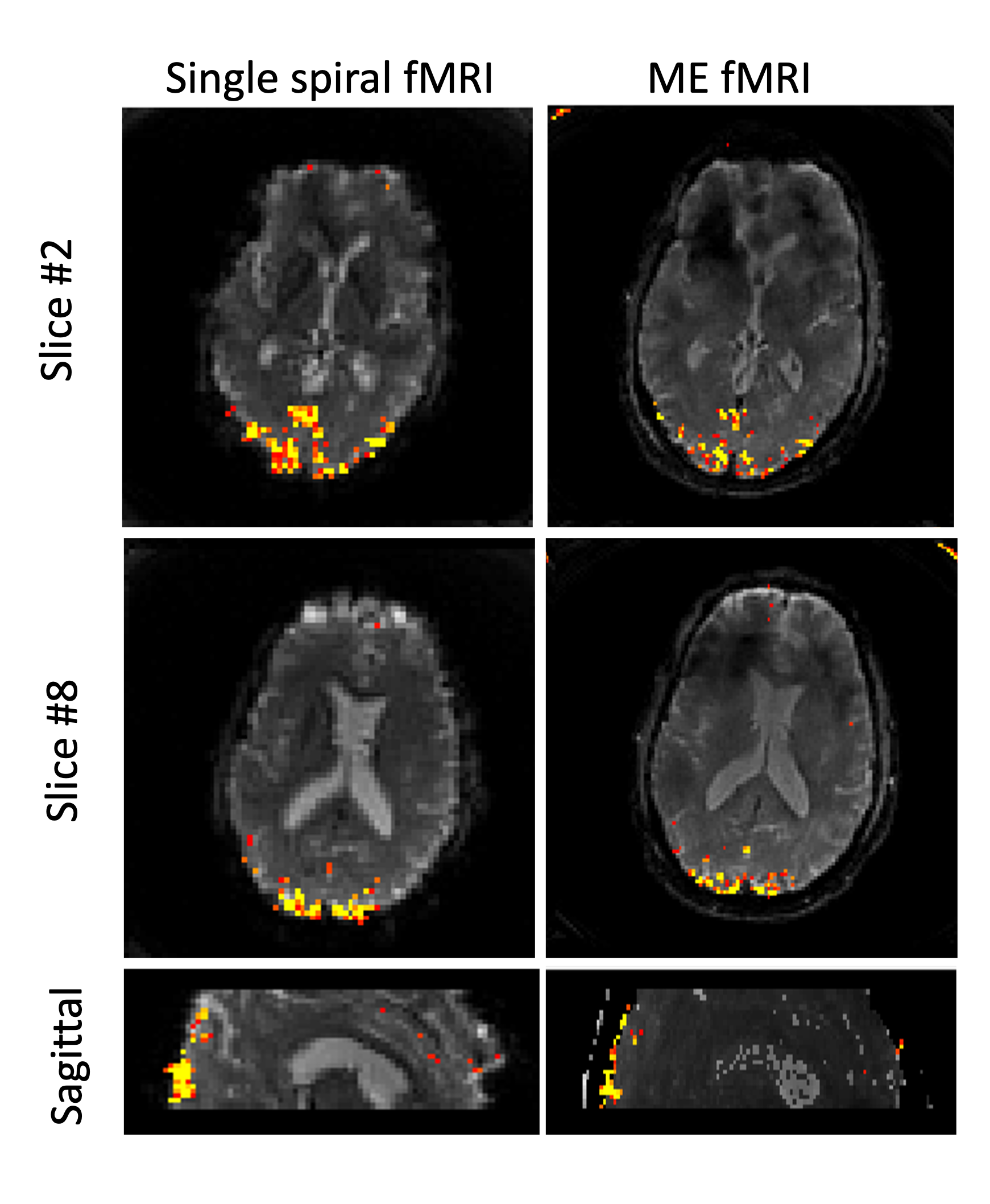
As shown in Fig.3, the images reconstructed by CG-SENSE are clearly not applicable for BOLD analysis. SSDU show decent quality images however there are some residual artifacts which may be caused by the mismatch of the TR lengths between the training data and the fMRI data. BOLD signal has been well contained with SSDU reconstruction as shown in Fig.4. The BOLD sensitivity from SSDU results was not as strong as the single shot fMRI as a result of the difference between the under-sampling rate and the resolution. The SNR of the single spiral fMRI is about 4.7($$$=\sqrt{10}\times\frac{3}{2}$$$) times higher than the SSDU, not to mention it has a longer TR.Discussion& Conclusion
The SSDU reconstruction has not only enabled the possibility for multi-echo spiral fMRI to use a short readout time, but also preserved the BOLD sensitivity with the highly under-sampled data. Future plans include optimization of the acquisition parameters and fine tune the DL model using more rigorous training/testing datasets, in order to improve the performance of the DL methods.Acknowledgements
Acknowledgment: NIH R01EB028627, NIH R01EB032830, NIH P41 EB027061, NIH 1P20GM139753-01A1. The first three authors contributed equally.References
[1] Kundu, P. et al. Multi-echo fMRI: A review of applications in fMRI denoising and analysis of BOLD signals. NeuroImage 154, 59–80 (2017).
[2] Pruessmann KP, Weiger M, Börnert P, Boesiger P. Advances in sensitivity encoding with arbitrary k-space trajectories. Magn Reson Med. 2001;46(4):638-51.
[3] Feng L, Grimm R, Block KT, Chandarana H, Kim S, Xu J, Axel L, Sodickson DK, Otazo R. Golden-angle radial sparse parallel MRI: combination of compressed sensing, parallel imaging, and golden-angle radial sampling for fast and flexible dynamic volumetric MRI. Magn Reson Med. 2014;72(3):707-17.
[4] Nam S, Akçakaya M, Basha TA, Stehning C, Manning WJ, Tarokh V, Nezafat R. Compressed sensing reconstruction for whole-heart imaging with 3D radial trajectories: a graphics processing unit implementation. Magn Reson Med. 2013;69(1):91-102.
[5] Akçakaya M, Nam S, Basha TA, Kawaji K, Tarokh V, Nezafat R. An augmented Lagrangian based compressed sensing reconstruction for non-Cartesian magnetic resonance imaging without gridding and regridding at every iteration. PLoS One. 2014;9(9):e107107.
[6] Baron CA, Dwork N, Pauly JM, Nishimura DJ. Rapid compressed sensing reconstruction of 3D non-Cartesian MRI. Magn Reson Med. 2018;79(5):2685-2692.
[7] Hammernik K, Klatzer T, Kobler E, Recht MP, Sodickson DK, Pock T, Knoll F. Learning a variational network for reconstruction of accelerated MRI data. Magn Reson Med. 2018;79(6):3055-3071.
[8] Aggarwal HK, Mani MP, Jacob M. MoDL: Model-Based Deep Learning Architecture for Inverse Problems. IEEE T Med Imaging. 2019;38(2):394-405.
[9] Hosseini SAH, Yaman B, Moeller S, Hong M, Akçakaya M, Dense Recurrent Neural Networks for Accelerated MRI: History-Cognizant Unrolling of Optimization Algorithms. IEEE J Sel Top Signal Process. 2020;14(6):1280-1291.
[10] Poser, Benedikt A., et al. "BOLD contrast sensitivity enhancement and artifact reduction with multiecho EPI: parallel‐acquired inhomogeneity‐desensitized fMRI." Magnetic Resonance in Medicine: An Official Journal of the International Society for Magnetic Resonance in Medicine 55.6 (2006): 1227-1235.
[11] Yaman B, Gu H, Hosseini SAH, Demirel OB, Moeller S, Ellermann J, Uğurbil K, Akçakaya M. Multi-mask self-supervised learning for physics-guided neural networks in highly accelerated magnetic resonance imaging. NMR Biomed. 2022;35(12):e4798.
[12] Fessler JA, Sutton BP. Nonuniform fast Fourier transforms using min-max interpolation. IEEE Trans. Signal Process. 2003;51(2):560-574.
[13] Pipe JG, Menon P. Sampling density compensation in MRI: rationale and an iterative numerical solution. Magn Reson Med. 1999;41(1):179–186.
[14] Yaman B, Hosseini SAH, Moeller S, Ellermann J, Uğurbil K, Akçakaya M. Self-supervised learning of physics-guided reconstruction neural networks without fully sampled reference data. Magn Reson Med. 2020;84(6):3172-3191.
[15] Ramani S, Liu Z, Rosen J, Nielsen JF, Fessler JA. Regularization parameter selection for nonlinear iterative image restoration and MRI reconstruction using GCV and SURE-based methods. IEEE Trans Image Process. 2012;21(8):3659-72.
Figures