2779
Noisy-Signal2Parameter(S2P): Structure-adaptative parameter map reconstruction for filter-exchange imaging without clean data1College of Information Science and Electronic Engineering, Zhejiang University, Hangzhou, China, 2Key Laboratory of Biomedical Engineering of Ministry of Education, College of Biomedical Engineering and Instrument Science, Zhejiang University, Hangzhou, China, 3Siemens Healthineers Ltd, Shanghai, China, 4School of Medicine, Zhejiang University, Hangzhou, China
Synopsis
Keywords: Image Reconstruction, Machine Learning/Artificial Intelligence, Filter-exchange imaging, water exchange rate, parameter map reconstruction, denoise
Motivation: Water exchange measured by filter-exchange imaging (FEXI) is expected to serve as an important biomarker for several brain diseases. However, its estimation accuracy is easily affected by noise.
Goal(s): To develop an approach for reconstruction of FEXI parameters from noisy signals.
Approach: An end-to-end framework was constructed to achieve parameter reconstruction without corresponding labels. An adaptative deformable convolutional network was introduced to explore structural information. A loss function was designed to enhance network denoising performance.
Results: Simulation results under SNR=30~50 showed that the S2P achieved optimal results in the reconstruction of apparent water exchange rate, with PSNR of 27.44 and SSIM of 0.9050.
Impact: The S2P, an end-to-end framework, reconstructs high-quality FEXI parameter maps from only a single scan when it has been trained with noisy pairs, which can provide efficient and reliable medical images for clinical diagnosis.
Introduction
Filter-exchange imaging (FEXI)1-3, a technique developed from diffusion exchange spectroscopy4 for clinical applications, is suitable for the detection of subtle blood-brain barrier impairment in patients with Alzheimer’s disease (AD)5. In some brain regions, the apparent exchange rate (AXR) measured by FEXI has shown a significant correlation with impairment of the respective cognitive domains5, indicating great potential to serve as a biomarker for early diagnosis of AD. However, the current FEXI technique has three limitations:1. The current FEXI technique improves the SNR by averaging the acquired multiple scans under the same configuration2,3, which is time-consuming and causes patient discomfort.
2. FEXI parameters are estimated by performing non-linear least squares (NLLS) model fit on a pixel-by-pixel basis2,3, which is inefficient and cannot capture spatially continuous information.
3. The effects of noise in FEXI signals can be mitigated by Block matching 3D denoising (BM3D)6 or other traditional denoisers. However, signal denoising and parameter estimation are conducted in two separate steps3, resulting in remaining noise in the final reconstructed parameter map.
To address these issues, we proposed a deep learning-based method termed S2P (i.e., Noisy-Signal2Parameter), which directly achieves FEXI parameter map reconstruction from noisy signals.
Theory
To quantitatively assess the methods, we generated simulation dataset based on a Sheep-Logan Phantom (Fig.1A) and reference distributions (Fig.1B)3,7. Corresponding FEXI parameter maps and MR signals were obtained based on the FEXI technique. The FEXI sequence (Fig.1C) contains filter, mixing, and detection blocks. The diffusion weighting $$$b_f$$$ in the filter block is $$$250 s/mm^2$$$. At the fixed $$$b_f$$$, the acquired MR signal is formulated as:$$S(b_f) = S^{eq}(1-f_i^{eq})e^{-b_fD_e}+S^{eq}f_i^{eq}e^{-b_fD_i}$$
$$S'(t_m) = S(b_f)f_i(t_m) + S(b_f)f_e(t_m) = S(b_f)$$
$$S(b_d,t_m) = S'(t_m)e^{-b_dADC'(t_m)}$$
where $$$S^{eq}$$$ denotes the signal before filter block and is randomly sample in [500,1500], $$$ADC'(t_m) = ADC(1-{\sigma}e^{-t_mAXR})$$$. The labels of the FEXI parameters can be calculated as:
$$ADC = f_i^{eq}D_i+(1-f_i^{eq})D_e$$
$$AXR = k_{ie} + k_{ei} = \frac{k_{ie}}{1-f_i^{eq}}$$
$${\sigma} = 1 - \frac{ADC'(t_m=0)}{ADC} = 1 - \frac{f_i(t_m=0)D_i+(1-f_i(t_m=0))D_e}{ADC}$$
where $$$f_i(t_m=0) = \frac{f_i^{eq}e^{-b_fD_i}}{(1-f_i^{eq})e^{-b_fD_e}+f_i^{eq}e^{-b_fD_i}}$$$. The $$$ADC'(t_m)$$$ can be computed from two $$$b_d$$$ values, $$$b_d1$$$ and $$$b_d2$$$, at each $$$t_m$$$:
$$ADC'(t_m) = -\frac{1}{b_{d2}-b_{b1}}ln(\frac{S(b_{d2},t_m)}{S(b_{d1},t_m)})$$
To simulate a real signal acquisition scenario, we introduced Rician noise (SNR=30~50 for $$$S^{eq}$$$) and applied random rotations, translations, and scaling to the signal map.
Methods
The proposed S2P (Fig.2) consists of two parts, parameter reconstruction (PRec) and signal reconstruction (SRec), which achieves simultaneous denoising and parameter reconstruction. The PRec is a conventional downsample-upsample architecture8, which has achieved substantial success in visual tasks. However, its key module, a convolutional neural network, performs poorly in geometric transformation modeling because of the rigid geometric structures of the convolutional kernel9, especially for noisy inputs. Therefore, an adaptative deformable convolution network10 (ADCN) (Fig. 3) is used in the second layer of S2P to enable the framework to focus on pertinent image contents, which can be formulated as:$$y(p) = \sum_{k=1}^{K}w_k{\cdot}x(p+AD_k{\cdot}p_k+{\Delta}p_k){\cdot}((1-AD_k){\cdot}{\Delta}m_k) $$
where $$$x(p)$$$ and $$$y(p)$$$ denote feature representation at location $$$p$$$ of the input and output feature maps, respectively; $$$AD_k$$$ is the adaptative dilation factor; $$$p_k$$$ and $$${\Delta}p_k$$$ are fixed and learnable offsets; and $$${\Delta}m_k$$$ is the modulation scalar.
Considering that there are no labels but multiple scans for the same image in our dataset, we defined the loss function based on Noise2Noise(N2N)11 as:
$$Loss = MSE(f_{\theta}(S)-S')$$
where MSE denotes Mean-Squared-Error, $$$f_{\theta}(\cdot)$$$ denotes the network, $$$S$$$ and $$$S'$$$ denote two independent scans of the same image. It has been proved that a network trained to map a noisy image to another noisy image is equivalent to supervised training11,12.
Results
To evaluate the effectiveness of the N2N-based loss function, end-to-end framework, and ADCN, we compared the S2P with NLLS, N2N-NLLS, S2Avg, S2P_wo_ADCN, and S2P (Details of these methods are explained in Fig.4). The results (Fig.4) were assessed by the peak signal-to-noise ratio (PSNR) and structural similarity index (SSIM)13. The S2P achieved optimal results in the reconstruction of AXR, with PSNR of 27.44 and SSIM of 0.9050. Additionally, visualization of the parameter reconstruction results in Fig. 5 revealed that the S2P performed equally well in AXR reconstruction.Discussion
As shown in Figs.4&5, the NLLS exhibited the worst performance, demonstrating the effects of noise on parameter reconstruction. Compared with S2P_wo_ADCN, S2Avg showed inferior performance, indicating that targeting the average of multiple scans limits the denoising capacity of the network. For ADC and sigma, N2N-NLLS achieved the highest PSNR&SSIM, presumably because of the excellent denoising performance of N2N.Conclusion
The S2P provides a novel approach for reconstructing parameter maps from noisy signals without labels. The optimized network can achieve parameter reconstruction with a single scan, substantially reducing the time required for clinical data acquisition.Acknowledgements
This work is supported in part by the National Natural Science Foundation of China (NSFC) (Grant Nos. 82111530201, 82222032, 82172050), the STI2030-Major Projects Q22 of China (Grant No. 2022ZD0206000).
References
1. Åslund I, Nowacka A, Nilsson M, et al. Filter-exchange PGSE NMR determination of cell membrane permeability. J Magn Reson, 2009, 200(2): 291-295.
2. Nilsson M, Lätt J, van Westen D, et al. Noninvasive mapping of water diffusional exchange in the human brain using filter-exchange imaging. Magn Reson Med, 2013, 69(6): 1572-1580.
3. Bai R, Li Z, Sun C, et al. Feasibility of filter-exchange imaging (FEXI) in measuring different exchange processes in human brain. Neuroimage, 2020, 219: 117039.
4. Callaghan P T, Furo I. Diffusion-diffusion correlation and exchange as a signature for local order and dynamics. J Chem Phys, 2004, 120(8): 4032-4038.
5. Zhang Y, Wang Y, Li Z, et al. Vascular-water-exchange MRI (VEXI) enables the detection of subtle AXR alterations in Alzheimer's disease without MRI contrast agent, which may relate to BBB integrity. NeuroImage, 2023, 270: 119951.
6. Dabov K, Foi A, Katkovnik V, et al. Image denoising by sparse 3-D transform-domain collaborative filtering. IEEE Trans Image Process, 2007, 16(8): 2080-2095.
7. Bisdas S, Koh T S, Roder C, et al. Intravoxel incoherent motion diffusion-weighted MR imaging of gliomas: feasibility of the method and initial results. Neuroradiology, 2013, 55: 1189-1196.
8. Ronneberger O, Fischer P, Brox T. U-net: Convolutional networks for biomedical image segmentation. Proc. Int. Conf. Med. Image Comput. Comput.-Assisted Intervention, 2015: 234-241.
9. Dai J, Qi H, Xiong Y, et al. Deformable convolutional networks. Proc. of IEEE/CVF Int. Conf. on Comput. Vis. 2017: 764-773.
10. Chen F, Wu F, Xu J, et al. Adaptive deformable convolutional network. Neurocomputing, 2021, 453: 853-864.
11. Lehtinen J, Munkberg J, Hasselgren J, et al. Noise2Noise: Learning Image Restoration without Clean Data. Proc. Int. Conf. Mach. Learn. PMLR, 2018: 2965-2974.
12. Huang T, Li S, Jia X, et al. Neighbor2neighbor: Self-supervised denoising from single noisy images. Proc. IEEE Conf. Comput. Vis. Pattern Recognit. 2021: 14781-14790.
13. Wang Z, Bovik A C, Sheikh H R, et al. Image quality assessment: from error visibility to structural similarity. IEEE Trans Image Process, 2004, 13(4): 600-612.
Figures
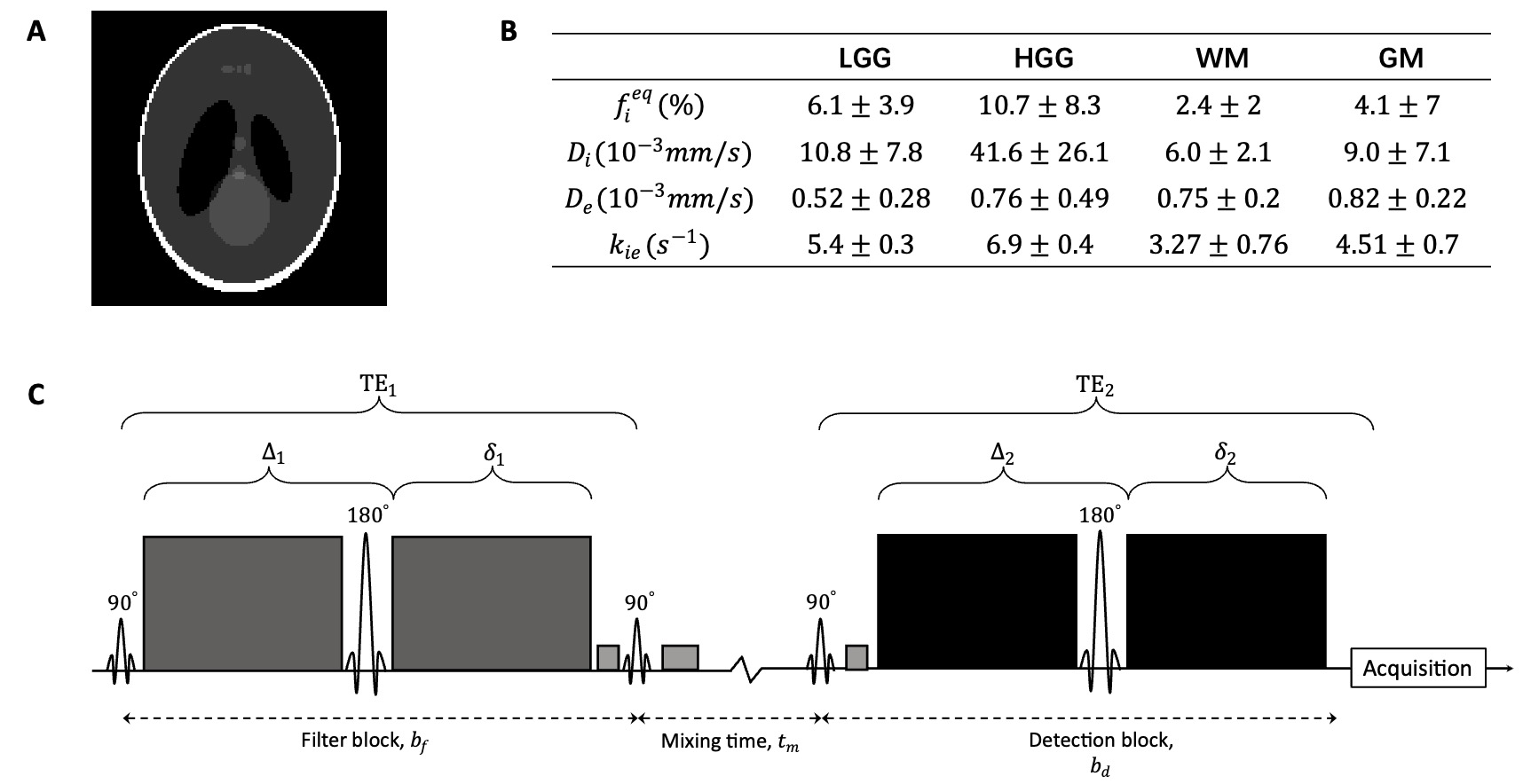
Figure1. Generation of simulated data. (A) Sheep-Logan Phantom. (B) Statistical distributions of low- and high-grade gliomas (LGG and HGG), white matter (WM), and gray matter (GM). $$$f_i^{eq}$$$ denotes volume fractions, $$$D_{i(e)}$$$ denotes diffusivities, $$$k_{ie}$$$ denotes exchange rate from intravascular to extracellular. (C) FEXI pulse sequence.
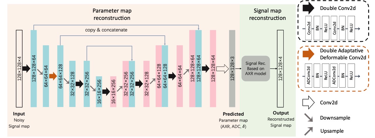
 according to the noisy signal. The signal map reconstruction part is constructed based on the FEXI technique and contains no trainable parameter, it reconstructs the signal based on predicted parameters.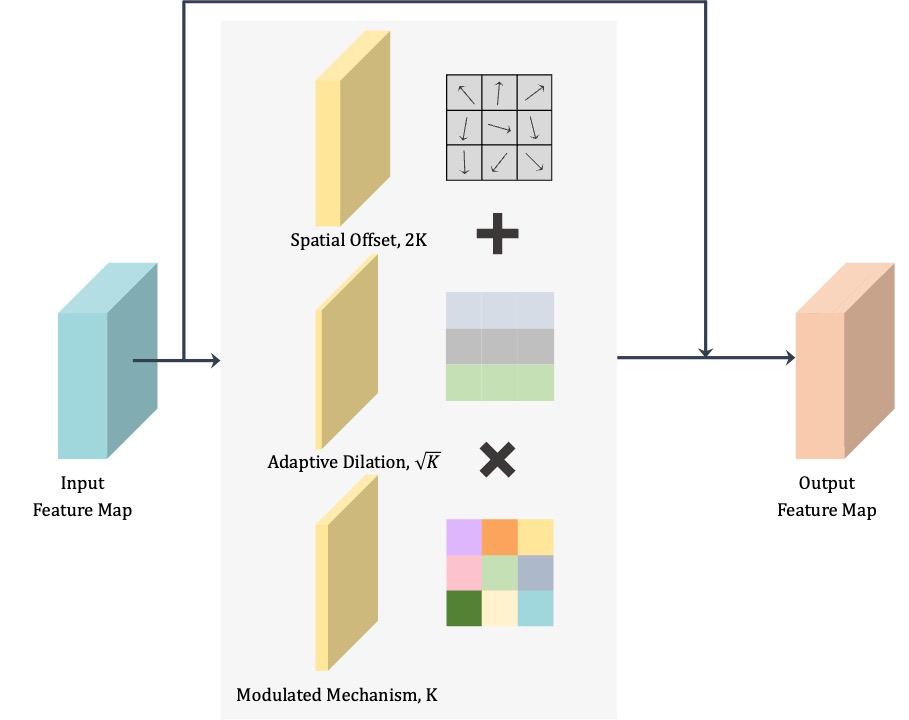
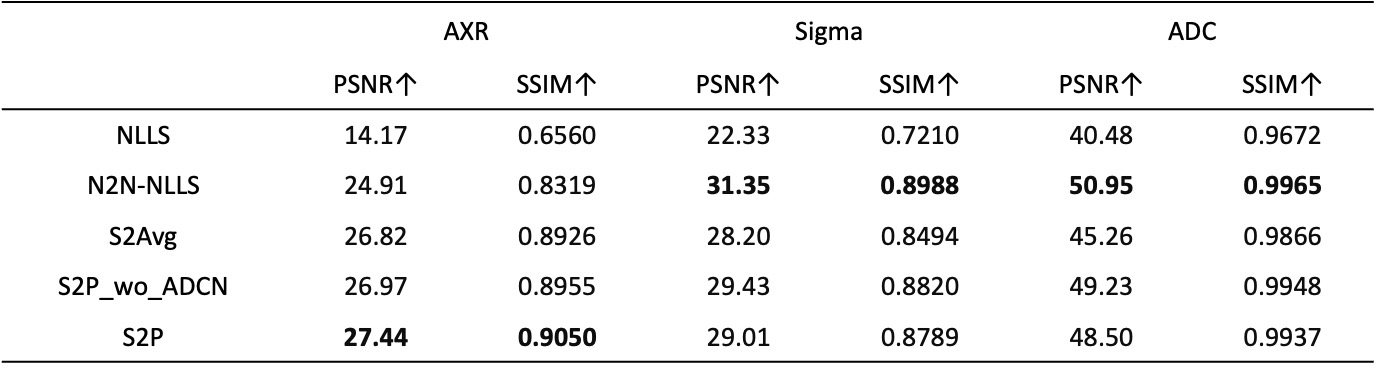
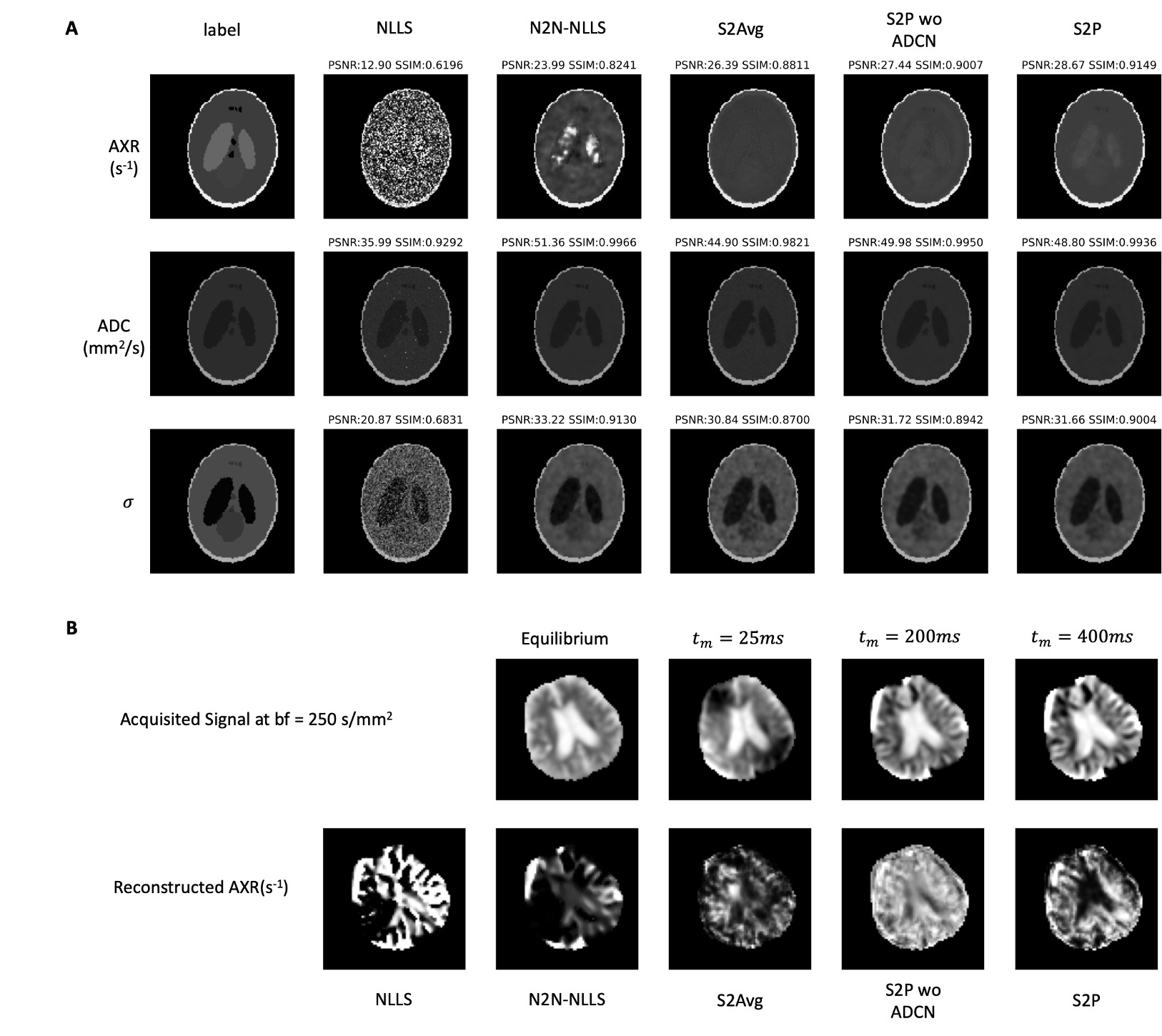
Figure 5. Results of reconstructed parameter maps. (A) Reconstruction results of simulation data. Presented from top to bottom are AXR, ADC and sigma maps. Presented from left to right are labels and reconstructed results based on NLLS, N2N-NLLS, S2Avg, S2P_wo_ADCN, and S2P. (B) Reconstruction results of one healthy subject. The first row are the $$$ADC’(t_m)$$$ with $$$b_f=250 s/mm^2$$$ at equilibrium ($$$b_f=0s/mm^2$$$ with shortest $$$t_m$$$) and three $$$t_m$$$. The second row denotes the reconstructed AXR map based on NLLS, N2N-NLLS, S2Avg, S2P_wo_ADCN and S2P.