2777
fastMRI Breast: A publicly available radial k-space dataset of breast dynamic contrast-enhanced MRI1Radiology, Weill Cornell Medicine, New York, NY, United States, 2Center for Advanced Imaging Innovation and Research (CAI2R), Department of Radiology, New York University, New York, NY, United States, 3Department Artificial Intelligence in Biomedical Engineering, Friedrich Alexander University Erlangen-Nuernberg, Erlangen, Germany
Synopsis
Keywords: Machine Learning/Artificial Intelligence, Cancer
Motivation: There is a lack of publicly available k-space data of breast dynamic contrast-enhanced (DCE) MRI that can be used for development of image reconstruction and machine learning methods for breast MRI.
Goal(s): We aim to make a publicly available radial k-space dataset of breast DCE-MRI which will promote development of fast and quantitative breast imaging methods.
Approach: Data of women undergoing routine diagnostic breast DCE-MRI exams have been acquired using a stack-of-stars radial imaging at 3T.
Results: Our fastMRI breast dataset includes radial k-space data and case-level labels for 275 cases (70 malignant, 158 benign and 47 no-lesion cases).
Impact: This work introduces the first large-scale dataset of radial k-space data for breast DCE-MRI acquired in diagnostic breast MRI exams. Having this dataset and accompanying reconstruction code publicly available, will support research and development of fast and quantitative breast DCE-MRI.
Introduction
Recently, a number of publicly available MRI datasets have been released, such as RNet (1), HCP (2), and ADNI (3). However, these datasets are limited to reconstructed images and do not include raw k-space data. Due to the limited availability of k-space datasets, the fastMRI initiative has recently been taken to make the raw k-space data of brain, knee (4) and prostate (5) publicly available. The current fastMRI datasets are all cartesian sequences. fastMRI+ (6) also include labels for various pathologies, enabling the development of clinically relevant reconstruction and detection methods. In this abstract, we present a new dataset for breast MRI, particularly with radial k-space data of Dynamic Contrast-Enhanced (DCE)-MRI acquired during diagnostic MRI exams. Breast DCE-MRI is widely used for detecting increased risk screening (7), cancer staging (8), and prognosis with chemotherapy (9,10). However, it presents significant challenges in image reconstruction, given the inherent trade-off between temporal resolution and image quality. To address this challenge, acquisition of radial DCE-MRI with a golden-angle has been proposed combined with compressed sensing and parallel imaging, offering high spatial resolution and flexible temporal resolution, while maintaining comparable diagnostic performance to conventional DCE (11). By making the fastMRI Breast dataset publicly available, we aim to facilitate the development of fast and quantitative breast DCE-MRI methods.Description of dataset and code
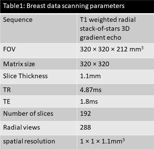
Patient population: The dataset includes raw k-space data for 275 breast cancer patients scheduled for clinical breast MRI. The patients were scanned between Dec-2018 and June-2022. 70 cases were found to have a malignant lesion, 158 benign and 47 cases had no suspicious lesions. The mean age of subjects is 43±11. The data was acquired with approval from the Institutional Review Board.Breast DCE-MRI study: All subjects underwent a diagnostic breast MRI exam on a whole-body 3T scanner (MAGNETOM TimTrio, Siemens Healthcare, Erlangen, Germany) with a 16-channel breast coil. The DCE-MRI was conducted with a golden-angle radial stack-of-stars 3D gradient echo sequence known has GRASP (Golden-angle RAdial Sparse Parallel imaging) (12). A total of 288 spokes were acquired continuously with 83 partitions for 2.5 min, resulting in 192 slices after partial Fourier (see scan parameters in Table 1). Spectrally selective Adiabatic Inversion Recovery (SPAIR) was used for fat suppression. A GRASP scan of 2.5 min was first acquired before the administration of contrast (Seg 1) and a second scan was acquired during contrast injection (Seg 2). For the second GRASP scan, a single dose of Gadobutrol (Gadavist, Bayer Healthcare Pharmaceuticals) at 0.1 mM/kg body weight was injected at 2 mL/s intravenously at the 1-min mark into the scan. Both scans can be used for image reconstruction or the second one alone as an ultrafast breast MRI scan.
Raw k-space dataset: We provide the raw k-space data for Seg 1 and Seg 2, along with GRASP reconstructions for all 275 cases. Raw data were exported from the scanners, anonymized, and saved into a Hierarchical Data Format (HDF5) file which can be easily manipulated in Python. The corresponding reconstructed images are also included in these files. Our dataset also includes case-level labels describing, for each case, whether it is malignant, benign, or has no lesion. Dataset will be hosted at: https://fastmri.med.nyu.edu/.
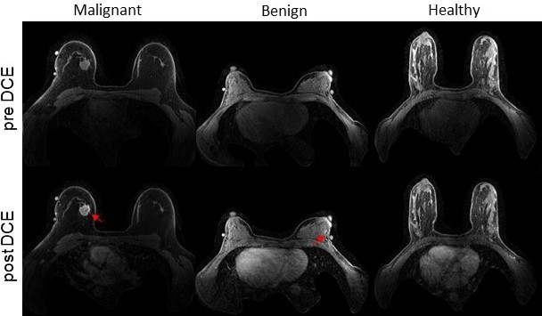
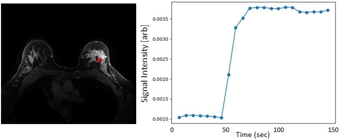
Reconstruction code: With this dataset, we provide a GPU-based GRASP reconstruction code implemented in python. The reconstruction code uses SigPy implementations of NUFFT (13), coil sensitivity estimation(14) and iterative reconstruction with temporal total variation (TV) regularization. Radial data can be used to reconstruct dynamic images with a flexible temporal resolution (aka, number of radial views per frame). Representative reconstructed images of malignant, benign and healthy datasets are shown in Fig. 1. In the presence of a cancer lesion, one can appreciate rapid time to enhancement following contrast injection using the provided DCE-MRI data (Fig.2).
Discussion
To our knowledge this is the first large-scale publicly available radial k-space data of breast DCE-MRI from a clinical patient population. While public breast DCE datasets do exist, they are generally focused on testing particular research question such as segmentation and classification (15) or machine learning performance (16). The fastMRI breast data can serve not only for qualitative examination of the post-contrast images, but also for the calculation of quantitative pharmacokinetic kinetic parameters to improve the diagnostic performance of breast DCE-MRI. It can also be used independently as a dataset for reconstruction of static images without contrast change. The availability of this dataset will further promote research of breast DCE image reconstruction and quantification of breast lesions.Acknowledgements
RSNA Research Seed Grant RSD1830, NIH P41EB017183, NIH R01CA160620, R01CA219964, and UH3CA228699References
- Bien N, Rajpurkar P, Ball RL, Irvin J, Park A, Jones E, Bereket M, Patel BN, Yeom KW, Shpanskaya K, Halabi S, Zucker E, Fanton G, Amanatullah DF, Beaulieu CF, Riley GM, Stewart RJ, Blankenberg FG, Larson DB, Jones RH, Langlotz CP, Ng AY, Lungren MP. Deep-learning-assisted diagnosis for knee magnetic resonance imaging: Development and retrospective validation of MRNet. PLoS Med 2018;15(11):e1002699.
- Elam JS, Glasser MF, Harms MP, Sotiropoulos SN, Andersson JLR, Burgess GC, Curtiss SW, Oostenveld R, Larson-Prior LJ, Schoffelen JM, Hodge MR, Cler EA, Marcus DM, Barch DM, Yacoub E, Smith SM, Ugurbil K, Van Essen DC. The Human Connectome Project: A retrospective. Neuroimage 2021;244:118543.
- Mueller SG, Weiner MW, Thal LJ, Petersen RC, Jack CR, Jagust W, Trojanowski JQ, Toga AW, Beckett L. Ways toward an early diagnosis in Alzheimer's disease: the Alzheimer's Disease Neuroimaging Initiative (ADNI). Alzheimers Dement 2005;1(1):55-66.
- Knoll F, Zbontar J, Sriram A, Muckley MJ, Bruno M, Defazio A, Parente M, Geras KJ, Katsnelson J, Chandarana H, Zhang Z, Drozdzalv M, Romero A, Rabbat M, Vincent P, Pinkerton J, Wang D, Yakubova N, Owens E, Zitnick CL, Recht MP, Sodickson DK, Lui YW. fastMRI: A Publicly Available Raw k-Space and DICOM Dataset of Knee Images for Accelerated MR Image Reconstruction Using Machine Learning. Radiol Artif Intell 2020;2(1):e190007.
- Tibrewala R, Dutt T, Tong A, Ginocchio L, Keerthivasan M, Baete S, Chopra S, Lui Y, Sodickson D, Chandarana H, Johnson P. FastMRI Prostate: A Publicly Available, Biparametric MRI Dataset to Advance Machine Learning for Prostate Cancer Imaging. 2023.
- Zhao R, Yaman B, Zhang Y, Stewart R, Dixon A, Knoll F, Huang Z, Lui YW, Hansen MS, Lungren MP. fastMRI+, Clinical pathology annotations for knee and brain fully sampled magnetic resonance imaging data. Sci Data 2022;9(1):152.
- Mann RM, Kuhl CK, Moy L. Contrast-enhanced MRI for breast cancer screening. J Magn Reson Imaging 2019;50(2):377-390.
- Rizzo V, Moffa G, Kripa E, Caramanico C, Pediconi F, Galati F. Preoperative Staging in Breast Cancer: Intraindividual Comparison of Unenhanced MRI Combined With Digital Breast Tomosynthesis and Dynamic Contrast Enhanced-MRI. Front Oncol 2021;11:661945.
- Heacock L, Lewin A, Ayoola A, Moccaldi M, Babb JS, Kim SG, Moy L. Dynamic Contrast-Enhanced MRI Evaluation of Pathologic Complete Response in Human Epidermal Growth Factor Receptor 2 (HER2)-Positive Breast Cancer After HER2-Targeted Therapy. Acad Radiol 2020;27(5):e87-e93.
- Kim SY, Cho N, Park IA, Kwon BR, Shin SU, Kim SY, Lee SH, Chang JM, Moon WK. Dynamic Contrast-enhanced Breast MRI for Evaluating Residual Tumor Size after Neoadjuvant Chemotherapy. Radiology 2018;289(2):327-334.
- Heacock L, Gao Y, Heller SL, Melsaether AN, Babb JS, Block TK, Otazo R, Kim SG, Moy L. Comparison of conventional DCE-MRI and a novel golden-angle radial multicoil compressed sensing method for the evaluation of breast lesion conspicuity. J Magn Reson Imaging 2017;45(6):1746-1752.
- Feng L, Grimm R, Block KT, Chandarana H, Kim S, Xu J, Axel L, Sodickson DK, Otazo R. Golden-angle radial sparse parallel MRI: combination of compressed sensing, parallel imaging, and golden-angle radial sampling for fast and flexible dynamic volumetric MRI. Magn Reson Med 2014;72(3):707-717.
- Fessler JA, Sutton BP. Nonuniform fast Fourier transforms using min-max interpolation. Ieee T Signal Proces 2003;51(2):560-574.
- Uecker M, Lai P, Murphy MJ, Virtue P, Elad M, Pauly JM, Vasanawala SS, Lustig M. ESPIRiT--an eigenvalue approach to autocalibrating parallel MRI: where SENSE meets GRAPPA. Magn Reson Med 2014;71(3):990-1001.
- Zhao X, Liao Y, Xie J, He X, Zhang S, Wang G, Fang J, Lu H, Yu J. BreastDM: A DCE-MRI dataset for breast tumor image segmentation and classification. Comput Biol Med 2023;164:107255.
- Saha A, Harowicz MR, Grimm LJ, Kim CE, Ghate SV, Walsh R, Mazurowski MA. A machine learning approach to radiogenomics of breast cancer: a study of 922 subjects and 529 DCE-MRI features. Br J Cancer 2018;119(4):508-516.
Figures