2776
Evaluation of blood flow changes in iliac vein compression: a hemodynamic model based on magnetic resonance multimodal imaging and CFD simulation1School of Life Science and Technology,Xi'an jiaotong University, Xi'an, China, 2The first affiliated Hospital of Xi'an jiaotong University, Xi'an, China, 3School of Energy and Power Engineering,Xi'an jiaotong University, Xi'an, China, 4Philips Healthcare, Xi'an, China, 5Philips Healthcare, Beijing, China
Synopsis
Keywords: Vessels, Velocity & Flow, iliac venous compression
Motivation: Hemodynamic changes are important for evaluating Iliac venous compression syndrome. CFD simulation can obtain individualized and quantitative hemodynamic information that traditional imaging methods cannot obtain.
Goal(s): Qbtain hemodynamic parameters through the establishment of patient individualized vascular model and CFD simulation. By observing hemodynamic parameters, the changes in blood flow caused by iliac venous compression can be more accurately evaluated.
Approach: The 3D model is constructed based on the MRA images. After CFD simulation, changes of flow dynamics parameters observed due to the influence of iliac venous compression.
Results: The CFD simulation shows that iliac venous compression can cause significant changes in hemodynamics
Impact: Hemodynamic changes are the pathological basis of iliac venous compression. Accurate diagnosis of blood flow changes is helpful to guide the surgical treatment of iliac venous compression and provide powerful help for clinical decision-making.
Introduction
Iliac venous compression syndrome (IVCS) is an umbrella term that refers to a composite of clinical symptoms due to pathologic structural and hemodynamic changes in the iliac vein.1 IVCS is an important pathophysiologic factor in the development of chronic venous disease (CVD) and has received increasing attention owing to its extremely high prevalence.1 The probability of deep vein thrombosis (DVT) induced by IVCS is 18-48%, and the symptom recurrence rate was as high as 18.9% after iliac vein interventional stenting during 1 year follow-up, especially for the stenosis rate was more than 70%.2-3 Therefore, the exploration of accurate diagnosis and prognosis of iliac vein stenosis is a very important part in the diagnosis of CVD. Compared with CT and Doppler ultrasound, multimodal MR could clearly show the iliac vein compression and obstruction, meanwhile could perform hemodynamic assessment of the narrow site, which provides great help to the clinical significance of pathologic iliac vein stenosis and hemodynamic abnormalities.4-5 At present, multimodal magnetic resonance imaging (MRI) was less used to build a hemodynamic change computational fluid dynamics (CFD) model of iliac pressure. Therefore, the purpose of this study is to build a CFD model of iliac pressure using multimodal MRI to elaborate the pathological changes of its blood flow and achieve accurate diagnosis of blood flow changes.Methods
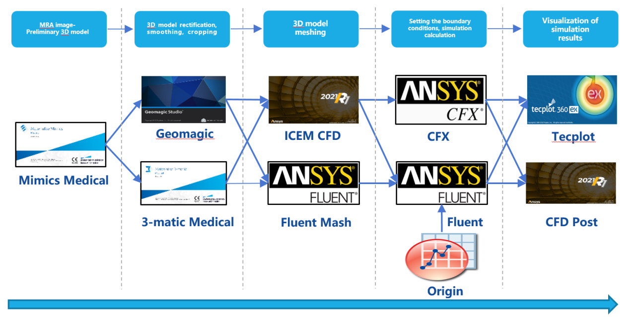
Two patients with iliac venous compression were selected. Philips 3.0T magnetic resonance imaging was used to perform pelvic coronal enhanced scanning. The top scan was carried out to the middle part of inferior vena cava, and the bottom contained bilateral external iliac veins. The Q-Flow scanning scheme was as follows: axial scanning was performed on inferior vena cava, bilateral common iliac vein and external iliac vein respectively. Preliminary 3D models of lower limb veins from inferior vena cava to external iliac vein and internal iliac vein were constructed from MRA original images. Geomagic Studio software was used to smooth the preliminary 3D model, and then discretization processing was performed on the 3D models by using Ansys Fluent. The blood flow velocity data of iliac vein extracted by Q-Flow were used as inlet boundary conditions for CFD simulation, and hemodynamic parameters such as blood vessel wall pressure, wall shear stress and flow velocity curve were obtained. The changes of flow dynamics parameters from iliac vein to inferior vena cava were observed due to the influence of iliac venous compression.The CFD simulation process is shown in the flow chart (Figure 1)Results
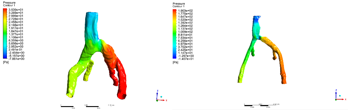
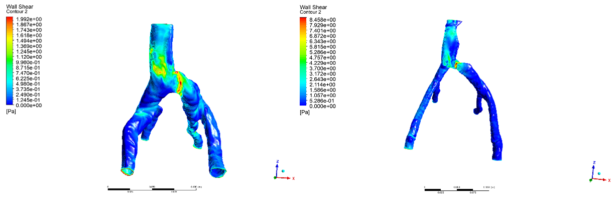
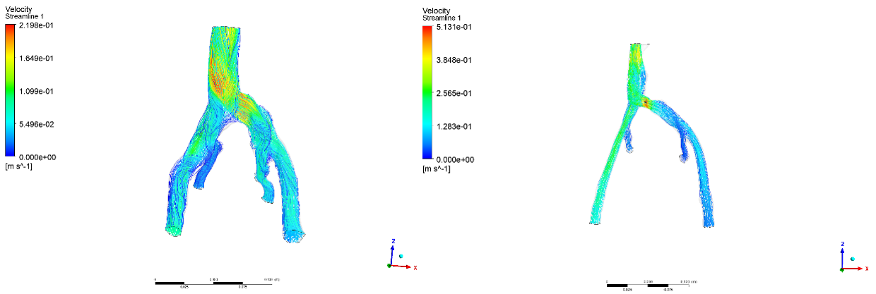
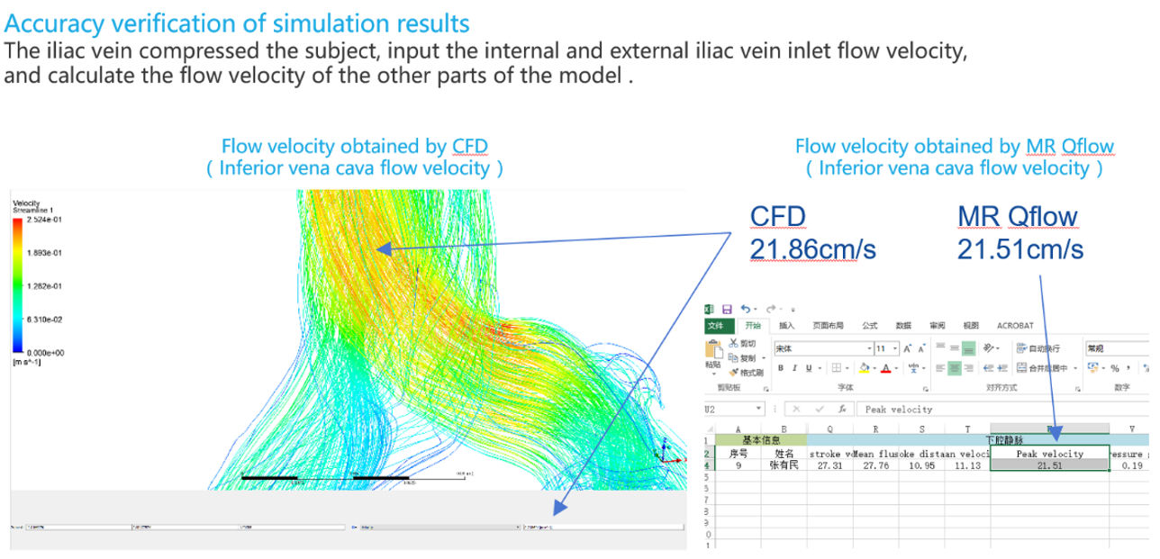
The CFD simulation results showed that the pressure of the blood vessel wall of patients 1 and 2 increased significantly from the compression stenosis site of the iliac vein to the distal end of the external and internal iliac vein (Figure 2). The wall shear stress of the compression site increased significantly (Figure 3). At the same time, the blood flow velocity of the compression site and the blood flow velocity of the common iliac vein and inferior vena cava on the compressed side increased (Figure 4). The results show that the blood flow velocity obtained by CFD simulation is highly consistent with the blood flow velocity measured by Q-Flow (Figure 5)Discussion
It can be seen from the experimental results that the CFD simulation method can obtain individualized and detailed hemodynamic parameters that cannot be obtained by traditional imaging methods, and can more directly and quantitatively reflect the hemodynamic changes of the compression site, proximal and distal of the lower limb vein caused by iliac venous compression. The results of the two CFD simulations indicated that iliac venous compression would lead to a significant increase in the vessel wall pressure at the distal iliac vein starting from the iliac vein compression site, which was a clear evidence of obstruction of venous return due to vascular compression. Changes in wall pressure, flow velocity, and flow rate can provide more direct and accurate hemodynamic evidence for interpretation and evaluation of the obstruction of venous return caused by iliac venous compression. High consistency between CFD simulation results and clinical measurements makes CFD become an important supplement to traditional imaging examination and clinical hemodynamic measurement methods.Conclusion
The hemodynamic parameters such as wall pressure, wall shear stress, flow velocity and flux obtained by individualized CFD simulation can provide more direct and accurate evidence for interpretation and evaluating the changes in blood flow caused by iliac venous compression.Acknowledgements
No acknowledgement found.References
1. Chen ZH, Huang Y, Wang LP, Peng MY, Li C, Huang W. Preliminary study of hemodynamics of iliac venous compression syndrome using magnetic resonance imaging. J Vasc Surg Venous Lymphat Disord. 2022 Jan;10(1):131-138.e3.
2. Wang S, He Y, Xin S, Zhang J. Iliac vein stenting is a safe and effective treatment for iliac vein compression syndrome: A systematic review of Chinese data. Phlebology. 2020 Dec;35(10):752-770.
3. Han Y, Tian Y, Gao L, Tang J, Fan P, Cong L, Dong J, Yang L. Clinical outcomes of different endovenous procedures among patients with varicose veins and iliac vein compression: A retrospective cohort study. Int J Surg. 2022 May;101:106641.
4. Gloviczki P, Lawrence PF. JVS-VL is the leading journal in venous and lymphatic disorders. J Vasc Surg Venous Lymphat Disord. 2019 Jan;7(1):1-6.
5. Saleem T, Lucas M, Raju S. Comparison of intravascular ultrasound and magnetic resonance venography in the diagnosis of chronic iliac venous disease. J Vasc Surg Venous Lymphat Disord. 2022 Sep;10(5):1066-1071.e2.
Figures