2774
Three-dimensional Phase-contrast MRA of Foot Vessels at 5T: Visualization Improved by Warm Water Immersion1Radiology, Zhongshan Hospital, Fudan University, Shanghai, China, 2Institute of Science and Technology for Brain-Inspired Intelligence, Fudan University, Shanghai, China
Synopsis
Keywords: Vessels, Vessels, velocity; high-field MR
Motivation: Unenhanced MRA on feet has always been challenging due to slow blood flow. The ultra-high field strength MR systems have been proved to offer improved visualization of distal small vessels.
Goal(s): We aim to assess the visualization of foot vessels using three-dimensional phase contrast MRA (3D-PC-MRA) at 5T field-strength.
Approach: Participants were prospectively recruited and underwent 5T 3D-PC-MRA on feet with and without warm water immersion.
Results: Immersion of feet in warm water significantly improves visualization of distal small branches, and increases the average velocity of blood flow by 40%, resulting in the visualized enhancement in blood signal.
Impact: 5T 3D phase-contrast MRA is effective in visualizing lower-extremity vasculatures and small branches, and its visualization can be significantly improved by immersing feet in warm water.
INTRODUCTION
Non-contrast-enhanced MRA techniques have been used for the diagnosis of peripheral vascular diseases. However, their flow-dependency limits their ability to visualize distal vessels and tortuous anatomy with slow blood flow, especially in the case of distal foot vessels. This study proposes a flow-enhancing strategy to improve the visualization of foot vessels for phase-contrast (PC) MRA by immersing feet in warm water.METHODS
To conduct warm water immersed (WWI) PC-MRA, patients' feet were immersed in warm water before the MRI examination. The temperature of the water ranged from 40 to 45 ℃ and was adjusted based on the tolerance level of each patient. WWI acquisitions were conducted after normal acquisitions without WWI within two hours.Eighteen healthy volunteers (age 38 ± 9 years) were imaged using a 5T MRI system (uMR Jupiter, United Imaging Healthcare, Shanghai) with a 48-channel receiver head coil between October 2022 and March 2023. 3D PC-MRA was performed using three-directional velocity encoding (VENC) with value of 3cm/s for all subjects. Other parameters include FOV 220×220×78 mm³, spatial resolution of 0.6mm³, flip angle=15, TE=8.5ms, TR=54.7ms, and compressed sensing factor 5. The acquisition time was 9 min 4 sec.Images were independently graded by two experienced radiologists based on diagnostic image quality, visualization of vessel segments and depiction of the small branches. The SNR and CNR were calculated from the original PC images, with blood signal measured from the dorsalis pedis artery and distal posterior tibial artery and noise measured from plantar muscle tissue. Morphologic evaluation was performed using a semi-automatic analysis method [1,2]. All vessels were classified into large and small vessels by global thresholding based their radius distribution (threshold = 0.72 mm). Morphologic measurements, including the number of branches and the total branch length, were calculated for all vessels as well as for small vessels specifically. To investigate the impact of WWI on velocity, we computed the mean velocity from the phase information of PC-MRA based on five volunteer's data [3].RESULTS
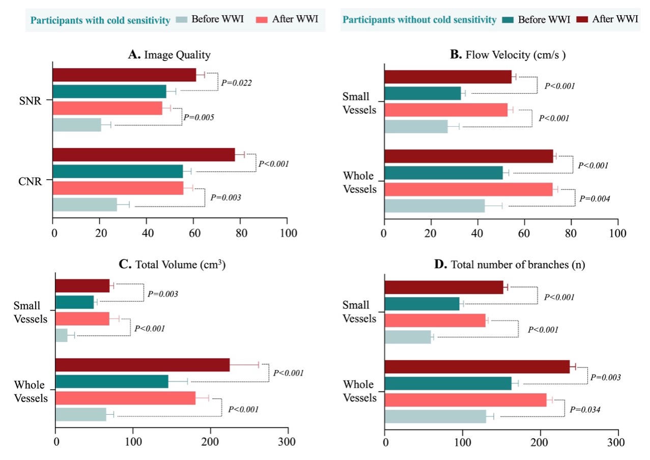
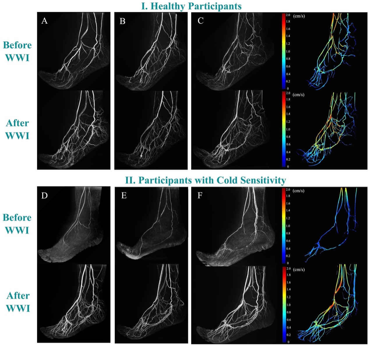
Twenty participants (mean age, 44.6 ± 6.1; males, 14) were included. The image quality scores for 5T scans were excellent (median score = 5.0). All measurements related to image quality and vessel visualization significantly improved after WWI, including the percentage of excellent depiction of vessel segments (score = 3, P = .008), total volume (118.95 ± 55.38 vs 209.79 ± 54.73 cm3; P = .008) and the total number of branches (151.92 ± 29.17 vs 225.63 ± 16.76; P < .001) (Table-1). The percentage of good collateral status (scale≥2) in both calcaneal and plantar branches also increased after WWI. Flow velocity was enhanced by WWI (P < .001). All the increases were particularly pronounced for small vessel and branches and for individuals with cold sensitivity (Figure-1). Figure 2 shows representative maximum intensity projection (MIP) and velocity maps of normal and WWI PC-MRA.DISCUSSION
Limb ischemia is a prevalent peripheral arterial disease that frequently affects the feet. Contrast-enhanced MRA and CTA are routinely used for diagnosis. However, for those with tibial disease, they have limitations in providing sufficient resolution of ankle and foot vessels for procedural planning. Unenhanced MRA techniques have been developed as an alternative for peripheral MRA, but their flow-dependency limits their ability to visualize distal vessels and tortuous anatomy with slow blood flow. This study proposes a flow-enhancing strategy to improve the visualization of foot vessels for PC-MRA by immersing feet in warm water, and the findings showed immersing the feet in warm water prior to scans greatly improved the visualization of foot arteries for PC-MRA, particularly for small vessels and branches.Acknowledgements
NONE.References
[1] Shi Z et al., Radiology 2023.
[2] Zhang B et al., JMRI, 2021.
[3] Kang C et al., MRM 2015.
Figures