2770
Serum metabolic profiling of human valve diseases based on proton NMR spectroscopy1NMR, AIIMS, NEW DELHI, DELHI, India, 2CARDIOLOGY, AIIMS, NEW DELHI, NEW DELHI, India, 3Pathology, AIIMS, NEW DELHI, DELHI, India, 4CTVS, AIIMS, NEW DELHI, DELHI, India
Synopsis
Keywords: Vessels, Heart, NMR spectroscopy
Motivation: Heart valve disease (HVD) is a complex condition with a poorly known pathogenesis.
Goal(s): NMR-based serum metabolomics of HVD patients (aortic, mitral valve, and double valve replacement) and healthy controls to identify potential biomarkers for HVD.
Approach: Proton NMR spectroscopy
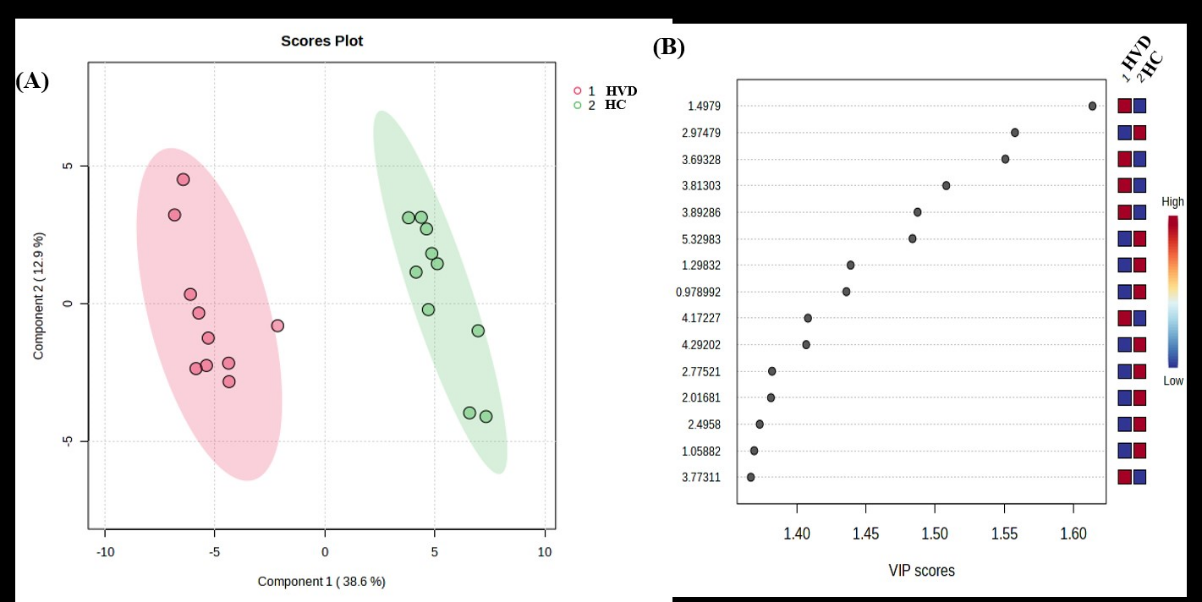
Results: The results obtained from PLS-DA and VIP score plots of metabolites in serum showed a separation between patients with HVD and HC.
Impact: To understanding the potential metabolic alteration such as BCAA, and fatty acids, amino acids, and carbohydrate metabolism associated with inflammation, oxidative stress, and tissue degradation of HVD.
Degenerative and rheumatic heart valve disease (HVD) is caused by the interaction of several risk factors such as infections, age-related changes, ischemic heart disease, congenital, heart failure, stroke, genetic, inflammatory, autoimmune, and oxidative stress(1-2). The diagnosis of heart valve disease is difficult due to the lack of sensitive and specific valvular dysfunction biomarker/s. Thus, the aim of the study is to investigate and compare NMR-based serum metabolomics of HVD patients (aortic, mitral valve, and double valve replacement) and healthy controls to identify potential biomarkers for HVD.
Methods
Blood samples were collected from HVD patients (n =10) and HC (n=10), in the morning pre-prandial and stored at -80°C until NMR experiments were performed. Proton spectra were acquired at a 700 MHz spectrometer. 300 µl blood serum, 30 µl formate (0.5 Mm), and 270 µl D2Owill be used to make a 600 µl overall volume of sample. one-dimensional spectrum was acquired by using a standard (1D) Carr-Purcell-Meiboom-Gill (CPMG) pulse sequence to suppress broad signals from bigger molecules, such as lipids and proteins. The parameter for spin echo (CPMG) was; no. of scan 64, tau value 16 ms, data point 64K, and relaxation delay 14 seconds with pre-saturation of water during relaxation delay. Some important metabolites were assigned using 1D and 2D NMR. The spectral regions at 0.5-10 ppm were segmented into bins with equal widths of 0.04 ppm. Thereafter, the serum spectral region of δ5.5–4.5 ppm (water resonance) was removed from the whole spectrum (δ10.0-0.5–0. ppm) to eliminate the distorted baseline from imperfect water saturation. P values were two-tailed and a value < 0.05 was considered significant. The data were normalized and subjected to multivariate pattern recognition analysis using MetaboAnalyst 5.0 software. Metabolites were considered significant at VIP >1.0 for further analysis of metabolomic data analysis. Partial least square discriminant analysis (PLS-DA) plots were carried out for significant metabolite identification.
Results
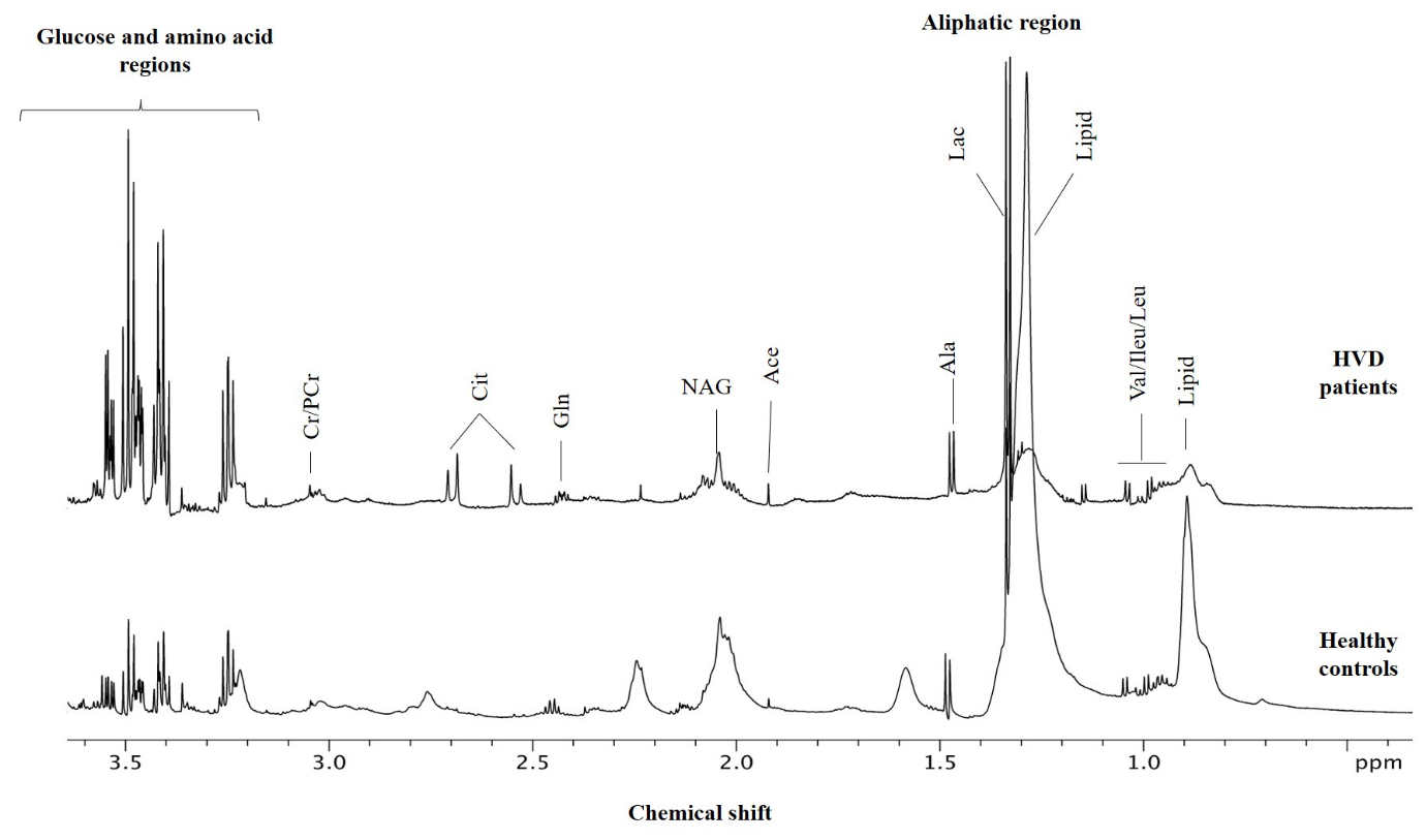
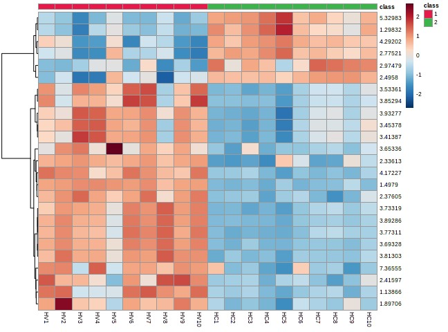
Figure 1 shows Representative CPMG 1H NMR spectra of blood serum obtained from patients with HVD and HC. The serum metabolomic profile of HVD patients is characterized by increased levels of alanine, glycerol, and β-hydroxybutyrate, lysine and decreased levels of asparagine, glucose, leucine, isoleucine valine glutamate, citrate, N-acetyl glycoprotein compared with HC. The results obtained from PLS-DA and VIP score plots of metabolites in serum showed a separation between patients with HVD and HC in Figure 2.
Discussion
The present study suggests that compared to HC, the blood metabolomic profile of HVD patients shows lower levels of asparagine, glucose, leucine, isoleucine, valine, glutamate, citrate, and N-acetyl glycoprotein and higher levels of alanine, glycerol, and β-hydroxybutyrate, lysine. In patients during disease progression in the serum sample, amino acids are now becoming more widely appreciated as cardioprotective substrates, as evidenced by the recent increase in excellent review articles detailing the importance of cardiac amino acid metabolism (3). Other amino acids, such as asparagine, may also be important metabolic indicators due to their ability to remove amine groups and excess Krebs cycle intermediates downstream from the conversion to organic acids (4-5). 1H-NMR-based metabolomics may offer insight into analyzing the potential metabolic alteration such as BCAA and fatty acid, amino acid, and carbohydrate metabolism associated with inflammation, oxidative stress, and tissue degradation of HVD, the current study demonstrated the ability to distinguish metabolic profiling of serum of HVD patients from HC. Findings of the PLS-DA and VIP score plots of blood metabolites demonstrated a difference between patients with HVD and HC. These novel serum biomarkers may improve the management of human valve disease complications, thus reducing the use of valve replacement and improve medical care.
Conclusion The present study revealed discrimination of metabolic profiling of serum of HVD patients from HC implying that 1H-NMR-based metabolomics may provide an insight into understanding the potential metabolic alteration such as BCAA, and fatty acids, amino acids, and carbohydrate metabolism associated with inflammation, oxidative stress, and tissue degradation of HVD.
Acknowledgements
Thanks AIIMS New Delhi for Research grant.References
1. Fu B, Wang J, Wang L, Wang Q, Guo Z, Xu M, Jiang N. Integrated proteomic and metabolomic profile analyses of cardiac valves revealed molecular mechanisms and targets in calcific aortic valve disease. Front Cardiovasc Med. 2022 Oct 13;9:944521.
2. 26. Kamburov A, Cavill R, Ebbels TM, Herwig R, Keun HC. Integrated pathway-level analysis of transcriptomics and metabolomics data with IMPaLA. Bioinformatics. (2011) 27:2917–8.
3. 27. Surendran A, Edel A, Chandran M, Bogaert P, Hassan-Tash P, Asokan AK, et al. Metabolomic signature of human aortic valve stenosis. JACC Basic Transl Sci. (2020) 5:1163–77.
4. 28. Wang W, Wu J, Liu P, Tang X, Pang H, Xie T, et al. Urinary proteomics identifying novel biomarkers for the diagnosis and phenotyping of carotid artery stenosis. Front Mol Biosci. (2021) 8:714706.
5. 29. Gibb AA, Hill BG. Metabolic coordination of physiological and pathological cardiac remodeling. Circ Res. (2018) 123:107–28.
Figures