2769
Consistency of hemodynamics information measured by 4D Flow MRI and 2D phase contrast MRI in patients with mild to moderate carotid artery stenosis1Department of Radiology and Nuclear Medicine, Xuanwu Hospital, Capital Medical University, Beijing, China, 2Clinical & Technical Support, Philips Healthcare, Beijing, China
Synopsis
Keywords: Vessels, Vessels
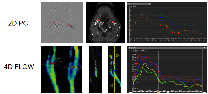
Motivation: The blood flow of carotid artory can be assessed by the standard 2D PC MRI, as well as the advanced 4D Flow MRI.
Goal(s): The aim of this study is to test the potential of 4D flow for accuracy of quantification of carotid flow parameters compared to the reference standards 2D PC MRI in patients.
Approach: 4D Flow MRI and 2D PC MRI were performed in patients with carotid artery stenosis. Quantitative hemodynamics of carotid arteries were measured in 2D PC and 4D Flow MRI.
Results: 4D Flow MRI and 2D PC MRI had a good consistency in the evaluation of carotid hemodynamics.
Impact: This study found that 4D Flow MRI and 2D PC MRI had a good consistency in the evaluation of carotid hemodynamics in patients with mild to moderate carotid artery stenosis.
Introduction
Carotid artery stenosis can result in a reduced supply of blood to the brain. 2D phase contrast (PC) MRI is considered as the standard for evaluating blood flow. However, it is limited by velocity analysis in 2D planes and by single-direction velocity measurement which may be inadequate to characterize the complex 3D hemodynamics. 4D Flow MRI provides simultaneous assessment of 3D blood flow characteristics of all vessels within a 3D volume and offers the ability to retrospectively quantify blood flow parameters at selectable regions of interest. The aim of this study is to test the potential of 4D flow for accuracy of quantification of carotid flow parameters compared to the reference standards 2D PC MRI in patients with mild to moderate carotid stenosis.Methods
30 patients with mild to moderate carotid artery stenosis were prospectively collected. 4D Flow MRI and 2D PC MRI (at 2-3 cm above the carotid artery bifurcation) scans for the the carotid artery were performed on a 3.0T MRI scanner (Ingenia, Philips Healthcare, Best, the Netherlands). The scan parameters are listed in Table 1. Quantitative hemodynamics data of internal and external carotid arteries, including velocity (cm/s), flow (mL/s), cross-aectional area (mm²) and maximum pressure gradient (mmHg), were mapped by two radiologists at 2-3 cm above the carotid artery bifurcations based on 2D PC and 4D Flow MRI, respectively, using the CVI42 platform (Version 5.6.6, Circle Cardiovascular Imaging, Canada). Kappa analysis was used to examine the consistency between two radiologists, paired sample t-test was used to compare the differences in hemodynamics measurements by 2D PC and 4D Flow, and Bland-Altman diagram was used to determine the consistency between 2D PC and 4D Flow MRI.Results
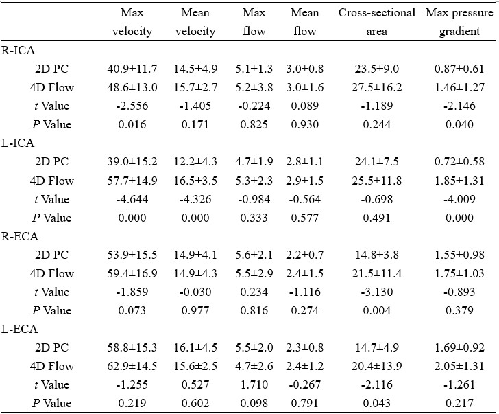
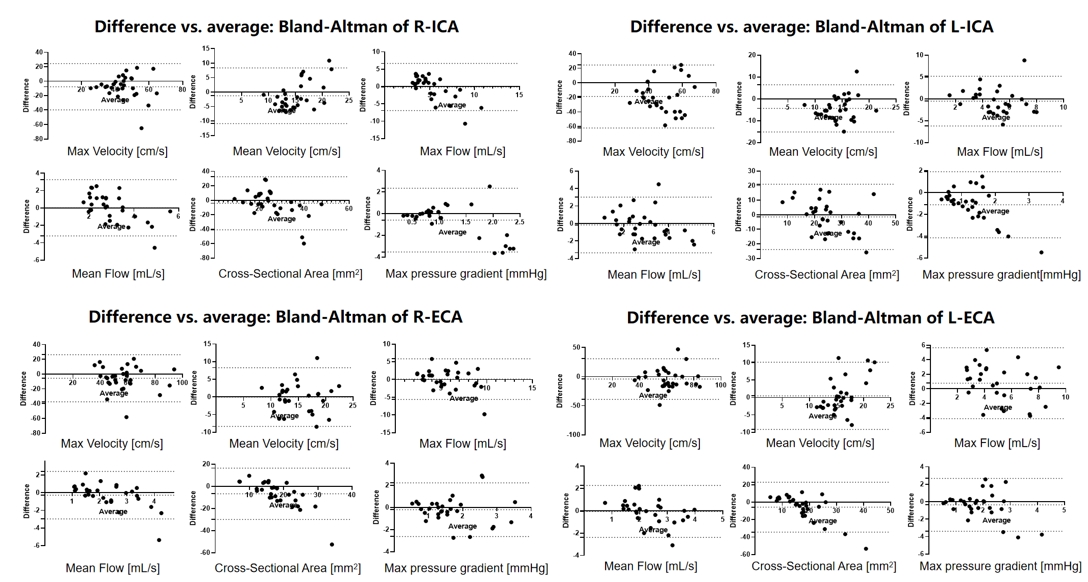
A total of 30 patients with mild to moderate carotid artery stenosis were collected, including 10 women (33.3%), 61±6 (50-74) years old, with a heart rate of 75±12 (55-102) bmp. In ICA vessels, the maximum velocity and pressure gradient measured by 2D-PC are significantly lower than those measured by 4D-Flow. In ECA vessels, the cross-sectional area measured by 2D-PC is significantly lower than that measured by 4D-Flow. The Bland-Altman diagram reveals the high consistency of hemodynamics measurements in carotid arteries between 2D PC and 4D Flow MRI, as evidenced by thant majority of data points were within the 95% confidence interval.Conclusion
2D PC and 4D Flow MRI showed a good consistency in the assessment of carotid hemodynamics in patients with mild to moderate carotid artery stenosis, and appropriate scan methods can be selected according to specific conditions in clinical work.Acknowledgements
No acknowledgement found.References
No reference found.Figures