2766
Oxygen-enhanced Ultrashort Echo Time MRI in Diagnosis of Benign and Malignant Solid Pulmonary Nodules: a preliminary study1Department of Radiology, Southwest Hospital, Army Medical University (Third Military Medical University), Chongqing, China, 27T Magnetic Resonance Imaging Translational Medical Center, Southwest Hospital, Army Medical University (Third Military Medical University), Chongqing, China, 3YuYue Laboratory, Chongqing, China, 4Jinfeng Laboratory, Chongqing, China, 5MR Collaboration, United Imaging Research Institute of Intelligent Imaging, Beijing, China
Synopsis
Keywords: Lung, Cancer, ultrashort echo time (UTE);oxygen-enhanced MRI (OE-MRI)
Motivation: People pay more and more attention to the high incidence and mortality of lung cancer.
Goal(s): To develop a non-invasive, radiation-free imaging scheme to distinguish between benign and malignant solid pulmonary nodules and to improve the management of patients with pulmonary nodules.
Approach: Two three-dimensional ultrashort echo time series scans of pulmonary nodules were performed on 3.0T MRI under the condition of 21% and 100% oxygen, respectively, and the percent signal enhancement was used as the evaluation index of benign and malignant nodules.
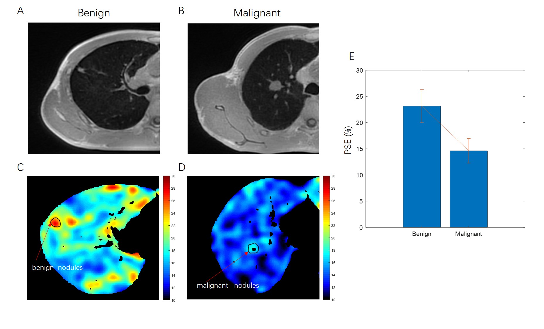
Results: The increase of the percent signal enhancement in benign pulmonary nodules was higher than that in malignant pulmonary nodules.
Impact: This imaging tool will reduce or eliminate the radiation damage caused by computed tomography, reduce clinicians' over-dependence on high resolution computed tomography, and bring new gospel for personalized and accurate diagnosis and treatment of patients.
Introduction
Lung cancer has the highest incidence rate among malignant tumors in the world, and its mortality rate ranks first. The Fleischner Society shows that nodule type and size assessment is an effective indicator of nodule management to reduce the specific mortality rate of lung cancer1. The main imaging screening method of pulmonary nodules is high resolution computed tomography (HRCT)2. However which HRCT has certain disadvantages in the detection and differentiation of solid pulmonary nodules, such as the presence of ionizing radiation, which limits its use in pregnant women and long-term follow-up patients.Because of the large amount of gas in the lungs, the display of pulmonary parenchyma by conventional magnetic resonance imaging (MRI) is poor. More recently, three-dimensional radial ultrashort echo time (3D-UTE) MRI has shown promise for noninvasively assessment of lung diseases3. It is comparable to HRCT in the detection rate of pulmonary nodules2,4-6. The 3D-UTE based oxygen enhanced (OE) MRI has also been used to assess regional ventilation abnormalities of the lung.
This preliminary study aims at investigating the utility of high-resolution 3D-UTE based OE-MRI to differentiate benign and malignant solid pulmonary nodules.
Methods
In this prospective study, we collected 8 patients with solid pulmonary nodules from the first affiliated hospital of Army Medical University. All patients underwent 3D-UTE OE-MRI scanning on 3.0T MRI (uMR 770, United Imaging Healthcare, Shanghai, China). Respiratory-gated 3D radial UTE pulse sequences (TR: 2.0 msec, TE: 0.09 msec, flip angle: 6, slice thickness: 2 mm, FOV: 360 × 360 mm2, matrix: 320 × 320) were acquired twice for each patient. The first was acquired during free-breathing with 21% oxygen (normoxic), while the second was acquired with 100% oxygen (hyperoxic). Percent signal enhancement (PSE) was used to quantify pulmonary ventilation. The pathology of the patients were obtained by puncture or operation.Results
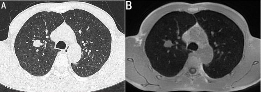
Of the 8 patients with solid pulmonary nodules, 6 were pathologically malignant and 2 were benign. The high-resolution 3D-UTE MRI achieved good lesion detectability of solid pulmonary nodules (Figure 1). We found that the mean PSE of malignant pulmonary nodule (14.61± 3.12%) was lower than that of the benign pulmonary nodule (23.15± 2.33%) (Figure 2). Due to the sample size of the data, although there was no significant difference between malignant and benign pulmonary nodule, it showed a good trend, and the P-value (P=0.07) was close to 0.05.Discussion
Our results suggest that the capability of 3D-UTE pulmonary MRI for the assessment of solid pulmonary nodules is potentially as good as that of standard thin-section CT. Further on, PSE was used to quantify malignant and benign pulmonary nodule ventilation. It was found that the average PSE of malignant pulmonary nodule was lower than that of the benign pulmonary nodule. This may be because the growth and spread of solid tumors require a large amount of oxygen and nutrition supply, resulting in the accumulation of neovascularization and the consumption of higher oxygen, resulting in a decrease in ventilation and a less obvious increase in PSE7.Conclusion
3D-UTE OE-MRI may become a new non-radiation imaging method in the differential diagnosis of benign and malignant solid nodules in the lung. This technique will bring good news to pregnant women, patients with iodine contrast allergy and patients who need long-term follow-up of pulmonary nodules.Key words
Magnetic resonance imaging (MRI); ultrashort echo time (UTE);oxygen-enhanced MRI (OE-MRI); solid pulmonary nodules; benign and malignantAcknowledgements
This work was funded by Chongqing Science and Health Joint key Project (2023ZDXM008) in 2022.References
1.MacMahon H, Naidich DP, Goo JM, et al. Guidelines for Management of Incidental Pulmonary Nodules Detected on CT Images: From the Fleischner Society 2017[J]. Radiology 2017;284(1):228-243.
2.Periaswamy G, Arunachalam VK, Varatharajaperumal R, et al. Comparison of Ultrashort TE Lung MRI and HRCT Lungs for Detection of Pulmonary Nodules in Oncology Patients[J]. Indian J Radiol Imaging 2022; 32(4): 497-504.
3.Ma Y, Jang H, Jerban S, et al. Making the invisible visible-ultrashort echo time magnetic resonance imaging: Technical developments and applications[J]. Appl Phys Rev 2022;9(4): 041303.
4.Ohno Y, Takenaka D, Yoshikawa T, et al. Efficacy of Ultrashort Echo Time Pulmonary MRI for Lung Nodule Detection and Lung-RADS Classification[J]. Radiology 2022; 302(3): 697-706.
5.Ohno Y, Koyama H, Yoshikawa T, et al. Standard-, Reduced-, and No-Dose Thin-Section Radiologic Examinations: Comparison of Capability for Nodule Detection and Nodule Type Assessment in Patients Suspected of Having Pulmonary Nodules[J]. Radiology 2017; 284(2): 562-573.
6.Burris NS, Johnson KM, Larson PE, et al. Detection of Small Pulmonary Nodules with Ultrashort Echo Time Sequences in Oncology Patients by Using a PET/MR System[J]. Radiology 2016; 278(1): 239-246.
7.Liu H, Zheng L, Shi G, et al. Pulmonary Functional Imaging for Lung Adenocarcinoma: Combined MRI Assessment Based on IVIM-DWI and OE-UTE-MRI[J]. Front Oncol 2021; 11: 677942.
Figures