2765
Comparative value of free-breathing DCE-MRI based on GRASP and conventional BH-VIBE sequence in evaluating the image quality in lung cancer1Department of Radiology, The first Hospital of LanZhou University, Lanzhou, China, 2Department of Medical Imaging, Anqing Municipal Hospital, Anqing, China, 3MR Research Collaboration, Siemens Healthineers Ltd, Shanghai, China
Synopsis
Keywords: Lung, DSC & DCE Perfusion, Lung
Motivation: Dynamic Contrast Enhancement-CT (DCE-CT) and Dynamic Contrast Enhancement-MRI (DCE-MRI) are among the most valuable imaging techniques for the noninvasive assessment of tumor vascular perfusion and trophoblast angiogenesis in lung cancer.
Goal(s): To evaluate the image quality value of free-breathing GRASP in MRI of lung cancer.
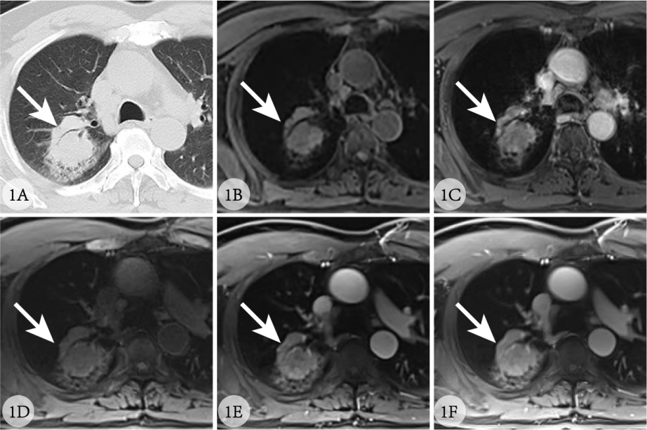
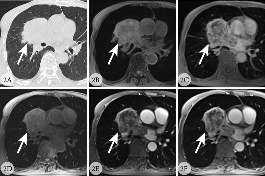
Approach: All patients underwent free-breathing DCE-MRI with breath-holding VIBE sequence before and after contrast injections. Two experienced thoracic radiologists independently evaluated the overall image quality, lesion contour, artifact level, and diagnostic confidence using the patient's simultaneous CT images as a reference.
Results: Free-breathing GRASP DCE-MRI technique is a reliable method for the high-resolution imaging of pulmonary lesions.
Impact: The study showcases GRASP's superiority over BH-VIBE in lung cancer imaging, offering clearer results with patient comfort. This breakthrough prompts investigation into GRASP's broader clinical applications and sets a new standard in patient-friendly MRI diagnostics.
Introduction
Lung cancer is one of the leading causes of cancer-related death from malignant tumors worldwide, with a high incidence 1. Tumor angiogenesis is crucial to its growth, invasion, and metastasis. DCE-CT is widely used in clinical lung cancer screening because of its high spatial resolution, high sensitivity, and rapid imaging. However, the cumulative ionizing radiation associated with repeated CT examinations is a concern. Recent studies have proved that GRASP can be used to optimize the free-breathing DCE-MRI of several organs sensitive to motion-induced artifacts in traditional Cartesian imaging, such as liver, esophagus, urinary system, and so forth [7-8], yielding satisfactory results. Therefore, this study aimed to explore the image quality of the free-breathing GRASP technique for high-resolution imaging of the lungs in a group of patients with lung cancer compared with the image quality of traditional Cartesian breath-holding volume interpolation scan (BH-VIBE).Materials and Methods
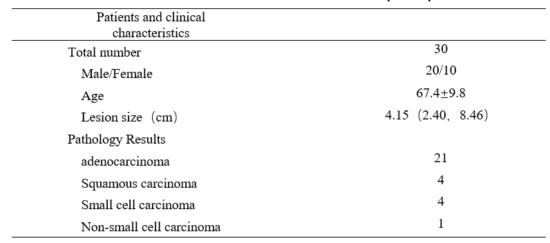
This study was approved by the Anqing Municipal Hospital Medical Ethics Committee (Approval Number: Medical Ethics Review [2022] No. 96). A total of 30 patients diagnosed with lung cancer through CT scans (20 male, 10 female; median age, 67 years; range, 48-91 years) using a 3T magnetic resonance imaging system (MAGNETOM Vida, Siemens Healthcare, Germany) with an 18-channel body phased-array coil from October 2022 to December 2022 were enrolled. Coronal T2-weighted imaging (HASTE sequence), axial diffusion-weighted imaging (EPI sequence), axial T1-weighted imaging (BH-VIBE sequence), axial GRASP (radial VIBE sequence), and axial T1-weighted contrast-enhanced imaging (BH-VIBE sequence) were performed. The total scan time for each patient was approximately 15 minutes. The specific imaging parameters of GRASP were as follows: FOV = 350 × 350 mm2; matrix size = 256 x 256; number of slices = 64; slice thickness = 3 mm; flip angle = 12°; in-plane spatial resolution = 1.2 x 1.2 mm2; TR/TE = 3.50 milliseconds/1.35 milliseconds; and total scan time = 366 seconds. Gadolinium-based contrast agent (GE Healthcare, Shanghai) was administered at a rate of 3.0 mL/s (0.2 mmol/kg body weight) 60 seconds after the start of data acquisition, followed by a 20-mL saline flush at the same rate. All participants were thoroughly informed regarding the examination process and provided informed consent before undergoing MRI scans. The inclusion criteria for patients were as follows: (1) solid lesion diameter greater than 10 mm; (2) absence of metallic implants; (3) patient cooperation with MR examination; and (4) availability of complete clinical and pathologic data. All selected GRASP images were visually assessed for quality compared with the corresponding BH-VIBE images and CT images. Two radiologists (each with 13 and 10 years of experience in chest MRI) blinded to the sequence information independently performed the visual image quality assessment. For each set of images, the readers assigned scores for overall image quality, lesion delineation, overall artifact level, and diagnostic confidence using a 4-point Likert scale: 4 = excellent; 3 = good; 2 = acceptable; and 1 = poor (nondiagnostic). Nonparametric paired-sample two-tailed Wilcoxon signed-rank tests were employed to compare readers’ scores between pre-contrast and post-contrast GRASP and BH-VIBE images. A P value less than .05 indicated a statistically significant difference. Cohen's kappa coefficient was used to assess the agreement between various image quality scores. Statistical analysis was performed using SPSS[Editor1] (version 25.0, Chicago). [Editor1]Please provide manufacturer details.Results
Finally, the study included 30 patients with lung cancer (20 male and 10 female; age 48-91 years; median age 67.4 years. The tumor sizes ranged from 1.5 to 7.1 cm. Histologically, the study was performed on 21 adenocarcinomas, 4 squamous cell carcinomas, 4 small cell lung carcinomas, and 1 non-small-cell lung carcinoma. The visual image quality scores for conventional Cartesian BH-VIBE and GRASP imaging indicated that all GRASP images had clear lesion delineation. Remarkable imaging performance was achieved before and after contrast injection, with all evaluation categories having P values less than .05. BH-VIBE image quality scores were consistently lower than those of GRASP, with considerable differences in the overall image quality and artifact level (P <.01).Discussion and conclusion
The study demonstrated the advantages of the GRASP sequence in effectively eliminating motion artifacts related to respiration and autonomous cardiac pulsation. The image quality of GRASP was remarkably superior to that of BH-VIBE (P <.01). Therefore, GRASP is a highly practical clinical lung MR imaging technique, which allows patients to breathe freely during MR scans, and hence is more patient-friendly and minimally impacts the image quality and diagnostic confidence.Acknowledgements
NoneReferences
[1]Wei W, Zeng H, Zheng R, et al. Cancer registration in China and its role in cancer prevention and control[J].Lancet Oncol,2020,21(7):e342-e349.DOI:10.1016/ S1470-2045 (20) 30073-5.
[2]Parikh N,Ream JM,Zhang HC,et al.Performance of simultaneous high temporal resolution quantitative perfusion imaging of bladder tumors and conventional multi-phase urography using a novel free-breathing continuously acquired radial compressed-sensing MRI sequence.Magn Reson Imaging 2016;34(5):694–8.DOI:10.1016/j.mri.2015.12.033.
[3]Yoon JH, Lee JM, Yu MH, et al.Evaluation of Transient Motion During Gadoxetic Acid-Enhanced Multiphasic Liver Magnetic Resonance Imaging Using Free-Breathing Golden-Angle Radial Sparse Parallel Magnetic Resonance Imaging[J]. Invest Radiol,2018,53(1):52-61.DOI:10.1097/RLI.0000000000000409.
Figures