2757
Virtual elastography based on an intravoxel incoherent motion for assessing chronic kidney disease1Department of Radiology, Shenzhen Traditional Chinese Medicine Hospital (The Fourth Clinical Medical College of Guangzhou University of Chinese Medicine), Shenzhen, China, 2Shenzhen University, Shenzhen, China, 3MR Research Collaboration, Siemens Healthineers, Shanghai, China, 4MR Research Collaboration, Siemens Healthineers, Guangzhou, China, 5MR Research Collaboration, Siemens Healthineers, Beijing, China
Synopsis
Keywords: Kidney, Data Analysis, Aging, chronic kidney disease, diagnosis,renal stiffness, intravoxel incoherent motion, shifted apparent diffusion coefficients ,virtual elastography
Motivation: Various b values of shifted apparent diffusion coefficients (sADC) and virtual magnetic resonance elastography (vMRE) were not validated for renal stiffness. Hence, a new method was required for assessing chronic kidney disease (CKD) noninvasively.
Goal(s): To investigate the relationship between sADC and vMRE and the clinical indicators and diagnostic efficacy of CKD.
Approach: The μdiff generated with sADC was used to quantify renal stiffness. The data were analyzed using Spearman, partial correlation, Mann–Whitney U test, and receiver-operating characteristic curves.
Results: sADC and vMRE were effective in assessing renal function and diagnosing CKD, but distinctly at various b values.
Impact: This study provides a reference b value for sADC and vMRE and suggests that vMRE could be used as a new noninvasive technique to assess chronic kidney disease.
Introduction
Magnetic resonance elastography (MRE) can quantify tissue stiffness and is feasible in assessing renal stiffness[1]. Intravoxel incoherent motion (IVIM) is a technique for noninvasively assessing renal function and chronic kidney disease (CKD) pathology[2-6]. An IVIM-based virtual MRE (vMRE) technique can help obtain elastography-like stiffness values by calculating shifted apparent diffusion coefficients (sADC) from 2 key b values in IVIM [7]. It can be used to assess liver fibrosis, few tumors, and placenta [ 8, 9, 10, 11]. sADC with a b value of 200-800 has clinical value for the noninvasive assessment of CKD[12]. This study investigated the relationship between multiple b values of ADC and vMRE and the clinical indicators of CKD, providing a new technique for the noninvasive clinical assessment of CKD.Methods
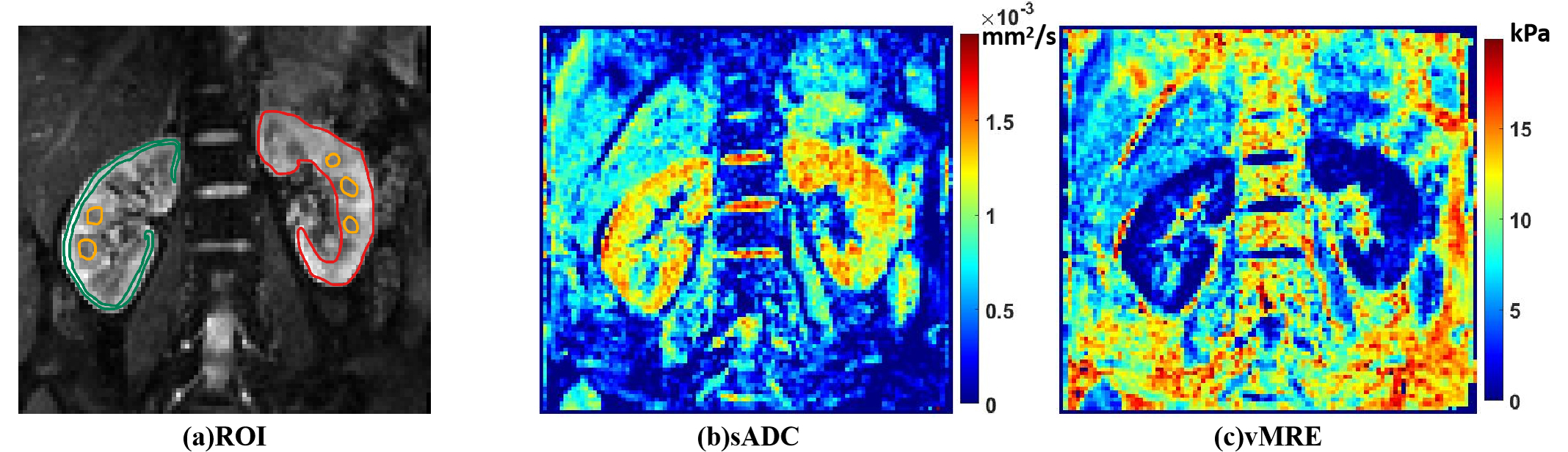
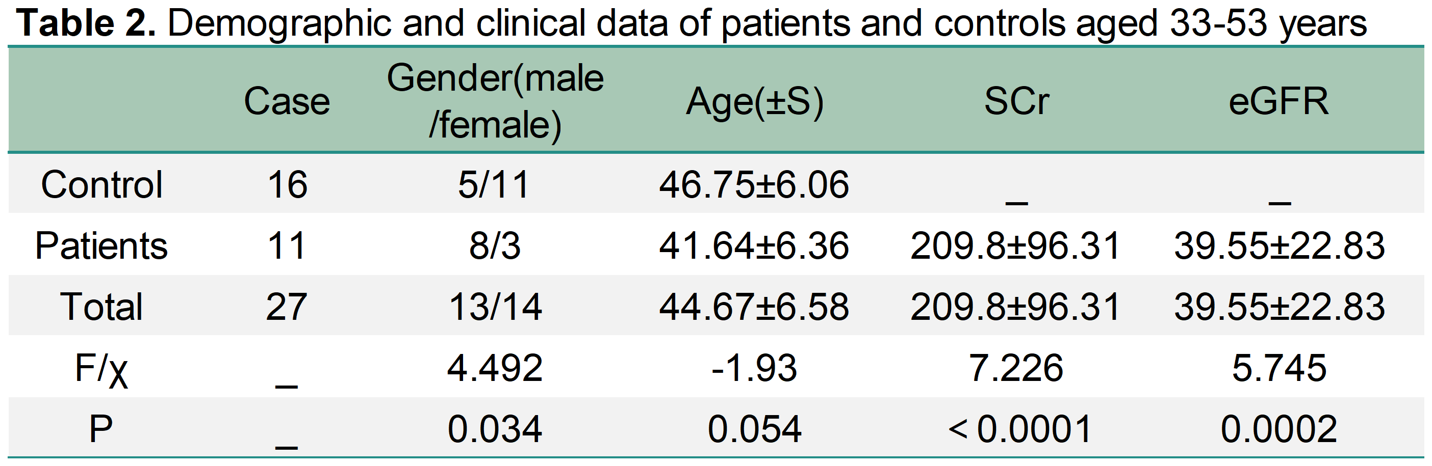
We recruited 21 patients and 27 healthy volunteers. All underwent an MRI examination on a 3T system (MAGNETOM Prisma; Siemens Healthineers AG. IVIM parameters were: TR/TE: 2700 milliseconds/48 milliseconds; voxel size: 2.5 × 2.5 × 4.0 mm3; FOV: 250 × 250 mm; b value: 0, 50, 100, 200, 400, 600, 800, 1000, 1200, 1500, and 2000 s/mm2. sADC was calculated from the IVIM sequence using: sADC = ln(SLKb/SHKb)/(HKb – LKb) [9, 13, 14]. Thus, the virtual shear stiffness value was estimated as follows: μdiff = αln (SLKb/SHKb ) + β. Based on previous calibration studies on the liver [ 8, 10], α and β were set to –9.8 and 14, respectively. The regions of interest were plotted, as depicted in Figure 1. Spearman's analysis was used to analyze the relationship between sADC and age in the 2 groups. The demographic and clinical data of 27 patients (33–53 years old) are exhibited in Table 2. The Mann–Whitney U test was used to analyze the difference in age between the 2 groups. Partial correlation analysis of the relationship between multigroup b values sADC, vMRE, SCr, and eGFR was performed in 11 patients. The receiver-operating characteristic (ROC) curve was used to assess the diagnostic efficacy of various b values of sADC and vMRE for CKD.Results
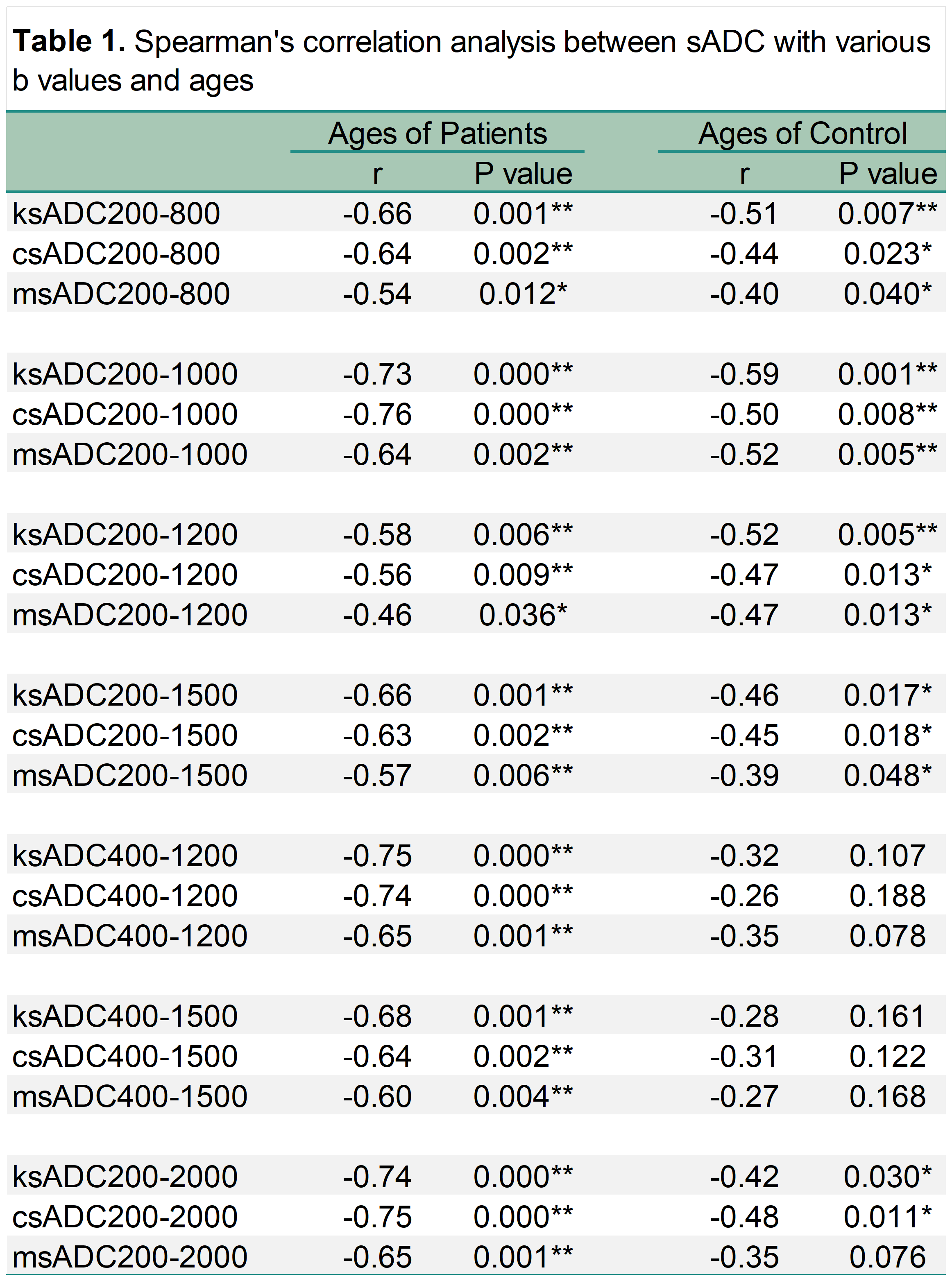
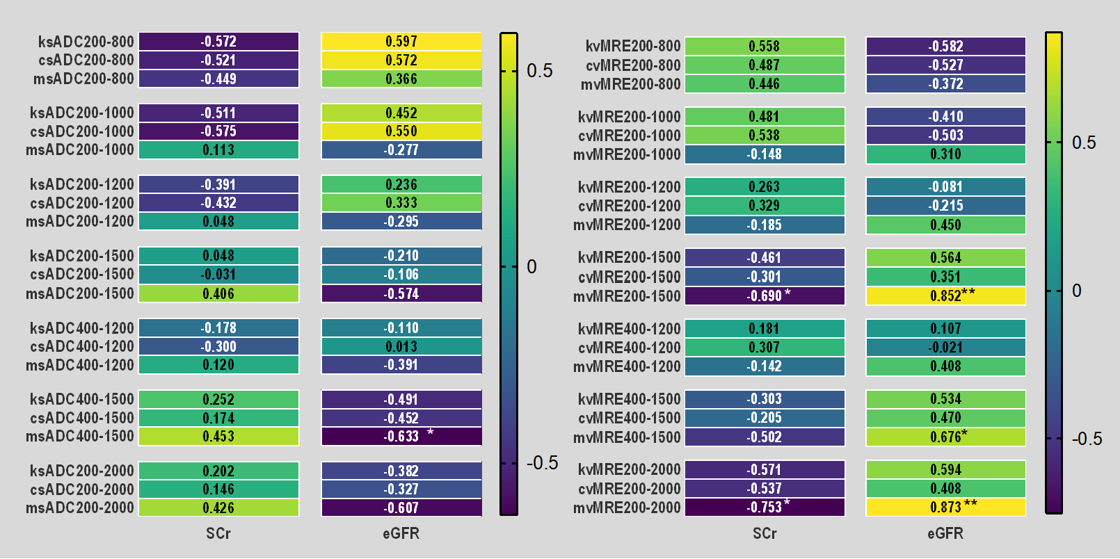
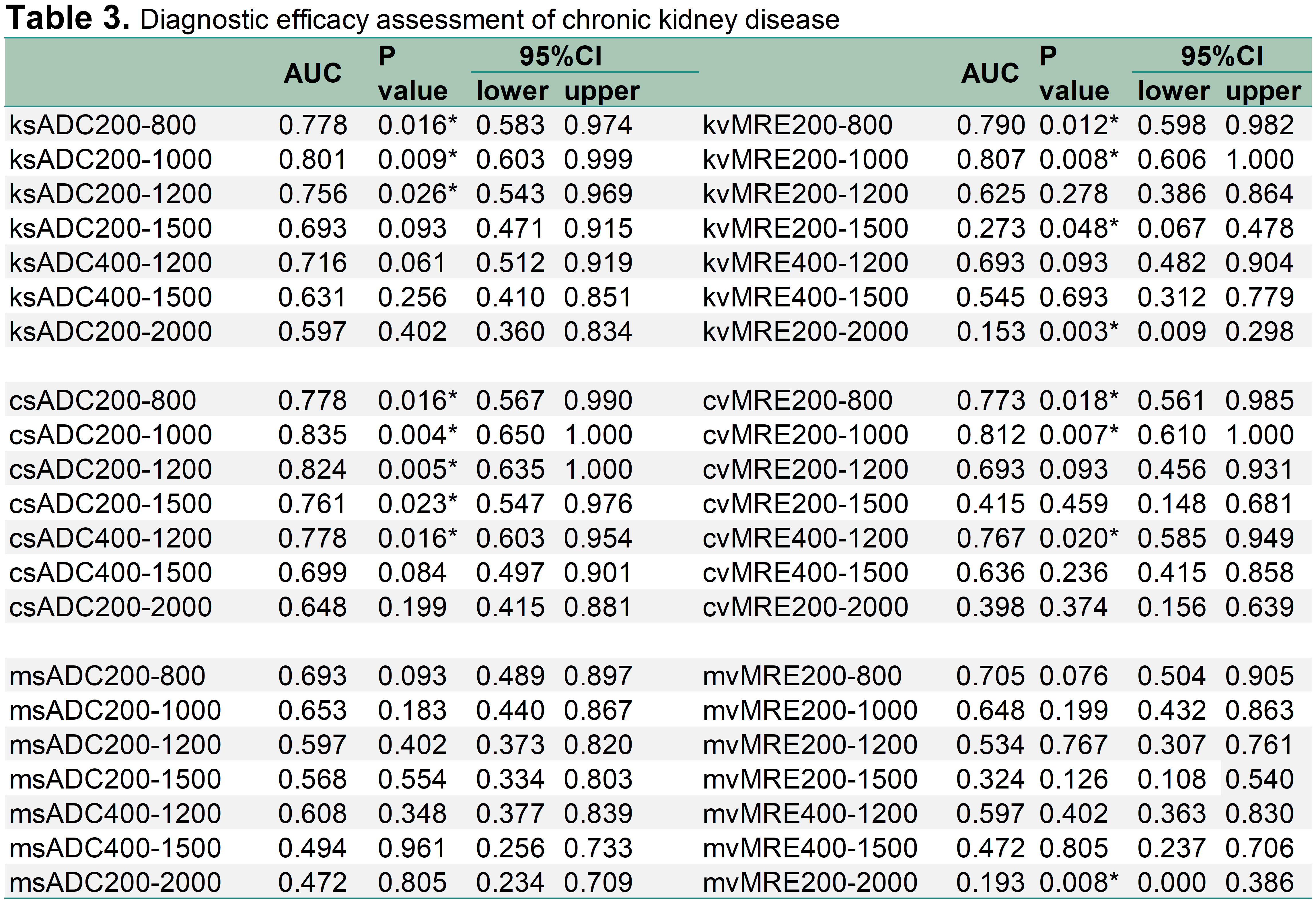
Various sADC b values were considerably negatively correlated with age, except for sADC400-1200, sADC400-1500 in both groups , and msADC200-2000 in the control group in Table1.In 11 patients, msADC400-1500, mvMRE200-1500, mvMRE200-2000, and mvMRE400-1500 were considerably correlated with eGFR, and mvMRE200-1500 and mvMRE200-2000 considerably negatively correlated with SCr. The detailed results are depicted in Figure 2. The ROC curves revealed that, at b values of 200-1000, the sADC and vMRE of the whole renal parenchyma and cortex had the highest diagnostic efficacy for CKD. At b values of 200-800, sADC and vMRE of the medulla had the best diagnostic results. The detailed results are illustrated in Table 3.Discussion
This study reported that age substantially affected sADC, as previously reported [15]. Age physiologically increases renal stiffness and reduces functional glomeruli [16]; pathologically, CKD mainly causes interstitial fibrosis, restricting water molecule diffusion. sADC compared with DWI-ADC, includes non-Gaussian diffusion and IVIM effect [13], offering heightened sensitivity to tissue microstructure [9, 14]. Thus, it better captures kidney structure, necessitating age consideration in the future study.A previous study investigated the correlation between sADC and clinical indicators and the diagnostic efficacy of CKD using DWI with b values less than 800 s/mm2[12]. This study analyzed 7 groups of sADC and vMRE with LKb-HKb of 200-800, 200-1000, 200-1200, 200-1500, 400-1200, 400-1500and 200-2000. In 11 patients, only medullary sADC400-1500(msADC400-1500), medullary vMRE200-1500 (mvMRE200-1500), mvMRE400-1500, and mvMRE200-2000 were considerably correlated with eGFR, contrary to previous results [12]. This suggested that choosing high b values of sADC and vMRE might be more appropriate when exploring the relationship between the medulla and the clinical indicators of CKD.A previous study reported a remarkable correlation of cortical sADC200-800(csADC200-800) with eGFR [12]. However, in our study, the correlations between various b values of csADC and cortical vMRE(cvMRE) and the clinical indicators were not statistically significant. The b -values (600-800 s/mm2) displayed non-Gaussian effects remarkably [17]. The renal cortex with richer perfusion than the medulla, correlated better with the clinical indexes at these values unlike the medulla.
csADC200-800 displayed remarkable diagnostic efficacy for CKD, echoing previous results. Previous studies overlooked the diagnostic efficacy of sADC and vMRE at high b values (≥1000 s/mm2) for CKD. However, our study found sADC and vMRE with b ≤1000 s/mm2 superior, as higher b values led to the increased likelihood of distortion and artifacts [18]. Exploring broader b-value ranges can refine combinations for future CKD studies.
Conclusion
IVIM-based virtual elastography could be used for the noninvasive assessment of renal function and fibrosis.Acknowledgements
No acknowledgement found.References
[1]Rouvière O, Souchon R, Pagnoux G, Ménager JM, Chapelon JY. Magnetic resonance elastography of the kidneys: feasibility and reproducibility in young healthy adults. J Magn Reson Imaging. 2011;34(4):880‐886. https://doi.org/10.1002/jmri.22670
[2]Mao W, Zhou J, Zeng M et al (2018) Chronic kidney disease: pathological and functional evaluation with intravoxel incoherent motion diffusionweighted imaging. J Magn Reson Imaging 47(5):1251–1259
[3]Mao W, Zhou J, Zeng M et al (2018) Intravoxel incoherent motion diffusion-weighted imaging for the assessment of renal fibrosis of chronic kidney disease: a preliminary study. Magn Reson Imaging 47:118–124
[4]Ebrahimi B, Rihal N, Woollard JR, Krier JD, Eirin A, Lerman LO (2014) Assessment of renal artery stenosis using intravoxel incoherent motion diffusion-weighted magnetic resonance imaging analysis. Invest Radiol 49:640–646
[5]Li Q, Li J, Zhang L, Chen Y, Zhang M, Yan F (2014) Diffusionweighted imaging in assessing renal pathology of chronic kidney disease: a preliminary clinical study. Eur J Radiol 83:756–762
[6] Mao W, Zhou J, Zeng M et al (2018) Chronic kidney disease: pathological and functional evaluation with intravoxel incoherent motion diffusion-weighted imaging. J Magn Reson Imaging 47:1251–1259
[7] D. Le Bihan, What can we see with IVIM MRI?, NeuroImage 187 (2019) 56-67.
[8] K. Lagerstrand, N. Gaedes, S. Eriksson, D. Farahmand, E. De Coursey, G. Johansson, L. Jönsson, T. Skoglund, Virtual magnetic resonance elastography has the feasibility to evaluate preoperative pituitary adenoma consistency, Pituitary 24(4) (2021) 530-541.
[9] M. Kromrey, D. Le Bihan, S. Ichikawa, U. Motosugi, Di usion-weighted MRI-based Virtual Elastography for the Assessment of Liver Fibrosis, Radiology 295(1) (2020) 127135.
[10] D. Le Bihan, S. Ichikawa, U. Motosugi, Di usion and Intravoxel Incoherent Motion MR Imaging-based Virtual Elastography: A Hypothesisgenerating Study in the Liver, Radiology 285(2) (2017) 609-619.
[11]Deng J, Cao Y, Lu Y, et al. Value of placental virtual magnetic resonance elastography and intravoxel incoherent motion-based diffusion and perfusion in predicting adverse outcomes of small-for-gestational-age infants. Insights Imaging. 2023;14(1):153. Published 2023 Sep 23. doi:10.1186/s13244-023-01503-9
[12]Hua C, Qiu L, Zhou L, et al. Value of multiparametric magnetic resonance imaging for evaluating chronic kidney disease and renal fibrosis. Eur Radiol. 2023;33(8):5211-5221.
[13] Iima M, Le Bihan D (2016) Clinical intravoxel incoherent motion and diffusion MR imaging: past, present, and future. Radiology 278:13–32
[14]Le Bihan D, Ichikawa S, Motosugi U (2017) Diffusion and intravoxel incoherent motion MR imaging-based virtual elastography: a hypothesis-generating study in the liver. Radiology 285:609–619
[15]Xu X, Fang W, Ling H, Chai W, Chen K. Diffusion-weighted MR imaging of kidneys in patients with chronic kidney disease: initial study. Eur Radiol. 2010;20(4):978-983. doi:10.1007/s00330-009-1619-8IF: 5.9
[16]Denic A, Glassock RJ, Rule AD. Structural and Functional Changes With the Aging Kidney. Adv Chronic Kidney Dis. 2016 Jan;23(1):19-28. doi: 10.1053/j.ackd.2015.08.004IF: 2.9 Q2. PMID: 26709059
[17]Pentang G, Lanzman RS, Heusch P et al (2014) Diffusion kurtosis imaging of the human kidney: a feasibility study. Magn Reson Imaging 32:413–420
[18]Wan Q, Deng YS, Lei Q, et al. Differentiating between malignant and benign solid solitary pulmonary lesions: are intravoxel incoherent motion and diffusion kurtosis imaging superior to conventional diffusion-weighted imaging?. Eur Radiol. 2019;29(3):1607-1615. doi:10.1007/s00330-018-5714-6
Figures