2753
Navigator-gated 2D radial MR fingerprinting of the kidney at 3T.1Radiology, Department of Radiology, Lausanne University Hospital (CHUV) and University of Lausanne (UNIL), Lausanne, Switzerland, 2CIBM Center for BioMedical Imaging, Lausanne, Switzerland, 3Radiology Service, Department of Diagnostics, Geneva University Hospital and University, Geneva, Switzerland
Synopsis
Keywords: Kidney, MR Fingerprinting
Motivation: A free-breathing renal joint T1-T2 MR fingerprinting technique with respiratory navigators would allow kidney function to be mapped non-invasively in patients that cannot hold their breath.
Goal(s): To assess the effect of rejected respiratory navigators on the relaxation times and their precision.
Approach: Joint kidney T1-T2 maps with four different navigator acceptance window widths (NAWW, from ±4mm to ±32mm) were acquired in 8 healthy volunteers and compared to clinical routine maps.
Results: Map accuracy and precision were high and did not change as a function of NAWW, suggesting that the NAWW can be chosen as a balance between navigator inefficiency and through-plane motion.
Impact: This work demonstrates the feasibility of a free-breathing 2D joint T1-T2 renal MR fingerprinting in healthy volunteers. The navigator allows a free breathing acquisition with limited through-plane motion.
Introduction
Quantitative magnetic resonance Imaging (qMRI) offers the possibility to non-invasively quantify tissue alteration and renal function, since a correlation between T1 cortico-medullary differentiation (CMD) and renal function has been demonstrated in several renal diseases 1,2. However, T1 mapping techniques usually require breath holding, which is not always feasible in these patients. In the current study, we aimed to demonstrate that an optimized version of the free-breathing navigator-gated magnetic resonance fingerprinting (cMRF) technique PARMA 3, (for PARametric Mapping), can be used to obtain accurate and precise native parametric maps of the kidney at 3T. The accuracy of the technique was previously validated versus gold-standard spin-echo techniques in the ISMRM-NIST phantom 3. The correlation of the T1 and T2 relaxation times with the reference techniques (MOLLI and T2-prepared (T2prep) bSSFP) was therefore established in both the kidney cortex and medulla in N=8 healthy volunteers (29±3y, 2F). The impact of the navigator acceptance window width (NAWW) on the T1 and T2 values as well as the CMD was quantified.Method
PARMA consists of a 2D single-shot gradient echo (RF excitation angle=12°, TR=3.49ms, TE=1.56ms ms, 45 lines/image, 800ms between each single-shot image) with a continuous radial golden angle trajectory that is preceded by 5 different magnetization preparations. These were an adiabatic inversion pulse, no preparation, and 3 different T2 preparation modules (TET2prep=23/45/70ms) to sensitize the contrast to both T1 and T2 relaxation 4,5 (Figure 1). This set of five preparations was then repeated five times. Respiratory motion was tracked using a lung-liver navigator before the preparation modules; when the navigator position was outside the acceptance window, neither the following preparation module nor the image acquisition were activated. The undersampled images were reconstructed using compressed sensing with local low rank regularization along the contrast dimension 6. Non-rigid registration was applied to account for residual in-plane motion.A dictionary was simulated for each acquisition using extended phase graphs (EPG 7) for a range of T1 (40:10:3000 ms) and T2 (20:1:100; 102:2:230 ms) values in MATLAB (The Mathworks) and included slice-profile and inversion-inefficiency corrections 8. The in-vivo study was approved by the local ethics committee and all participants provided written informed consent. PARMA maps of N=8 healthy volunteers were acquired in a 3T clinical scanner (Magnetom PrismaFit, Siemens Healthineers, Erlangen, Germany) with matrix size=192x192, pixel size=(1.56mm)2, slice thickness=8mm, TR=3.49ms, and TE=1.56ms. PARMA was repeated with four different NAWW (4mm, 8mm, 16mm, and 32mm) of the lung-liver navigator. Clinical routine T1 and T2 maps (pixel size=(1.8-1.9mm)2, slice thickness 8mm) were acquired using breath-held MOLLI 9 and T2-prepared bSSFP T2 mapping 10, respectively.
The T1 and T2 value of the visible cortex and medulla as well as the CMD (using the ratio T1cortex/T1medulla) were determined for each map in each volunteer by manually segmenting a region of interest in the corresponding tissue, and differences were tested with a repeated-measures ANOVA with post-hoc Tukey analysis. Bland-Altman analysis was performed to quantify biases.
Results
In all 8 healthy volunteer sharp and precise maps in the kidney were obtained (Figure 2). PARMA T1, T2 values and CMD were significantly different from routine techniques, which are known to underestimate T1 4 and overestimate T2 5, but there were no significant differences as a function of the different NAWW (Figure 3). Given this similarity and the need for balance between high navigator inefficiency (at small NAWW) and through-plane motion (at large NAWW), NAWW=8 mm mapping was retained for further analysis.Bland Altman analysis (Figure 4) then demonstrated a bias versus the reference techniques, which is consistent with the previous phantom results 3 and segmental averages (Figure 2).
Discussion
PARMA resulted in precise and robust free-breathing joint T1-T2 maps of the kidney. It was successfully acquired in all healthy volunteers with all tested NAWW, which resulted in similar mapped relaxation times.The efficacy of this technique needs to be confirmed in a large patient population. Further developments might include semi-automated segmentation, an extension to T2* mapping to more sensitively assess oxygenation, and diffusion modules to further characterize fibrosis.
In conclusion, we preliminarily demonstrated the feasibility of precise free-breathing joint T1-T2 mapping in the kidney at 3T, and that navigator rejection does not affect the accuracy of the mapped relaxation times.
Acknowledgements
This study was funded by a grant from the Swiss National Science Foundation (grant number CRSII5_202276).References
1. Marotti M, Hricak H, Terrier F, McAninch JW, Thuroff JW. MR in renal disease: importance of cortical-medullary distinction. Magn Reson Med. 1987 Aug;5(2):160-72. doi: 10.1002/mrm.1910050207. PMID: 3657504.
2. Hricak H, Terrier F, Demas BE. Renal allografts: evaluation by MR imaging. Radiology. 1986 May;159(2):435-41. doi: 10.1148/radiology.159.2.3515420. PMID: 3515420.
3. Calarnou, P., et al. Navigator acceptance window width does not affect accuracy and precision in free-breathing 2D cardiac MR fingerprinting of the myocardium at 3T. ISMRM 2023, #3999
4. Cruz G, Jaubert O, Qi H, et al. 3D free-breathing cardiac magnetic resonance fingerprinting. NMR Biomed. 2020;33:e4370 doi: 10.1002/nbm.4370.
5. Henningsson M. Cartesian dictionary-based native T1 and T2 mapping of the myocardium. Magn. Reson. Med. 2022;87:2347–2362 doi: 10.1002/mrm.29143.
6. Di Sopra L, Roy CW, Bastiaansen JAM, et al. Fully Self-Gated Cardiac and Respiratory Motion-Resolved Isotropic 5D T1 Mapping of the Heart: Preliminary Results. In: Proc Int Soc Magn Reson Med 27. Montréal QC, Canada; 2019.
7. Malik SJ, Padormo F, Price AN, Hajnal JV. Spatially resolved extended phase graphs: modeling and design of multipulse sequences with parallel transmission. Magn. Reson. Med. 2012;68:1481–1494 doi: 10.1002/mrm.24153.
8. Hamilton JI, Jiang Y, Ma D, et al. Investigating and reducing the effects of confounding factors for robust T1 and T2 mapping with cardiac MR fingerprinting. Magn. Reson. Imaging 2018;53:40–51 doi: 10.1016/j.mri.2018.06.018.
9. Messroghli DR, Radjenovic A, Kozerke S, Higgins DM, Sivananthan MU, Ridgway JP. Modified Look-Locker inversion recovery (MOLLI) for high-resolution T1 mapping of the heart. Magn. Reson. Med. 2004;52:141–6 doi: 10.1002/mrm.20110.
10. Giri S, Chung YC, Merchant A, et al. T2 quantification for improved detection of myocardial edema. J Cardiovasc Magn Reson 2009;11:56 doi: 1532-429X-11-56 [pii] 10.1186/1532-429X-11-56.
Figures
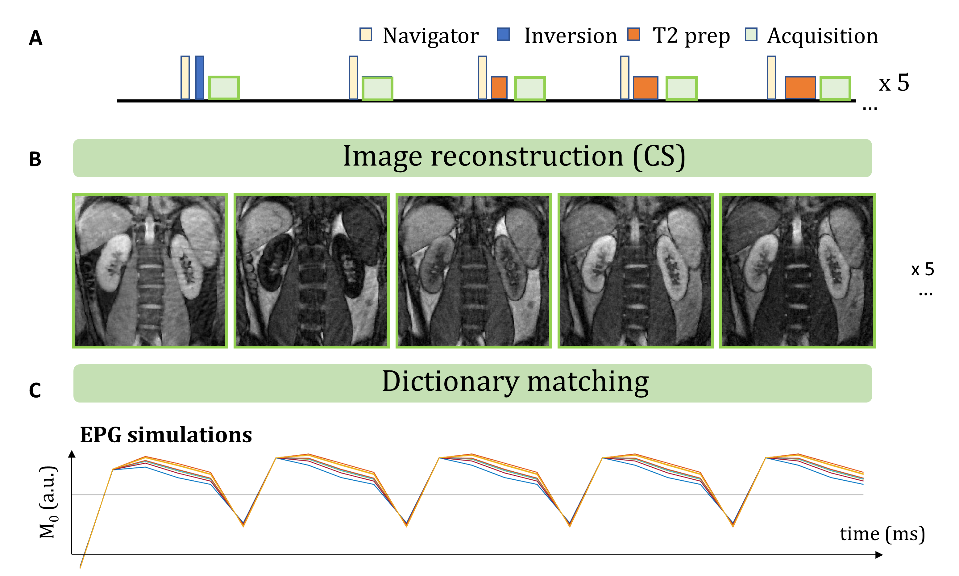
Figure 1. Pulse sequence diagram and example images for the proposed free-breathing radial 2D MR fingerprinting technique PARMA. A) Pulse sequence diagram. 25 single-shot images are acquired with inversion and three different T2-prep modules. B) Example first 5 images in a healthy volunteer kidney, reconstructed using compressed sensing with local low-rank regularization in the contrast dimension. C) Dictionary signal evolution across 25 images for different T1-T2 pairs obtained with EPG simulations.
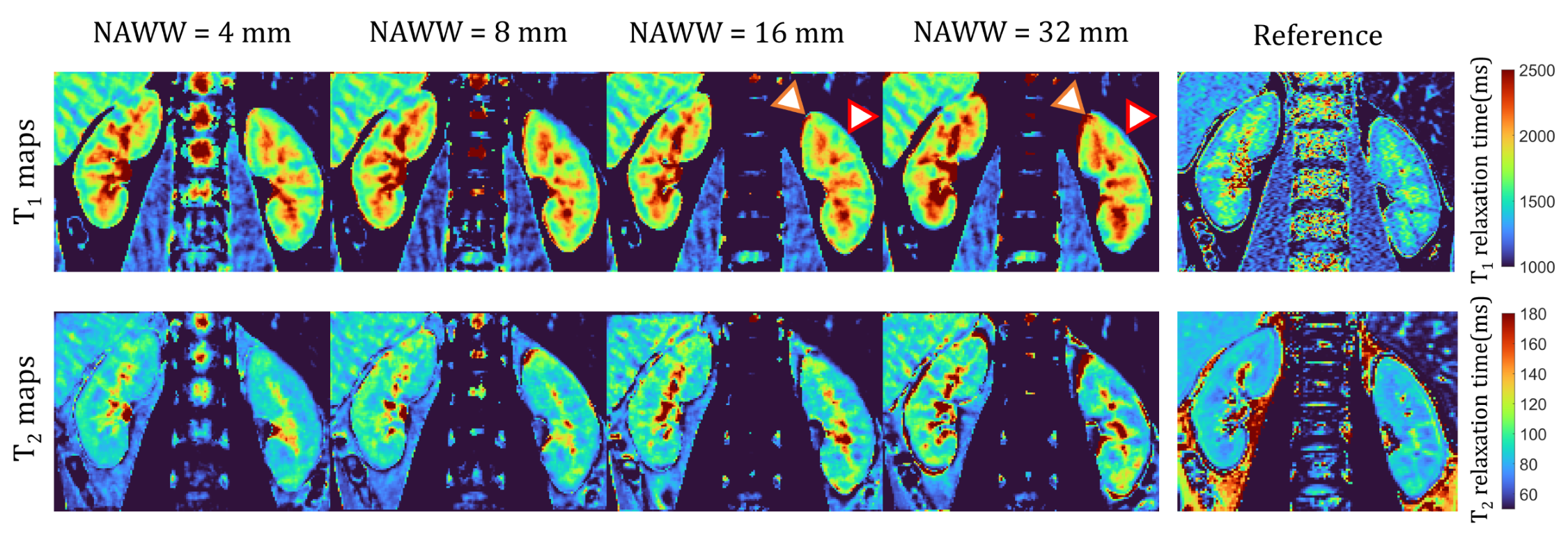
Figure 2. PARMA T1 and T2 maps for the different NAWW compared to the Reference techniques. The NAWW does not significantly change the relaxation times in the resulting PARMA T1 maps of the cortex. However, the shape and the T1 value of the medulla varies with the NAWW (red and orange arrowheads), likely due to through-plane motion. The PARMA T1 and T2 values are consistently higher than those obtained with the routine techniques.
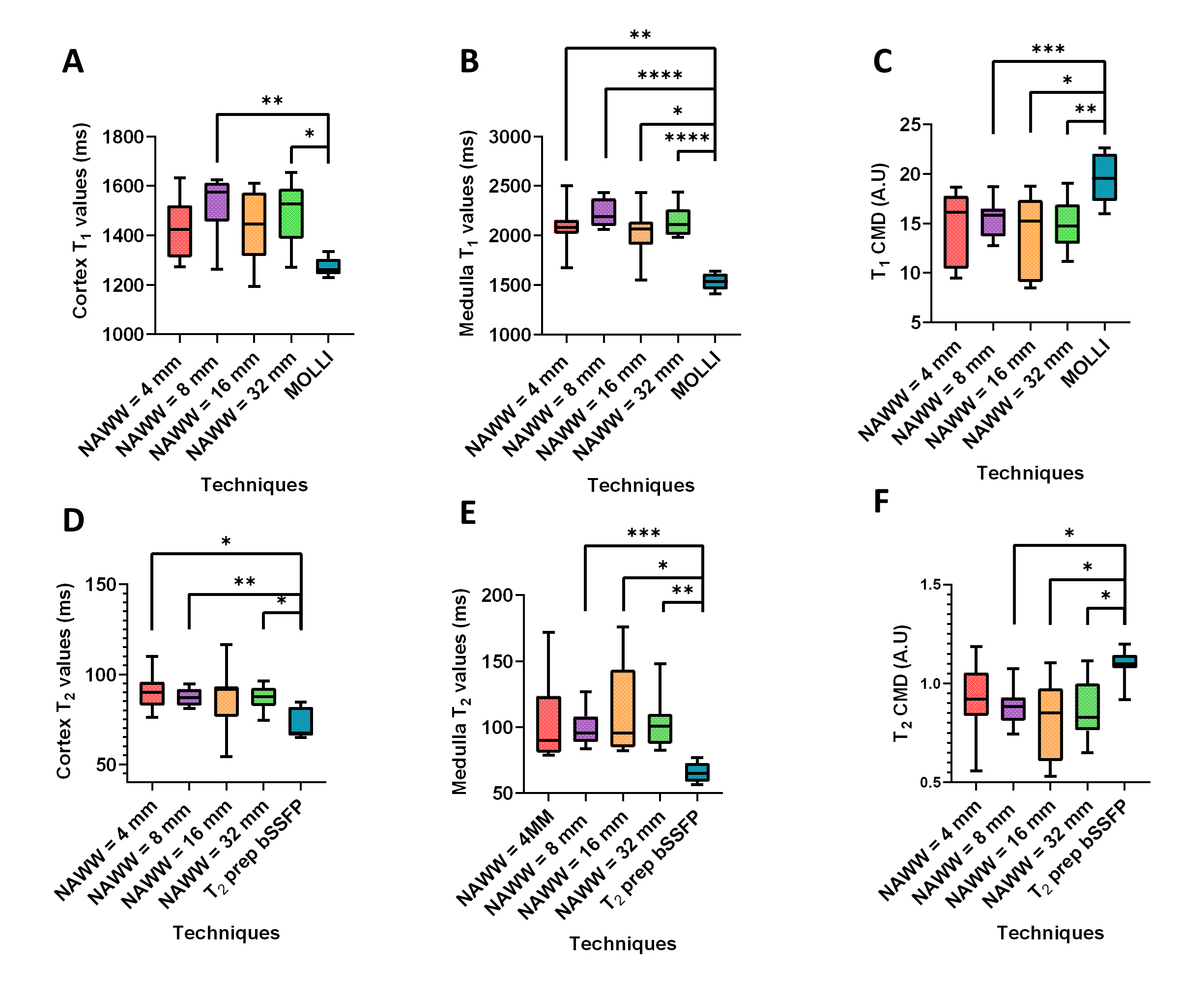
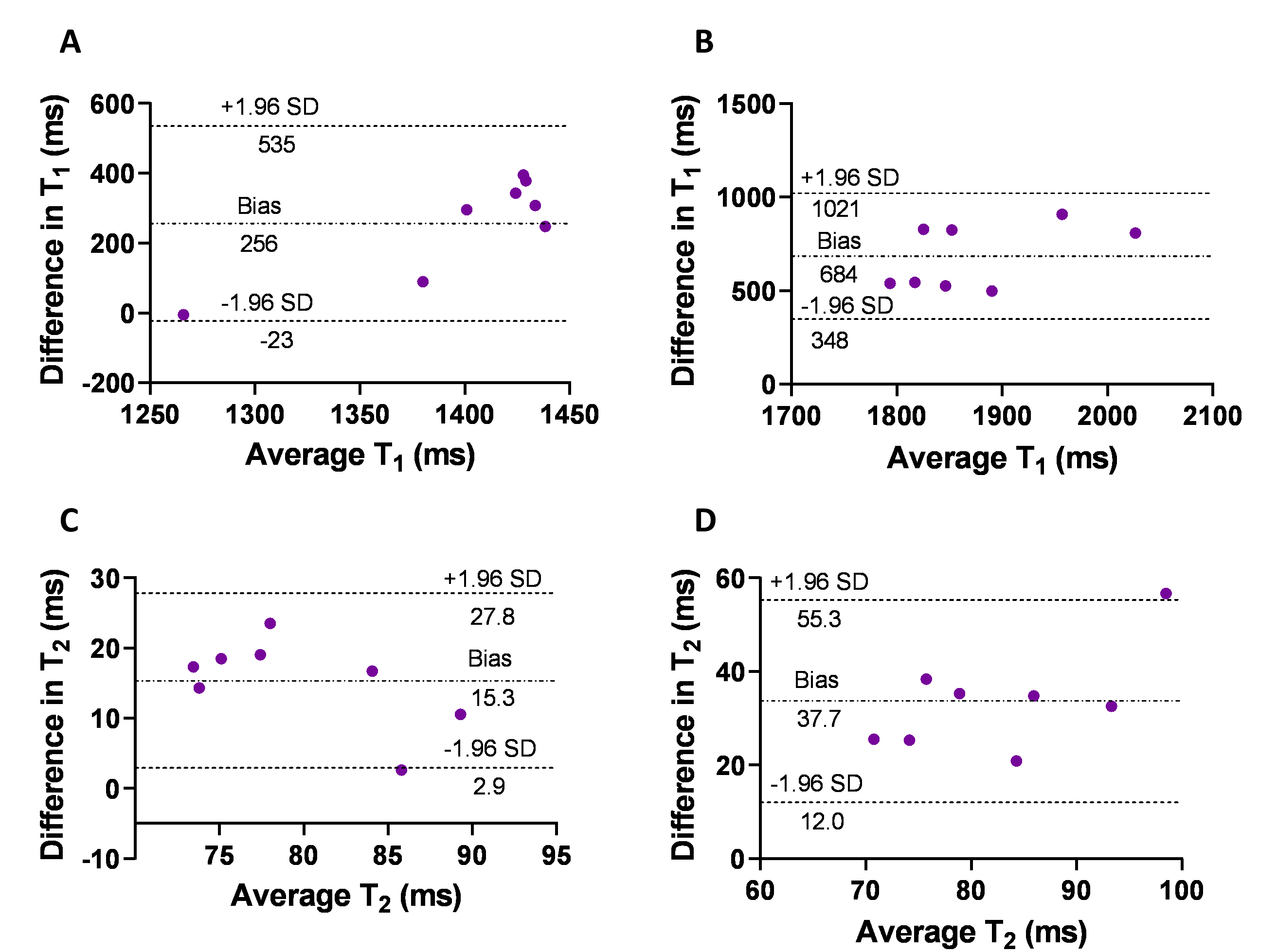
Figure 4. Bland-Altman analyses of the agreement between PARMA with NAWW=8mm and the reference techniques. A) Cortex T1 values. B) Medulla T1 values. C) Cortex T2 values. D) Medulla T2 values.