2747
Technical recommendations for renal DCE-MRI: Preliminary results from the consensus project of the ISMRM Renal MR study group1Division of Clinical Medicine, School of Medicine and Population Health, University of Sheffield, Sheffield, United Kingdom, 2Computational Radiology Laboratory, Department of Radiology, Boston Children's Hospital, Boston, MA, United States, 3Department of Radiology, Harvard Medical School, Boston, MA, United States, 4Department of Radiology and Surgery (Austin), Faculty of Medicine, Dentistry and Health Science, The University of Melbourne, Carlton, Australia, 5Department of Radiology, Austin Health, Heidelberg, Australia, 6School of Biomedical Engineering, ShanghaiTech University, Shanghai, China, 7Department of Radiology, Children's Healthcare of Atlanta, Atlanta, GA, United States, 8Mannheim Institute for Intelligent System, Medical Faculty Mannheim, Heidelberg University, Mannheim, Germany, 9Computer Assisted Clinical Medicine, Mannheim, Germany, 10Auckland Bioengineering Institute, The University of Auckland, Auckland, New Zealand, 11Radiology Department, Urology and Nephrology Center, Mansoura University, Mansoura, Egypt, 12Bernard and Irene Schwartz Center for Biomedical Imaging, Department of Radiology, New York University Grossman School of Medicine, New York, NY, United States, 13Comparative Medicine Lab, Department of Clinical Medicine, Aarhus University, Aarhus, Denmark, 14Radiology Research Unit, Department of Clinical Medicine, Odense University Hospital, Odense, Denmark, 15MedTech West, Chalmers University of Technology, Gothenburg, Sweden, 16Antaros Medical, BioVenture Hub, Mölndal, Sweden, 17Department of Radiology, University of Wisconsin, Madison, WI, United States, 18Biomedical Imaging, University of Leeds, Leeds, United Kingdom, 19Department of Radiology, Ghent University Hospital, Ghent, Belgium, 20Department of Diagnostic Sciences, Faculty of Medicine and Health Sciences, Ghent University, Ghent, Belgium, 21Ghent Institute of Functional and Metabolic Imaging (GIFMI), Ghent University, Ghent, Belgium, 22Department of Nuclear Medicine, Cambridge University Hospitals NHS Foundation Trust, Cambridge, United Kingdom, 23Department of Radiology, University of Cambridge, Cambridge, United Kingdom, 24Core Radiology Research Group, Department of Radiology, Children's Hospital of Philadelphia, Philadelphia, PA, United States, 25Perelman School of Medicine, University of Pennsylvania, Philadelphia, PA, United States, 26Department of Radiology, Leiden University Medical Center, Leiden, Netherlands
Synopsis
Keywords: Kidney, DSC & DCE Perfusion, kidney, renal blood flow, standardisation, consensus
Motivation: Clinical translation of renal functional and quantitative MRI is hindered by a lack in standardisation of scan protocols.
Goal(s): To develop expert consensus-based technical recommendations on the acquisition and post-processing of renal DCE-MRI.
Approach: Experts were recruited and surveyed following the Delphi method to create consensus-based technical recommendations in renal DCE-MRI. Preliminary results will be used to construct the second survey round.
Results: 16 experts responded to the preliminary survey. Consensus statements informed by the responses were drafted and will be circulated and refined in the next phase of the project.
Impact: The insights obtained from this work will be invaluable in delivering recommendations that are comprehensive and widely accepted. Consensus-based technical recommendations for renal DCE-MRI aim to contribute to harmonisation of MRI scan protocols across sites, facilitating clinical translation.
INTRODUCTION
Dynamic contrast-enhanced (DCE) MRI has potential to be a useful tool for non-invasively assessing renal hemodynamics and function, however clinical implementation is hindered by a lack of standardisation and concerns over safety and cost.A recent consensus project1 developed technical recommendations for renal ASL2, BOLD3, DWI4, T1 and T2 mapping5 and Phase Contrast6. Recently, the ISMRM renal MR study group initiated a similar initiative aiming to produce technical recommendations for renal DCE-MRI. The process follows the methodology outlined in1 and has currently completed the first round of questionnaires.
This abstract reports on these preliminary results with a view of promoting further discussion at the meeting and widening expert participation.
METHODS
Recruitment for the consensus panel began in August 2022, with an initial meeting conducted virtually to provide information on the initiative and to garner interest from a broad range of experts. A two-step modified Delphi method7,8 was used for defining the consensus recommendations, involving iterative survey rounds.Three working groups (A. participant preparation and hardware considerations; B. sequence parameters, including motion correction; C. data analysis and reporting) were identified and a list of items to be included for the first Delphi survey round were drafted. These were then combined and formulated into a survey circulated electronically to the panel (June 2023), the first iteration of the Delphi process.
This first survey consisted of a mixture of close-ended and open-ended questions, participants being encouraged to provide detailed responses based on their experiences and expertise. Results of the first electronic survey round were reviewed at the Fifth International Renal Imaging Meeting in Ghent, Belgium (September 10-11, 2023).
Based on these discussions and responses from the first survey round, a follow-up survey was constructed (second round). This second survey round was formulated as a set of ‘consensus statements’ and will be circulated electronically to the panellists as the next stage of the project. As in previous work, these statements must achieve ≥ 75% majority in response to reach consensus, and [60-74]% agreement to indicate a clear preference among the experts.
RESULTS
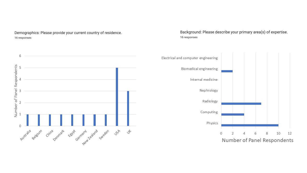
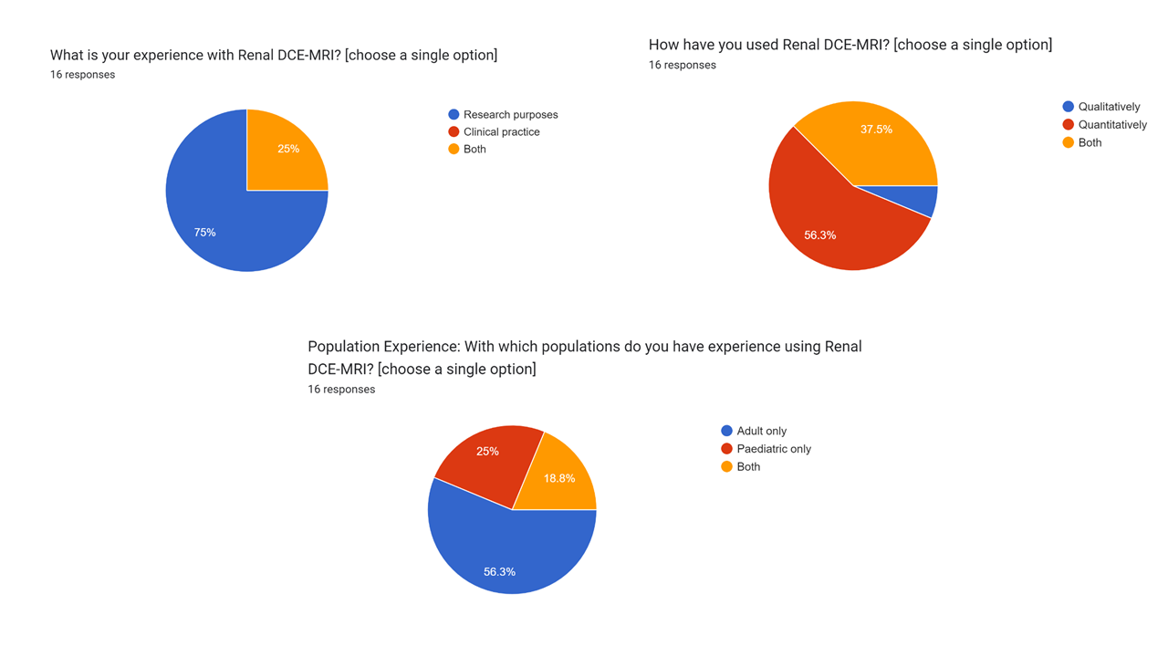
In total, 22 experts participated in the Delphi panel, of which 16 responded to the first survey round, meeting the required number of experts for content validation9. Ten experts of the Delphi panel have a physics background, seven in clinical radiology, and seven in computing or engineering disciplines. Additional demographics for the panel can be seen in Figure 1.The majority of experts were found to have experience in using renal DCE-MRI for quantitative research applications in adult populations, with only 25% of respondents using renal DCE-MRI in both clinical and research settings (Fig. 2). However, routine use in paediatric studies for clinical purposes was also reported. Populations with whom respondents reported having experience using renal DCE-MRI included healthy volunteers, paediatric patients, chronic kidney disease patients, diabetic patients, patients with congenital kidney abnormalities, patients with renal cell carcinoma, kidney transplant recipients, and animal models.
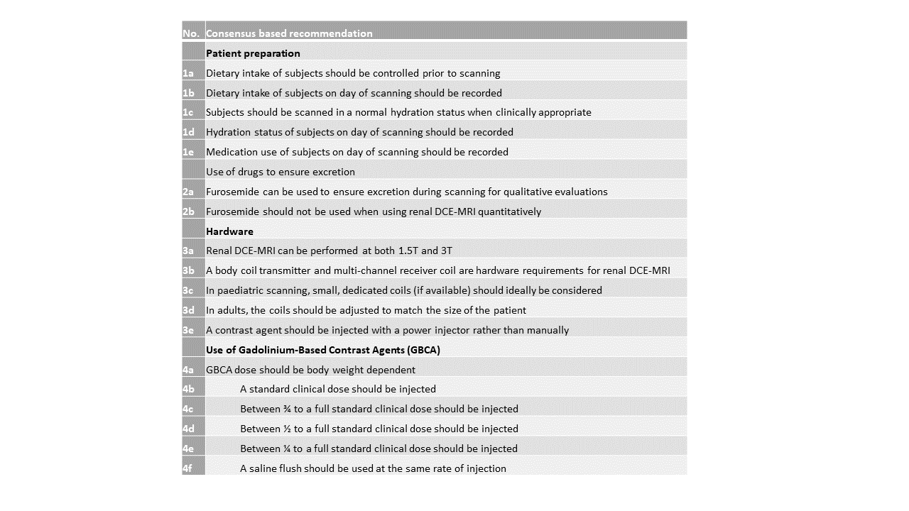
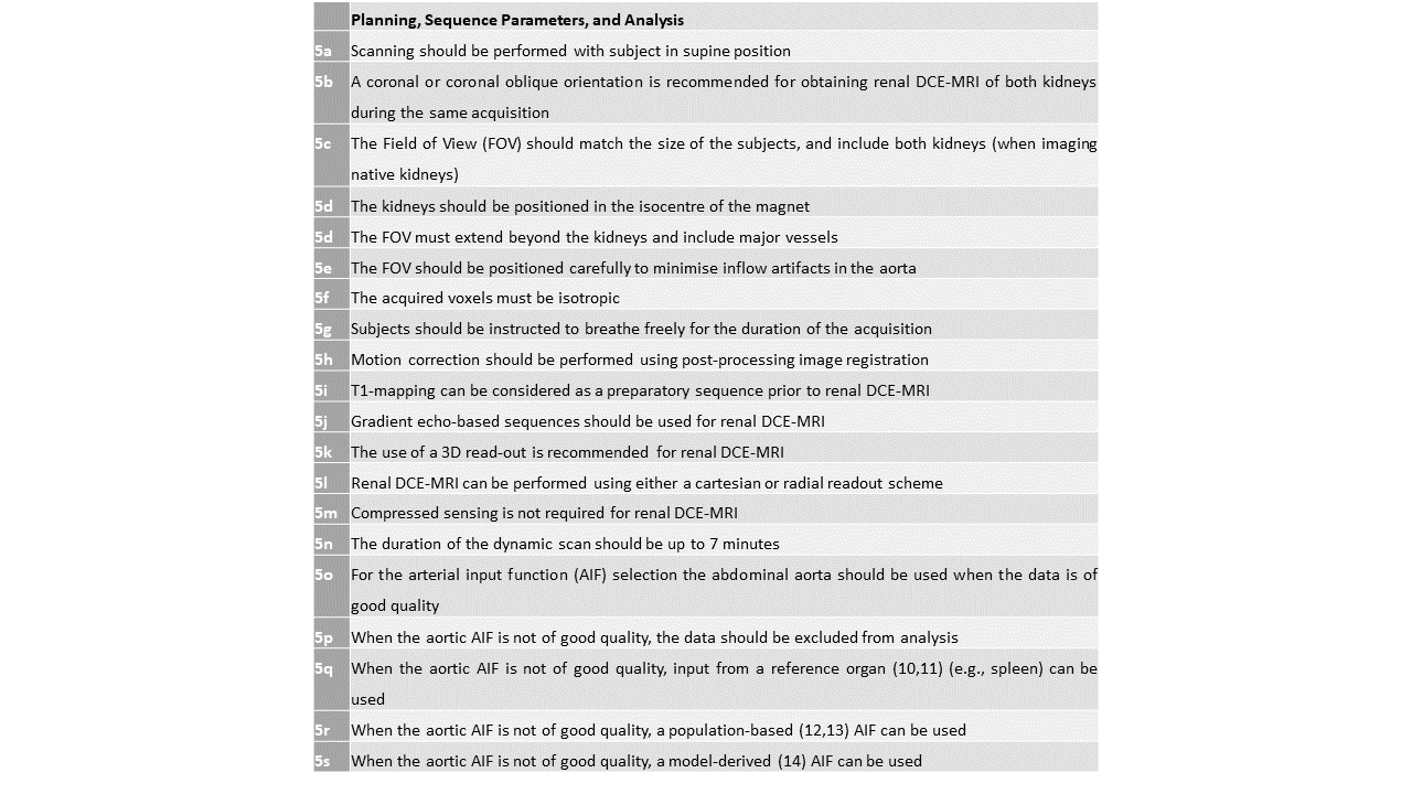
Formulated recommendations for the second survey round are shown in Table 1 and are aimed at determining consensus in the final reference paper.
DISCUSSION
By gathering expert opinions and experiences on renal DCE-MRI usage, this preliminary work identified key areas for consensus that will aid in harmonisation of this technique to promote technical uptake and implementation.A broad range of experts from multiple countries and disciplines contributed to this work, crucial for avoiding bias and minimising health disparities in clinical translation. However, while the panel size meets the minimal requirements for consensus validation, the number of experts that responded to the first survey round was of limited size (n=16), and due to the varied backgrounds, not all experts were confident in responding to every aspect of the survey.
Expert recommendations comprised a mixture of semi-automated and in-house analysis protocols, where solutions are dependent upon the metric. For instance, to measure single-kidney glomerular filtration rate (GFR) a different protocol will likely be needed than when measuring cortical perfusion. This resulted in methodology being established on a case-by-case basis, generating scenarios where experts may struggle to agree.
As with the previous consensus studies, the survey responses and discussions also highlighted a critically unmet need for dedicated, reliable, and automated post-processing software for accurate quantification of renal DCE-MRI imaging biomarkers.
CONCLUSION
Preliminary results on expert consensus-based technical recommendations for renal DCE-MRI were formulated and used to develop a set of consensus statements. This will provide a starting point for MRI centres worldwide wishing to use renal DCE-MRI and help to harmonise existing research MRI protocols, ultimately aiding in adoption of renal DCE-MRI into clinical practice.Acknowledgements
This abstract is based upon work from the COST Action CA16103 Magnetic Resonance Imaging Biomarkers for Chronic Kidney Disease (PARENCHIMA), funded by COST (European Cooperation in Science and Technology). For additional information, please visit www.renalmri.org.
References
1. Mendichovszky I, Pullens P, Dekkers I, Nery F, Bane O, Pohlmann A, de Boer A, Ljimani A, Odudu A, Buchanan C. Technical recommendations for clinical translation of renal MRI: a consensus project of the Cooperation in Science and Technology Action PARENCHIMA. Magnetic Resonance Materials in Physics, Biology and Medicine 2020;33:131-140.
2. Nery F, Buchanan CE, Harteveld AA, Odudu A, Bane O, Cox EF, Derlin K, Gach HM, Golay X, Gutberlet M. Consensus-based technical recommendations for clinical translation of renal ASL MRI. Magnetic Resonance Materials in Physics, Biology and Medicine 2020;33:141-161.
3. Bane O, Mendichovszky IA, Milani B, Dekkers IA, Deux J-F, Eckerbom P, Grenier N, Hall ME, Inoue T, Laustsen C. Consensus-based technical recommendations for clinical translation of renal BOLD MRI. Magnetic Resonance Materials in Physics, Biology and Medicine 2020;33:199-215.
4. Ljimani A, Caroli A, Laustsen C, Francis S, Mendichovszky IA, Bane O, Nery F, Sharma K, Pohlmann A, Dekkers IA. Consensus-based technical recommendations for clinical translation of renal diffusion-weighted MRI. Magnetic Resonance Materials in Physics, Biology and Medicine 2020;33:177-195.
5. Dekkers IA, de Boer A, Sharma K, Cox EF, Lamb HJ, Buckley DL, Bane O, Morris DM, Prasad PV, Semple SI. Consensus-based technical recommendations for clinical translation of renal T1 and T2 mapping MRI. Magnetic Resonance Materials in Physics, Biology and Medicine 2020;33:163-176.
6. de Boer A, Villa G, Bane O, Bock M, Cox EF, Dekkers IA, Eckerbom P, Fernández‐Seara MA, Francis ST, Haddock B. Consensus‐based technical recommendations for clinical translation of renal phase contrast MRI. Journal of Magnetic Resonance Imaging 2022;55(2):323-335.
7. Helmer O. Analysis of the future: The Delphi method. Rand Corporation Santa Monica; 1967.
8. Hsu C-C, Sandford BA. The Delphi technique: making sense of consensus. Practical assessment, research, and evaluation 2019;12(1):10.
9. Lynn MR. Determination and quantification of content validity. Nursing research 1986;35(6):382-386.
10. Ahmed Z, Levesque IR. Pharmacokinetic modeling of dynamic contrast‐enhanced MRI using a reference region and input function tail. Magnetic resonance in medicine 2020;83(1):286-298.
11. Ariyurek C, Afacan O, Chow J, Warfield SK, Kurugol S. Use of Reference Region Model to Improve Arterial Input Function Selection for Estimating Kidney Function with DCE-MRI [abstract]. In: Proceedings of the ISMRM & SMRT Annual Meeting & Exhibition, Online, 2021. Abstract nr 1107.
12. Parker GJ, Roberts C, Macdonald A, Buonaccorsi GA, Cheung S, Buckley DL, Jackson A, Watson Y, Davies K, Jayson GC. Experimentally‐derived functional form for a population‐averaged high‐temporal‐resolution arterial input function for dynamic contrast‐enhanced MRI. Magnetic Resonance in Medicine: An Official Journal of the International Society for Magnetic Resonance in Medicine 2006;56(5):993-1000.
13. Kim H. Modification of population based arterial input function to incorporate individual variation. Magnetic resonance imaging 2018;45:66-71.
14. van Osch MJ, Vonken EjP, Wu O, Viergever MA, van der Grond J, Bakker CJ. Model of the human vasculature for studying the influence of contrast injection speed on cerebral perfusion MRI. Magnetic Resonance in Medicine: An Official Journal of the International Society for Magnetic Resonance in Medicine 2003;50(3):614-622.
Figures