2745
A deep learning based super-resolution technique for MR image reconstruction in BLADE sequence1Siemens Shenzhen Magnetic Resonance Ltd., ShenZhen, China
Synopsis
Keywords: Machine Learning/Artificial Intelligence, Image Reconstruction, Super Resolution, BLADE sequence
Motivation: Conventional super-resolution techniques are not applicable to magnetic resonance images reconstructed from BLADE sequences, where four corners of the k-space are missing.
Goal(s): When BLADE data are fed into a common super-resolution model targeting ordinary Cartesian k-space data, strong Gibbs rings appear due to the truncation of k-space. On this basis, we propose a deep learning-based method specifically for the super-resolution task with BLADE data.
Approach: We mainly used the Residual Density Network (RDN) and designed the downsampling method based on the characteristics of BLADE data.
Results: Experimental results show that our model is able to predict high-resolution MR images with fewer artifacts.
Impact: By applying our RDN-based model specifically adapted to BLADE data, the image matrix size can be increased by a factor of 2 to produce sharper images without increasing acquisition time.
Introduction
The BLADE sequence samples k-space data in a rotating mode, with each blade block containing multiple phase-encoded lines in parallel. Due to the oversampling in the centre of the k-space, the data redundancy provides more possibilities for data correction1. As a result, the BLADE sequence is able to reduce motion artefacts caused by either body movements or physiological movements2. Since shorter scan times are more conducive to motion artefact suppression, we consider using super-resolution to speed up the scan time. The performance of traditional interpolation-based super-resolution methods is limited by their mathematical models, especially for complex image content3. However, deep learning-based super-resolution methods are able to capture more complicated image textures and structures, resulting in a more realistic and detailed image4. However, the current common deep learning based super-resolution technique in MRI is designed for Cartesian k-space5. The direct application of the BLADE data to the above-mentioned super-resolution neural networks leads to strong Gibbs ringing6. To fill the gap of the BLADE sequence in deep learning based super-resolution tasks, we propose an RDN-based super-resolution technique for MR image reconstruction in the BLADE sequence.Method
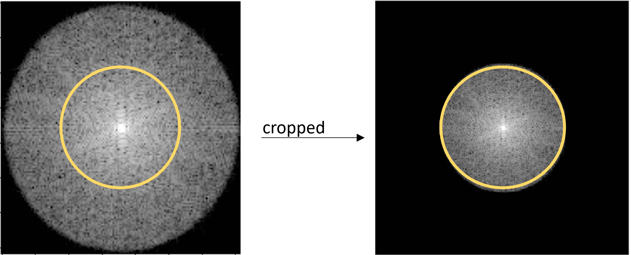
The dataset consists of 4308 images, collected from 130 volunteers on 0.55T and 1.5T scanners respectively. The matrix size of the images varies from each other, ranging from 96×96 to 320×320. In addition, the dataset is randomly divided into three parts: 80%, 10% and 10% of which belong to the training, validation and testing datasets respectively.First, all of the images are normalised using z-score normalization to avoid the effects of extreme values. In terms of downsampling, the central part of the k-space is cropped out to ensure that the low frequency information is complete, while leaving the image structure unchanged. It is worth noting that, in order to match the property that the k-space of the BLADE image is circular, the experiment also specifically takes a small circle in the center of the k-space to achieve downsampling. Figure 1 illustrates how this implementation accomplishes the downsampling of images from high to low resolution.
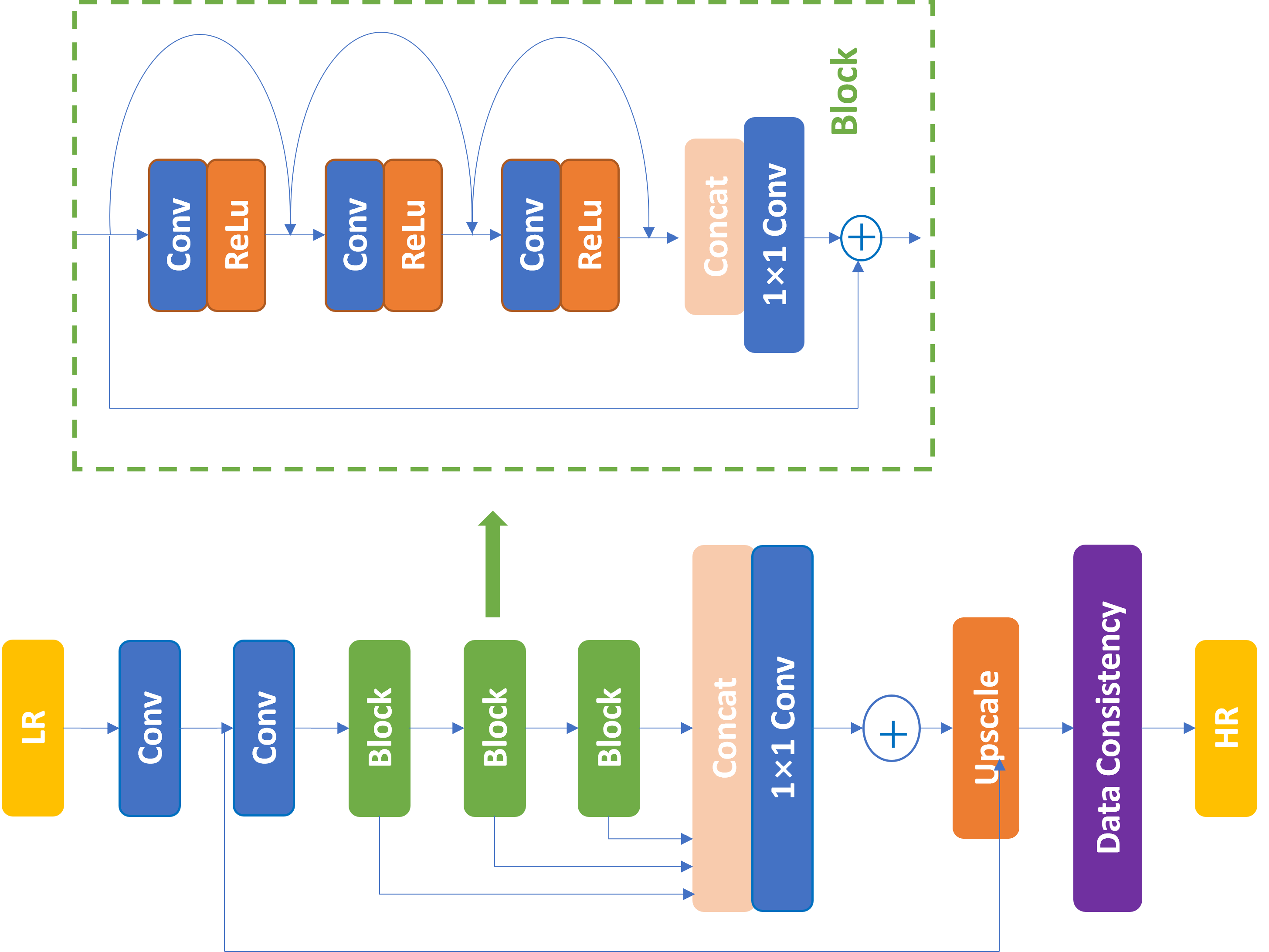
By comparison with the RDN7, the architecture of our network is adapted to the characteristics of MR data, the pipeline of which is shown in Figure 2. Dense connections between different layers allow the model to capture both shadow features and the dense features8. Based on the RDN, we add a data consistency step at the end of the model, i.e. the circle non-zero part of the low-resolution k-space replaces the incoming high-resolution k-space at the corresponding location.
Result and Discussion
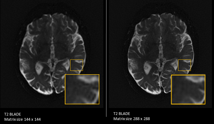
Table 1 displays the results of the image quality assessment metrics, including the structural similarity index measure (SSIM), the peak signal-to-noise ratio (PSNR), and the mean squared error (MSE). The values show that the difference between the predicted output image and the high resolution image is very small. The acceleration factor(Acc Factor) is taken to be 2 in this experiment. Overall, the model is able to increase the image resolution and improve the image quality.Figure 3 presents the qualitative comparison of the model on BLADE images. Compared to the low-resolution image, the predicted output image has sharper edges and enhances the detail of the original image, also reducing the Gibbs ring.
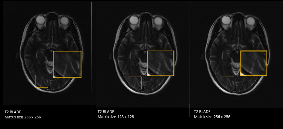
Figure 4 shows the ability of the model to recover high resolution images from low resolution images. When the model is used to predict an image from a downsampled image(middle), the resulting image (right) has a similar effect to the original image (left). This means that this model has potential for image acceleration.
Furthermore, this model works better on small images(sizes smaller than 300×300) than on large size images. One of the reasons for this is that the image size of the dataset is predominantly small.
Conclusion
The proposed model can increase the resolution of BLADE images while improving the image quality. It also has potential performance in reducing image artifacts such as Gibbs rings as well as accelerating image acquisition.Acknowledgements
Thanks to Dr. Dominik Nickel, Dr. Till Hülnhagen, Dr. Mario Zeller from Siemens Healthcare GmbH Ltd. for their code help with our work.
Thanks to Dr. Weng Dehe and Dr. Zhou Kun from Siemens Shenzhen Magnetic Resonance Ltd. for their guidance in our work.
References
1. Pipe JG. Motion correction with PROPELLER MRI: application to head motion and free-breathing cardiac imaging. Magn Reson Med 1999; 42:963-969.
2. Lavdas E, Mavroidis P, Kostopoulos S, et al. Improvement of image quality using BLADE sequences in brain MR imaging[J]. Magnetic resonance imaging, 2013, 31(2): 189-200.
3. Zhao C, Shao M, Carass A, et al. Applications of a deep learning method for anti-aliasing and super-resolution in MRI[J]. Magnetic resonance imaging, 2019, 64: 132-141.
4. Kaji S, Kida S. Overview of image-to-image translation by use of deep neural networks: denoising, super-resolution, modality conversion, and reconstruction in medical imaging[J]. Radiological physics and technology, 2019, 12(3): 235-248.
5. Li Y, Sixou B, Peyrin F. A review of the deep learning methods for medical images super resolution problems[J]. Irbm, 2021, 42(2): 120-133.
6. Zhang Q, Ruan G, Yang W, et al. MRI Gibbs‐ringing artifact reduction by means of machine learning using convolutional neural networks[J]. Magnetic resonance in medicine, 2019, 82(6): 2133-2145.
7. Zhang Y, Tian Y, Kong Y, et al. Residual dense network for image super-resolution[C]//Proceedings of the IEEE conference on computer vision and pattern recognition. 2018: 2472-2481.
8. Tong T, Li G, Liu X, et al. Image super-resolution using dense skip connections[C]//Proceedings of the IEEE international conference on computer vision. 2017: 4799-4807.
Figures