2744
Variational Network Meets Conjugate Gradient: Inline Reconstruction and Strain Analysis of Accelerated Cardiac Cine MRI1Computational Imaging Lab, Friedrich-Alexander-Universität Erlangen-Nürnberg, Erlangen, Germany, 2Magnetic Resonance, Siemens Healthcare GmbH, Erlangen, Germany, 3Institute of Radiology, University Hospital Erlangen, Friedrich-Alexander-Universität Erlangen-Nürnberg, Erlangen, Germany, 4Biomedical Engineering, The Ohio State University, Columbus, OH, United States
Synopsis
Keywords: Machine Learning/Artificial Intelligence, Image Reconstruction, Strain
Motivation: Accelerated cardiac cine MRI is prone to motion artifacts and underestimation of imaging biomarkers. Furthermore, many approaches lack prospective evaluation.
Goal(s): Improve data-driven reconstruction of cardiac cine MRI and enable inline reconstruction with improved estimation of strain parameters.
Approach: Training a neural network based on a Variational Network combined with intermediate conjugate gradient optimizations and evaluation on retrospectively undersampled data. Inline integration into scanner software using the FIRE framework and prospective evaluation in terms of image quality and cardiac strain parameters.
Results: The proposed network outperformed established compressed sensing approaches both retrospectively and prospectively, and in both image quality and cardiac strain estimation.
Impact: Our research enables inline reconstruction of highly accelerated (up to real-time) cardiac cine MRI with high motion fidelity and improved strain estimation compared to well-established compressed sensing approaches.
Introduction
Data-driven reconstruction of cardiac cine MRI is an active field of research. Many approaches, however, lack inline implementation and prospective evaluation. Furthermore, estimation of imaging biomarkers like cardiac strain may be biased in accelerated acquisitions1.We present a network architecture that combines the Variational Network2,3 (VN) with conjugate gradient descent for improved data consistency and reduced motion blurring. This network is implemented inline and strain parameters are evaluated on prospectively undersampled acquisitions.
Methods
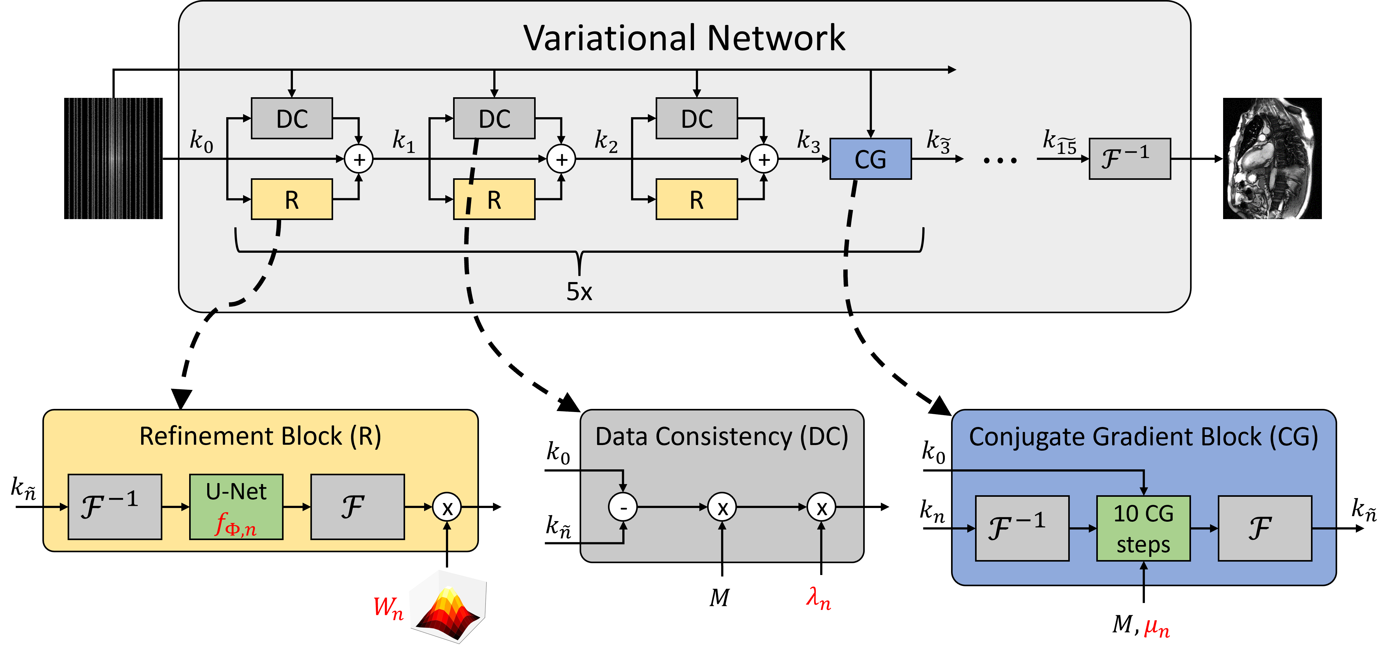
We trained a neural network to reconstruct a cardiac cine sequence $$$x$$$ from retrospectively undersampled multi-channel measurement data $$$k_0$$$. The network architecture (cf. Figure 1) is based on the Variational Network2,3 with 15 cascades, residual U-Nets $$$f_{\Phi,n}$$$ in each refinement block, and spatiotemporal convolutions4. Coil sensitivities are pre-computed using ESPIRiT5. The output of each refinement block is multiplied with a learnable piecewise linear function $$$W_n$$$ in $$$k$$$-$$$t$$$-space6. The update rule for cascade $$$n$$$ then becomes$$k_{n+1}=k_\tilde{n}-\lambda_nM(k_\tilde{n}-k_0)-W_n{\odot}Af_{\Phi,n}(A^{\dagger}k_\tilde{n})$$
$$$\lambda_n$$$ is a learnable data consistency weight, $$$M$$$ is the masking operator, $$$A$$$ is the forward operator consisting of coil sensitivities and Fourier transform and $$$A^\dagger$$$ is its adjoint. After every three cascades, we apply ten conjugate gradient (CG) descent steps to approximate the solution of the optimization problem:
$$x_\tilde{n}=arg\,min_{x}\lVert{MAx-k_0}\rVert_2^2+\mu_n\lVert{x-x_n}\rVert^2$$
where $$$\mu_n$$$ is a learnable scalar parameter and $$$x_n=A^{\dagger}k_n$$$. The input $$$k_\tilde{n}$$$ for Eq. 1 is then $$$Ax_\tilde{n}$$$ for $$$n\in\{3,6,9,12,15\}$$$ and $$$k_n$$$ otherwise.
The network was trained on fully sampled acquisitions from the OCMR dataset7, which includes samples of varying spatial and temporal resolution, cardiac views, and field strengths. A golden ratio variable density cartesian sampling mask8 was used for retrospective undersampling and the network was trained with a combination of SSIM-loss and $$$\perp$$$-loss9. Training/validation/testing splits were 186/44/38 slices.
For comparison, we trained a VN without the CG blocks. Furthermore, reconstructions with compressed sensing (CS) with temporal total variation (TTV) regularization were obtained using the Bart toolbox10. The TTV regularization weight was determined by optimization SSIM on the training dataset using a hyperparameter tuning framework11. Reconstructions were evaluated by computing peak signal-to-noise ratio (PSNR), structural similarity (SSIM), and spatiotemporal SSIM computed on spatiotemporal profiles through the center of the heart (SSIMst).
For prospective evaluation, the network was integrated inline on scanner reconstruction hardware using the FIRE framework12. Five healthy volunteers were scanned on three scanners at 1.5T and 3T (MAGNETOM Vida, Aera, and Sola, Siemens Healthineers AG, Erlangen, Germany). Written informed consent was obtained prior to data acquisition. bSSFP cines with readout resolution of $$$1.7\pm0.2\,mm$$$ along short- and long-axis views were acquired in real-time (acceleration rate $$$R\approx11$$$, temporal resolution $$$\Delta{t}=47\pm4\,ms$$$, phase resolution 60-70%) and over two heart cycles ($$$R\approx8$$$, $$$\Delta{t}=38\pm2\,ms$$$, phase resolution 90%). For reference strain values, short-axis slices were additionally measured using GRAPPA13 with $$$R=2$$$. Radial and circumferential strain was computed on mid-ventricular short-axis slices using a prototype software (Trufi Strain V3.0, Siemens Healthineers AG, Erlangen, Germany).
Results
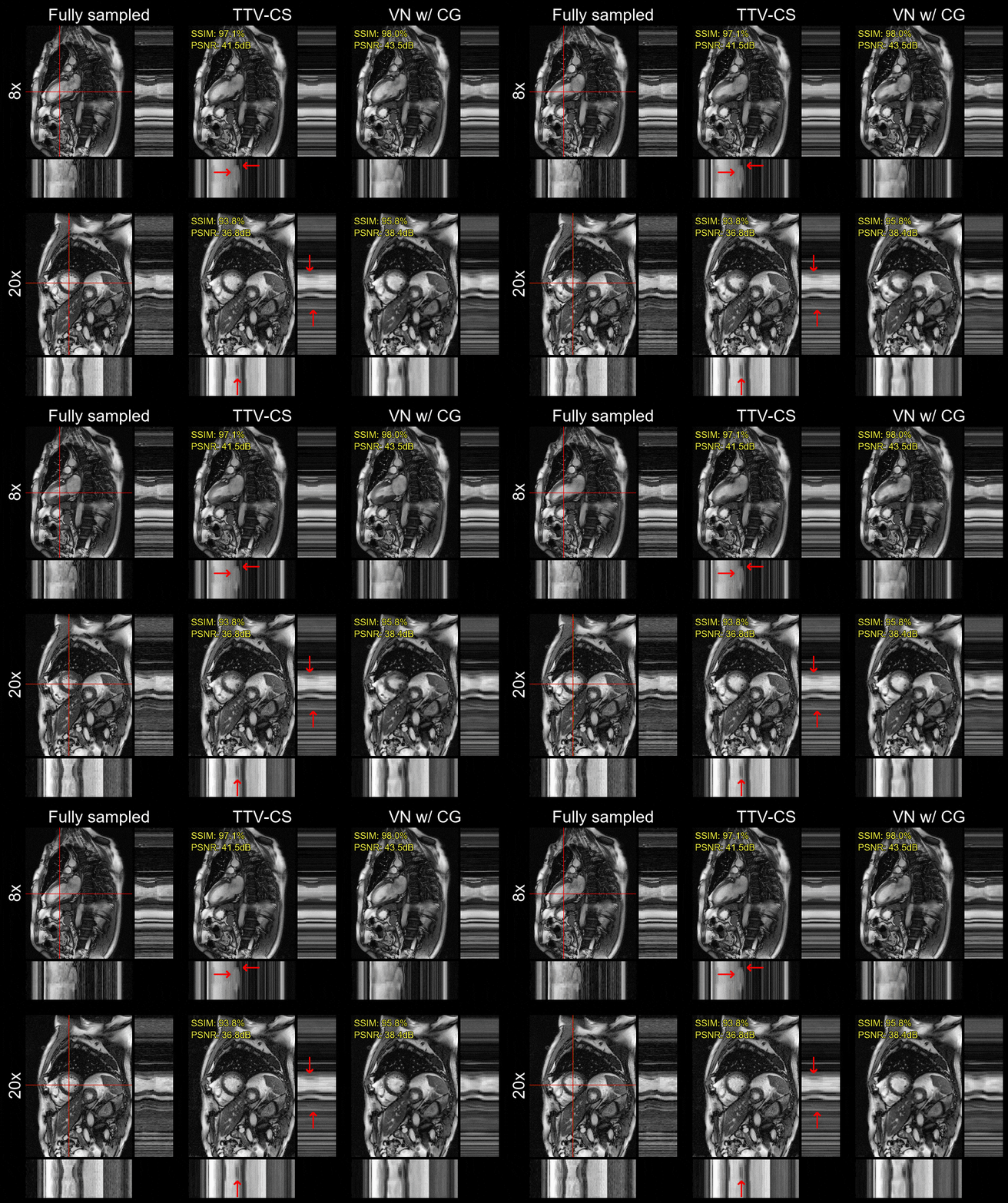
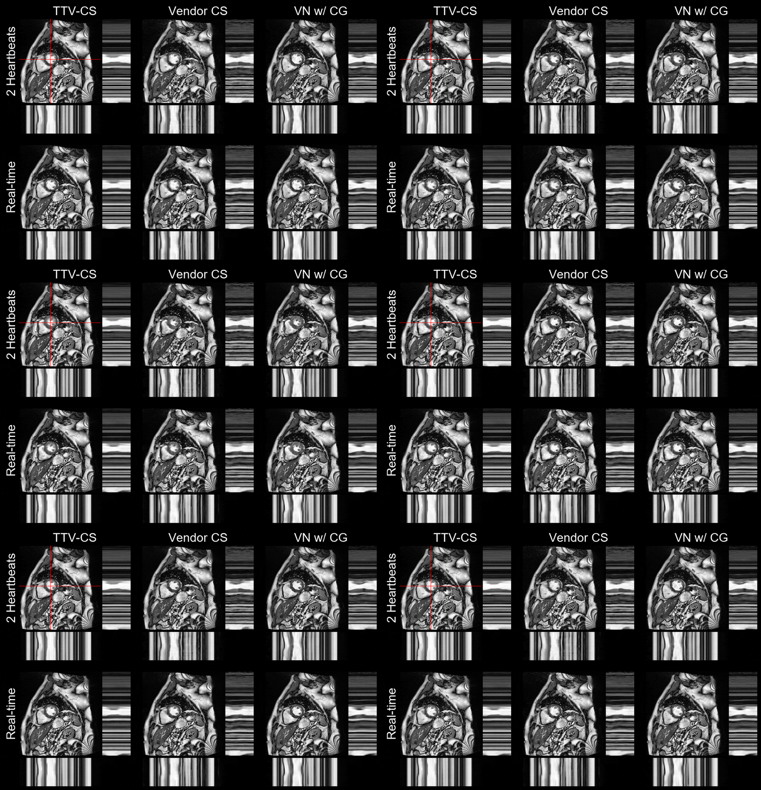
Quantitative results of the retrospectively undersampled test data are given in Figure 2. The VN with CG achieved significantly ($$$p<10^{-9}$$$) higher scores than VN without CG and TTV-CS in all metrics and for all acceleration rates. Exemplary reconstructions are shown in Figure 3.Prospectively undersampled acquisitions were reconstructed on scanner hardware using the FIRE framework and inference time was ~7-13s per slice with CG blocks and ~5-8s without. Exemplary reconstructions are shown in Figure 4.
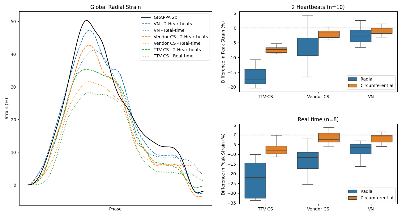
Agreement of peak strain with reference acquisitions was highest for the VN reconstructions, followed by the vendor CS reconstructions, and lowest for the TTV-CS reconstructions. Figure 5 shows radial strain curves for an exemplary slice and box plots comparing peak strain value agreement over all tested slices.
Discussion
The proposed VN yielded reconstructions with minimal noise or other artifacts and high motion fidelity. This is reflected in the presented time profiles and in the spatiotemporal SSIM on retrospective data. The network also had superior performance in a prospective evaluation. While TTV-CS and the vendor CS reconstructions display noticeable underestimation of peak strain parameters, this effect is minimized in the VN reconstructions.Inserting CG optimization into the network gave a substantial performance improvement. This suggests that the DC blocks in the VarNet cascades do not sufficiently enforce data consistency. This may be because the DC weights $$$\mu_n$$$ may converge to arbitrary values during training, while CG ensures convergence to the measurement data. Although this considerably increased inference time, it was still feasible for inline reconstruction.
Conclusion
The proposed reconstruction method outperformed CS both on retrospectively and prospectively accelerated data. Inline application and high agreement with reference strain values were demonstrated, highlighting the method’s feasibility for further clinical evaluation.Acknowledgements
This research was supported by NIH/NIBIB grant R01EB029957. In addition, we gratefully acknowledge the scientific support and HPC resources provided by the Erlangen National High Performance Computing Center (NHR@FAU) of Friedrich-Alexander-Universität Erlangen-Nürnberg (FAU) under the NHR project b143dc. NHR funding is provided by federal and Bavarian state authorities. NHR@FAU hardware is partially funded by the German Research Foundation (DFG) – 440719683. We furthermore thank Kelvin Chow for providing the FIRE framework and for his support.References
- Oberle R, Klimzak T, Forman C, et al. Comparison of Feature Tracking strain parameters derived from highly accelerated CINE images versus conventional CINE images. In: Proc ESCR Annual Meeting. Berlin; 2019:P-0045.
- Hammernik K, Klatzer T, Kobler E, et al. Learning a Variational Network for Reconstruction of Accelerated MRI Data. Magn Reson Med. 2018;79(6):3055-3071.
- Sriram A, Zbontar J, Murrell T, et al. End-to-End Variational Networks for Accelerated MRI Reconstruction. In: Martel AL, Abolmaesumi P, Stoyanov D, et al., eds. Medical Image Computing and Computer Assisted Intervention – MICCAI 2020. Vol 12262. Lecture Notes in Computer Science. Cham: Springer International Publishing; 2020:64-73.
- Vornehm M, Wetzl J, Giese D, Ahmad R, Knoll F. Spatiotemporal variational neural network for reconstruction of highly accelerated cardiac cine MRI. European Heart Journal - Cardiovascular Imaging. 2022;23(Supplement_2):jeac141.018.
- Uecker M, Lai P, Murphy MJ, et al. ESPIRiT—an Eigenvalue Approach to Autocalibrating Parallel MRI: Where SENSE Meets GRAPPA. Magn Reson Med. 2014;71(3):990-1001.
- Vornehm M, Wetzl J, Giese D, Knoll F. k-t adaptive Regularization in Variational Networks for Cardiac Cine Reconstruction. In: Proc ISMRM Workshop on Data Sampling and Image Reconstruction. Sedona, AZ; 2023.
- Chen C, Liu Y, Schniter P, et al. OCMR (v1.0)—Open-Access Multi-Coil k-Space Dataset for Cardiovascular Magnetic Resonance Imaging. arXiv:2008.03410 [eess.IV]. 2020.
- Joshi M, Pruitt A, Chen C, Liu Y, Ahmad R. Technical Report (v1.0)—Pseudo-random Cartesian Sampling for Dynamic MRI. arXiv:2206.03630v1 [eess.SP]. 2022.
- Terpstra ML, Maspero M, Sbrizzi A, van den Berg CAT. ⊥-loss: A Symmetric Loss Function for Magnetic Resonance Imaging Reconstruction and Image Registration with Deep Learning. Medical Image Analysis. 2022;80:102509.
- Blumenthal M, Holme C, Roeloffs V, et al. mrirecon/bart: version 0.8.00. 2022.
- Akiba T, Sano S, Yanase T, Ohta T, Koyama M. Optuna: A Next-generation Hyperparameter Optimization Framework. arXiv:1907.10902v1 [cs.LG]. 2019.
- Chow K, Kellman P, Xue H. Prototyping Image Reconstruction and Analysis with FIRE. In: Proc SCMR Virtual Scientific Sessions; 2021:838972.
- Griswold MA, Jakob PM, Heidemann RM, et al. Generalized Autocalibrating Partially Parallel Acquisitions (GRAPPA). Magn Reson Med. 2002;47(6):1202-1210.
Figures