2742
Deep Learning-based Distortion Correction of Diffusion-weighted Imaging1Department of Radiology, Mayo Clinic, Rochester, MN, United States
Synopsis
Keywords: Machine Learning/Artificial Intelligence, Diffusion/other diffusion imaging techniques, Diffusion acquisition, distortion reduction, deep learning
Motivation: Diffusion-weighted imaging (DWI) is typically based on single-shot echo-planar imaging (EPI), which is prone to magnetic field inhomogeneities-induced artifacts, such as geometric distortion and blurring. The multi-shot diffusion sequence, DIADEM, employing a dual spin-warp (SW) and EPI phase-encoding strategies, can produce distortion-free images at the cost of extended scan times.
Goal(s): We proposed a deep learning-based distortion correction method for conventional DWI, using DIADEM as reference.
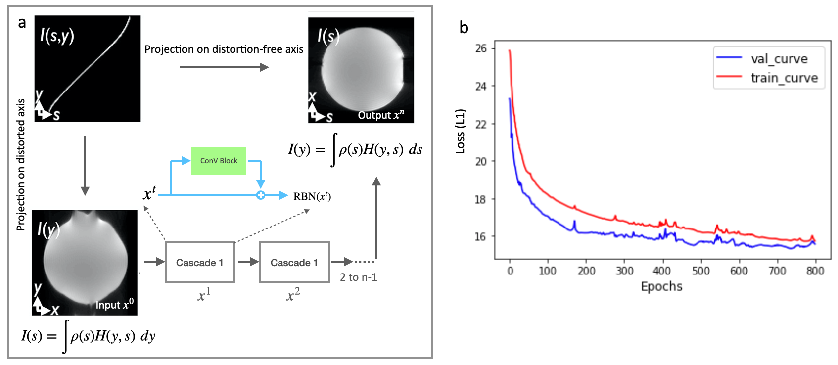
Approach: The 3D neural network was trained to learn the mapping between the projections of the point-spread-function, PSF H(y,s) along the EPI phase-encoding (y) direction and the PSF-encoding (s) direction, respectively.
Results: It demonstrated reduced geometric distortion.
Impact: Conventional DWI sequence suffers from distortion caused by susceptibility. We proposed a deep learning-based distortion correction method, leveraging distortion-free DIADEM images as reference. Our method was demonstrated to reduce geometric distortion and imaging blurring without distortion calibration.
Introduction
Diffusion-weighted imaging (DWI) has been a key clinical sequence utilizing molecular diffusion to probe tissue micro-architecture. Standard DWI typically employs single-shot echo-planar imaging (EPI), which is prone to magnetic field inhomogeneities-induced artifacts, such as geometric distortion and blurring. To address this challenge, a multi-shot diffusion sequence, DIADEM [1], which combines dual spin-warp (SW) and EPI phase-encoding strategies, has been shown to successfully produce distortion-free images, albeit with a longer scan time. In this study, we proposed a deep-learning-based distortion-correction method for conventional DWI, using DIADEM images as ground truth for training. The neural network was trained to learn the mapping between the projections of the point-spread-function (PSF), denoted as H(y,s), along the EPI phase-encoding (y) direction and the PSF-encoding (s) direction, respectively.Methods
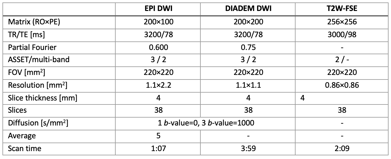
In this retrospective study, we analyzed scans from 41 subjects obtained from a 3T GE scanner (GE 750W, GE Healthcare, Waukesha, WI) under an IRB-approved protocol. In addition to DIADEM diffusion imaging, the conventional diffusion and anatomical T2W-TSE images were acquired. The imaging protocol is outlined in Table 1. A lower resolution imaging was employed in the conventional diffusion imaging to assess the improvement in image blurring with the proposed model. After collecting DIADEM diffusion raw data, offline reconstruction was performed using MatLab (MathWorks, Natick, MA). Images with b = 0 s/mm2 were used to train the model, which also incorporated T2W-TSE images to enhance the results.A 3D convolutional neural network was redesigned from a previous implemented super-resolution model [2] to learn the residual information between the distorted and distortion-free images. Instead of training on patches, we input the whole 3D images. Thus, the input has a dimension of 200 x 200 x 2, and the output is 200 x 200 x 1, while the intermediate layers have 128 channels. The number of convolutional layers is selected as 8, considering both performance and computational effort. L1 norm is used for loss function, which was minimized by Adam Minimizer. The network was trained in Keras with a NVIDIA GPU of A100 for about 5 hours.
Results
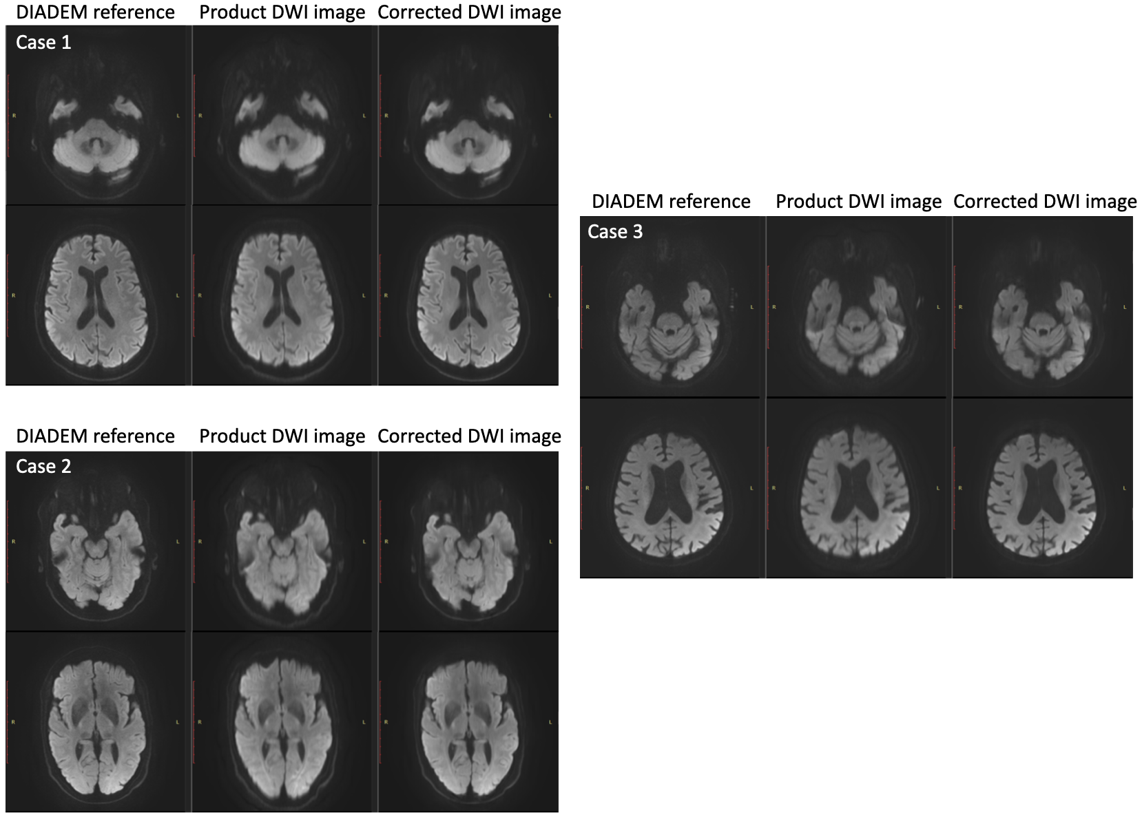
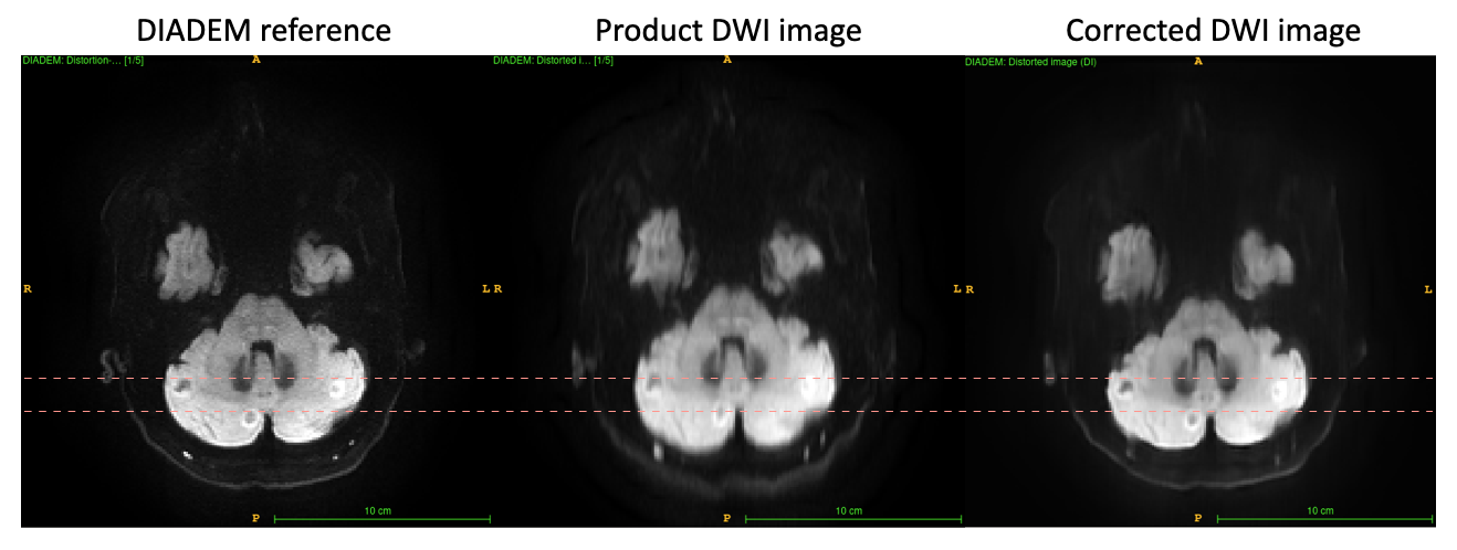
After the model was trained over 800 epochs, the best model’s evaluation was a L1 loss of 15.80 with a mean-square-error (MSE) of 1873.4. The learning curves were shown in Figure 1(c). Three representative testing examples were shown in Figure 2 that each includes the DIADEM reference, the product DWI image and the predicted image from the AI model. In addition, Figure 3 shows a case with brain tumors, which were misaligned in conventional DWI, and corrected by the model. The results demonstrated the model’s capability in correcting both geometrical distortion and blurring.Discussion
Previous research aimed to expedite DIADEM by employing subsampling within the spin-warp (SW)-PE dimension for its acceleration [1]. Parallel imaging and simultaneous multiple-slice techniques have been employed to expedite DIADEM. Moreover, further acceleration has been explored using high slew-rates gradient system such as that on the compact 3T [3]. Despite these efforts, DIADEM’s acquisition time remained suboptimal for swift clinical workflows. Our current study used a novel deep learning-based, end-to-end approach to learn the mapping between the SW-PE and EPI-PE dimensions of the DIADEM, offering distortion correction without sacrificing image quality and scan time.In contrast to slice-by-slice analysis, we trained a 3D convolutional model for comprehensive volumetric learning. Comparing to the super-resolution task [2], which focuses on enhancing local image details, our study aims to simultaneously deblur (via L1-norm optimization) and correct the geometrical distortions along the SW-PE axis. A holistic 3D registration of brain anatomy, as opposed to patch-based or 2D methodologies, is more beneficial for accurately repositioning tissues and amending distortions in this study. Feeding T2W-TSE to the ancillary channel of the input enriched this process, as exemplified by the marked improvements in brain tumor evaluations, yielding superior localization and clarity of lesions.
To further enhance image quality, perceptual loss functions or AI-generative diffusion model could be leveraged to refine deblurring techniques. Moreover, a direct learning approach for the PSF $H(y,s)$ could potentially unveil additional image information compared to the current methodology of mapping its orthogonal projections along the y-axis (EPI phase-encoding) and s-axis (PSF-encoding). Our further work aims to develop a physics-informed model to further recover the fidelity of DIADEM imaging.
Conclusion
We have introduced a novel deep learning-based, end-to-end method for distortion-correction for conventional DWI using distortion-free DIADEM images as training dataset. Preliminary in-vivo results demonstrated reduction of geometric distortion and image blurriness without the need for calibration approaches such as field mapping or gradient reversal method, and with inherent time savings compared to DIADEM-based acquisition.Acknowledgements
No acknowledgement found.References
[1] In, M.H., Posnansky, O., Speck, O., 2017. High-resolution distortion-free diffusion imaging using hybrid spin-warp and echo-planar PSF-encoding approach. NeuroImage. 148, 20-30.
[2] Zhang, K., Hu, H., Philbrick, K., Conte, G.M., Sobek, J.D., Rouzrokh, P., Erickson, B.J., 2022. SOUP-GAN: super-resolution MRI using generative adversarial networks. Tomography. 8, 905-919.
[3] In, M.H., Tan, E.T., Trzasko, J.D., Shu, Y., Kang, D., Yarach, U., Tao, S., Gray, E.M, Huston, J., Bernstein, M.A., 2020. Distortion-free imaging: a double encoding method (DIADEM) combined with multiband imaging for rapid distortion-free high-resolution diffusion imaging on a compact 3T with high-performance gradients. J Magn Reson Imaging. 51, 296-310.
Figures