2739
Advancing Ultralow-field Brain MRI through K-space Undersampling and Deep Learning Image Reconstruction1Laboratory of Biomedical Imaging and Signal Processing, The University of Hong Kong, Hong Kong, China, 2Department of Electrical and Electronic Engineering, The University of Hong Kong, Hong Kong, China
Synopsis
Keywords: Image Reconstruction, Low-Field MRI
Motivation: While the emerging ULF MRI shows potential of low-cost and point-of-care imaging applications, its image quality is poor and the scan time is long.
Goal(s): To reduce the ULF brain MRI scan time through deep learning image reconstruction from partial Fourier and uniformly undersampled data.
Approach: We proposed a DL reconstruction method for fast 3D brain MRI at 0.055T by applying the 3D DL image reconstruction to undersampled 3D k-space data, achieving speed up of 2x over our newly developed partial Fourier reconstruction and superresolution (PF-SR) method.
Results: Our preliminary results show the proposed method could reduce noise, artifacts, and enhance spatial resolution.
Impact: Our model can work with uniformly undersampled data, leading to acceleration factor of 2, and a PF sampling of at a fraction of 0.7. Our development enables fast and quality whole-brain MRI at 0.055T, indicating potential for widespread biomedical applications.
Introduction
High-field MRI are costly and highly inaccessible worldwide, this stimulates the development of MRI scanners at ultra-low-field (ULF) strength for low-cost and point-of-care clinical applications1-6. However, imaging at ULF suffers from a markedly low signal-to-noise ratio (SNR), which hinders its widespread clinical adoption.Deep learning (DL) has shown its potential in various high-field MR image processing application, including artifact reduction, denoising, and reconstruction from undersampled k-space7-10. The increasing availability of large-scale anatomy-specific and protocol-specific high-quality high-field MRI datasets will boost the ability of DL on the reconstruction of ULF MRI k-space data and enable low-cost ULF MRI for accessible health care11-14. Some DL attempts have been made recently to improve ULF MRI image quality, but with limit success so far15-18.
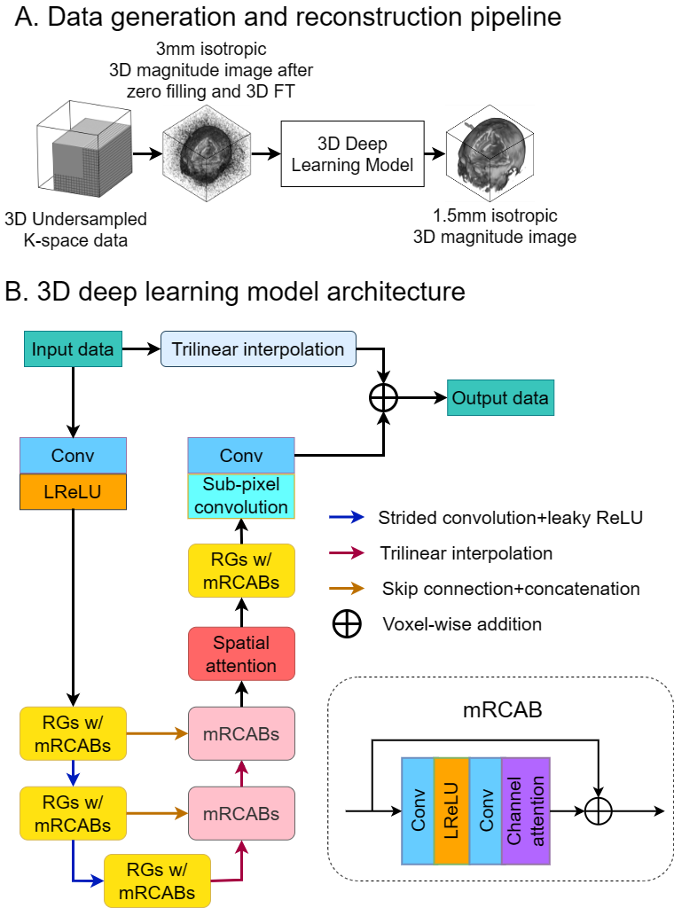
In this study, we adopted our recently developed partial Fourier (PF) reconstruction and superresolution (SR) model and proposed a DL reconstruction method on PF sampled plus uniformly undersampled k-space data for fast ULF 3D brain MRI.
Methods
We adopted the model architecture of PF-SR19 for the reconstruction of 2D PF-sampled plus 2D uniformly undersampled low-resolution noisy ULF data. The 2D uniform undersampling pattern contains 40 × 28 center fully-sampled lines with an acceleration factor of 2. The overall architecture of the DL model is shown in Figure 1. The model was trained using the AdamW optimizer with β1=0.9, β2=0.999, weight decay of 0.01, and minimizing the L1 loss. The model was trained with 350 epochs on four Nvidia A100 GPUs.A T2W dataset containing 1182 subjects from the HCP S1200 dataset20 were used for model training. The following steps were applied to prepare the undersampled data: 1) Local mean was applied to downsample the original 0.7mm isotropic high resolution 3T data to approximately 1.5mm isotropic resolution to generate the model training target 2) Symmetric k-space truncation in all three dimensions was performed to further downsample the model training target data to 3mm isotropic solution. 3) Retrospective 2D PF sampling of a fraction of 0.7 was applied along two PE (left-right and superior-inferior) directions in the k-space, followed by retrospective 2D uniform undersampling of an acceleration factor of 2 along the same PE directions. The total acceleration factor is 4. 4) Fourier transform was performed on the undersampled k-space, and the resulting image was further degraded by the addition of Rician noise.
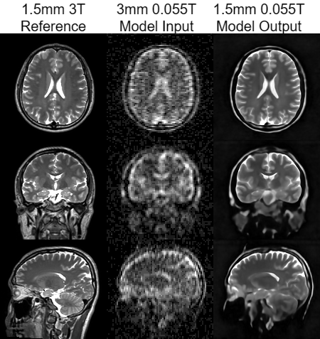
We tested our model on the synthetic data and experimental ULF data. The experimental ULF data was acquired with prospective 2D PF sampling at a fraction of 0.7 along two PE directions. The retrospective 2D uniform undersampling of an acceleration factor of 2 along the same PE directions was later applied.
Results
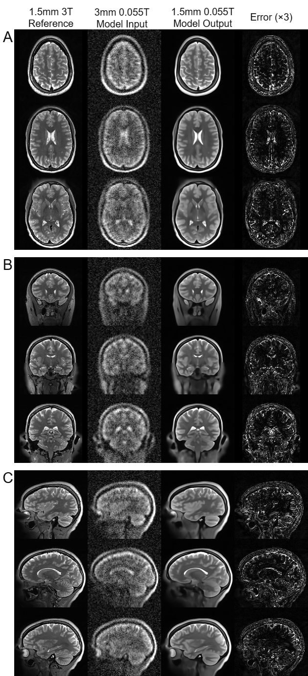
Figure 2 shows the reconstruction results using the proposed model on synthetic ULF data simulated from high-field data of a healthy subject with 2D uniform undersampling at an acceleration factor of 2 and 2D PF sampling at a fraction of 0.7 along two PE directions, which produced a total acceleration factor of 4. The preliminary results of experimental ULF data from one young volunteer are shown in Figure 3. The noise, blurring and ringing artifacts are largely reduced.Discussion and conclusion
In this study, we proposed a DL reconstruction method to further accelerate the 3D MR brain data acquisition by a factor of 2 through retrospective 2D uniform undersampling with central k-space lines. The preliminary results demonstrate the proposed method can reduce noise, artifacts resulted from both PF and uniform undersampling, and enhance spatial resolution to 1.5mm isotropic resolution in both synthetic and experimental ULF results.However, minor blurring could still be seen. In future study, we could include more publicly available high-field 3D brain data to expand the training data size, together with further refining the preprocessing steps of training data to match the characteristics of experimental ULF data to minimize the domain gap between synthetic and experimental ULF data.
Acknowledgements
This
work was supported in part by Hong Kong Research Grant Council (R7003-19F,
HKU17112120, HKU17127121, HKU17127022 and HKU17127523 to E.X.W).
References
[1] Cooley CZ, McDaniel PC, Stockmann JP, et al. A portable scanner for magnetic resonance imaging of the brain. Nat Biomed Eng. 2021;5(3):229-239. doi:10.1038/s41551-020-00641-5
[2] He Y, He W, Tan L, et al. Use of 2.1 MHz MRI scanner for brain imaging and its preliminary results in stroke. J Magn Reson. 2020;319:106829. doi:10.1016/j.jmr.2020.106829
[3] O'Reilly T, Teeuwisse WM, de Gans D, Koolstra K, Webb AG. In vivo 3D brain and extremity MRI at 50 mT using a permanent magnet Halbach array. Magn Reson Med. 2021;85(1):495-505. doi:10.1002/mrm.28396
[4] Liu Y, Leong ATL, Zhao Y, et al. A low-cost and shielding-free ultra-low-field brain MRI scanner. Nat Commun. 2021;12(1):7238. Published 2021 Dec 14. doi:10.1038/s41467-021-27317-1
[5] Kimberly WT, Sorby-Adams AJ, Webb AG, et al. Brain imaging with portable low-field MRI. Nat Rev Bioeng. 2023;1(9):617-630. doi:10.1038/s44222-023-00086-w
[6] Hennig J. An evolution of low-field strength MRI. MAGMA. 2023;36(3):335-346. doi:10.1007/s10334-023-01104-z
[7] Zhu B, Liu JZ, Cauley SF, Rosen BR, Rosen MS. Image reconstruction by domain-transform manifold learning. Nature. 2018;555(7697):487-492. doi:10.1038/nature25988
[8] Lin DJ, Johnson PM, Knoll F, Lui YW. Artificial Intelligence for MR Image Reconstruction: An Overview for Clinicians. J Magn Reson Imaging. 2021;53(4):1015-1028. doi:10.1002/jmri.27078
[9] Qu L, Zhang Y, Wang S, Yap PT, Shen D. Synthesized 7T MRI from 3T MRI via deep learning in spatial and wavelet domains. Med Image Anal. 2020;62:101663. doi:10.1016/j.media.2020.101663
[10] Chen L, Schär M, Chan KWY, et al. In vivo imaging of phosphocreatine with artificial neural networks. Nat Commun. 2020;11(1):1072. Published 2020 Feb 26. doi:10.1038/s41467-020-14874-0
[11] Miller KL, Alfaro-Almagro F, Bangerter NK, et al. Multimodal population brain imaging in the UK Biobank prospective epidemiological study. Nat Neurosci. 2016;19(11):1523-1536. doi:10.1038/nn.4393
[12] Knoll F, Murrell T, Sriram A, et al. Advancing machine learning for MR image reconstruction with an open competition: Overview of the 2019 fastMRI challenge. Magn Reson Med. 2020;84(6):3054-3070. doi:10.1002/mrm.28338
[13] Littlejohns TJ, Holliday J, Gibson LM, et al. The UK Biobank imaging enhancement of 100,000 participants: rationale, data collection, management and future directions. Nat Commun. 2020;11(1):2624. Published 2020 May 26. doi:10.1038/s41467-020-15948-9
[14] Babayan A, Erbey M, Kumral D, et al. A mind-brain-body dataset of MRI, EEG, cognition, emotion, and peripheral physiology in young and old adults. Sci Data. 2019;6:180308. Published 2019 Feb 12. doi:10.1038/sdata.2018.308
[15] Koonjoo N, Zhu B, Bagnall GC, Bhutto D, Rosen MS. Boosting the signal-to-noise of low-field MRI with deep learning image reconstruction. Sci Rep. 2021;11(1):8248. Published 2021 Apr 15. doi:10.1038/s41598-021-87482-7
[16] de Leeuw den Bouter ML, Ippolito G, O'Reilly TPA, Remis RF, van Gijzen MB, Webb AG. Deep learning-based single image super-resolution for low-field MR brain images. Sci Rep. 2022;12(1):6362. Published 2022 Apr 16. doi:10.1038/s41598-022-10298-6
[17] Manso Jimeno M, Ravi KS, Jin Z, Oyekunle D, Ogbole G, Geethanath S. ArtifactID: Identifying artifacts in low-field MRI of the brain using deep learning [published correction appears in Magn Reson Imaging. 2022 Apr 5;:]. Magn Reson Imaging. 2022;89:42-48. doi:10.1016/j.mri.2022.02.002
[18] Ayde R, Senft T, Salameh N, Sarracanie M. Deep learning for fast low-field MRI acquisitions. Sci Rep. 2022;12(1):11394. Published 2022 Jul 6. doi:10.1038/s41598-022-14039-7
[19] Man C, Lau V, Su S, et al. Deep learning enabled fast 3D brain MRI at 0.055 tesla. Sci Adv. 2023;9(38):eadi9327. doi:10.1126/sciadv.adi9327
[20] Van Essen DC, Smith SM, Barch DM, et al. The WU-Minn Human Connectome Project: an overview. Neuroimage. 2013;80:62-79. doi:10.1016/j.neuroimage.2013.05.041
Figures