2734
Deep-ECCENTRIC: Deep Learning Reconstruction of whole-brain non-Cartesian Compressed-Sensing MR Spectroscopic Imaging1Athinoula A. Martinos Center for Biomedical Imaging, Massachusetts General Hospital, Boston, MA, United States, 2Department of Radiology, Massachusetts General Hospital, Harvard Medical School, Boston, MA, United States, 3Computational Imaging Research Lab - Department of Biomedical Imaging and Image-guided Therapy, Medical University of Vienna, Vienna, Austria, 4High Field MR Center - Department of Biomedical Imaging and Image‐Guided Therapy, Medical University of Vienna, Vienna, Austria, 5Computer Science and Artificial Intelligence Lab, MIT, Cambridge, MA, United States, 6Pappas Center for Neuro-Oncology, Department of Neurology, Massachusetts General Hospital, Boston, MA, United States, 7Department of Neurology, Beth-Israel Deaconess Medical Center, Boston, MA, United States, 8Department of Neurology, Brigham and Women’s Hospital, Boston, MA, United States, 9Department of Neurosurgery, Massachusetts General Hospital, Boston, MA, United States, 10Advanced Clinical Imaging Technology, Siemens Healthineers International AG, Lausanne, Switzerland, 11Center for Biomedical Imaging (CIBM), Geneva, Switzerland
Synopsis
Keywords: Image Reconstruction, Spectroscopy, Brain, High-Field MR, Image Reconstruction
Motivation: Magnetic resonance spectroscopic imaging (MRSI) is a unique method for non-invasive mapping of brain neurochemistry. While the latest advancements in acquisition enable whole-brain high-resolution metabolic imaging, these methods have lengthy reconstruction times that limit the clinical use.
Goal(s): To realize a fast end-to-end reconstruction pipeline for high-resolution whole-brain MRSI compatible with online processing and clinical use.
Approach: We developed a rapid deep-learning reconstruction pipeline for 3D non-Cartesian Compressed-Sensing MRSI (ECCENTRIC).
Results: Our approach reconstructs in a few minutes high-resolution ECCENTRIC (k,t) data. We demonstrate a 60-fold speed-up in reconstruction time, facilitating the use in clinical routine.
Impact: We present Deep-ECCENTRIC: a deep-learning pipeline for end-to-end reconstruction of 3D non-Cartesian Compressed-Sensing MRSI. We showcase spatially precise reconstructions with high spectral consistency, at a 60-fold speed-up over conventional reconstructions, which facilitates the clinical use of fast high-resolution MRSI.
Authorship
*Sharing senior authorship.INTRODUCTION
Magnetic resonance spectroscopic imaging (MRSI) allows non-invasive mapping of in-vivo metabolism [1].While high resolution whole-brain MRSI has great value to study healthy tissues and diseases [2], its clinical use is limited by both long acquisition and reconstruction times. ECCENTRIC [3] combines non-Cartesian trajectories and sparse k-space sampling to accelerate the MRSI acquisition by a factor of 50-100. However, ECCENTRIC reconstruction requires solving iteratively a constrained optimization problem, which is computationally expensive and takes several hours.Recent advances in deep learning (DL) enables fast GPU-accelerated reconstructions of MRI with high accuracy [4]. Here, we propose a rapid DL-based pipeline for end-to-end reconstruction of whole-brain non-Cartesian compressed-sensing MRSI.
METHODS
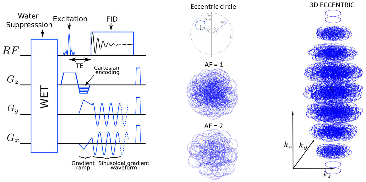
Data Acquisition:3D 1H-FID-ECCENTRIC[3] was acquired with the pulse sequence from Figure 1 on a 7T scanner (MAGNETOM Terra, Siemens Healthcare, Germany) and a 32Rx/1Tx head coil (NovaMedical, USA) using: 0.9 ms echo-time, 27° excitation flip-angle, 275 ms repetition-time, field-of-view 220x220x105 mm3, matrix size 64x64x31, 3.4x3.4x3.4 mm3 voxel size. The ECCENTRIC circle radius was set to kmax/8 with spectral bandwidth of 2326 Hz that did not require temporal interleaving. The acquisition was further accelerated (AF=2-4) by random undersampling ECCENTRIC, resulting in acquisition times between 4:11-9:21 min. A low-resolution water reference was acquired for coil combination with the same sequence but a smaller matrix size (22x22x11) in 1:16 minutes. For DL training, fully sampled water-unsuppressed MRSI data were acquired with the same ECCENTRIC sequence, but shorter TR=100ms in 6:46 minutes.
DL Reconstruction:
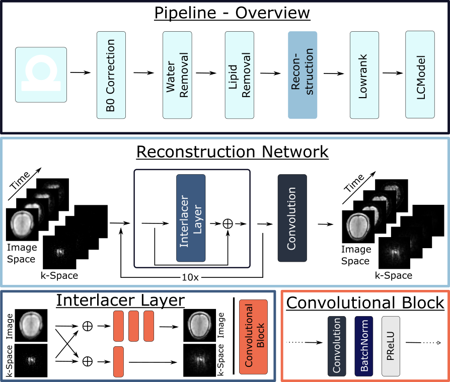
Figure 2 shows the MRSI reconstruction pipeline consisting of: A) (k,t) data are pre-processed by applying Hamming filter, density compensation, inverse non-uniform fast Fourier transformation (iNUFFT), and coil combination, resulting in the initial 4D image-time MRSI data. B) The pipeline further performs B0-correction, water removal, lipid removal, DL-reconstruction, low-rank denoising, and spectral fitting of metabolites.
DL-reconstruction of 4D (k,t) spatial-spectral ECCENTRIC data is performed by a recurrent Interlacer network [4]. Gridded k-space data and coil-combined iNUFFT image data are provided as input. Each time point is processed individually. To enhance reconstruction capability the channels are increased and reduced by applying three convolutional blocks in image space with feature size of 2-64-2. Only a single convolutional block with feature size 64 is applied in k-space. The network applies 10 Interlacer layers in a recurrent manner. The mean squared error (MSE) loss was optimized using Adam with a learning rate of 10^(-5) for 500 epochs. .
DL Lipid Removal:
Before the metabolite reconstruction, the nuisance lipid signal was removed by a convolutional neural network. The architecture for lipid removal is a Y-Net [5] with 2 encoders and 1 decoder. The encoders and decoder consist of 4 convolutional blocks.
Human Subjects:
27 subjects were scanned with informed consent, including 22 healthy volunteers and 5 patients with glioma tumors. 21 subjects were used for training/validation and 6 subjects for testing.
RESULTS
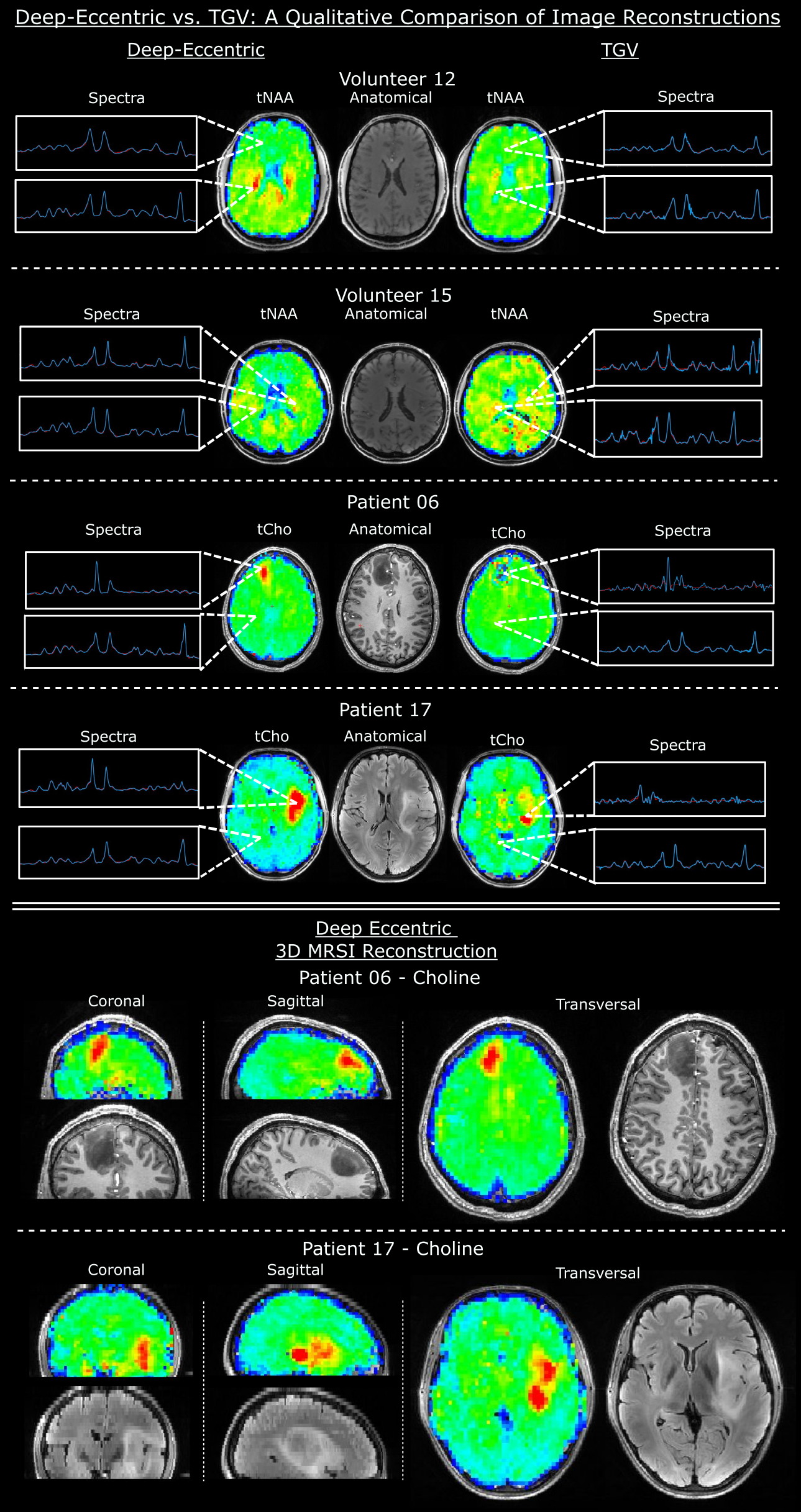
The reconstruction of the ECCENTRIC data by the Interlacer network is performed in 3 minutes, which is ~60 times faster to conventional reconstruction.Figure 3 compares metabolic maps in 2 glioma patients and 2 healthy volunteers obtained by DL reconstruction and conventional Compressed Sensing-SENSE LowRank model (CS-SENSE-LR) TGV reconstruction[3]. Metabolic maps of total Choline show better delineation of glioma tumors by Deep-ECCENTRIC. Total NAA shows sharper brain structure in healthy volunteers by Deep-ECCENTRIC. Selected spectra show lower noise and less lipid contamination by DL reconstruction.
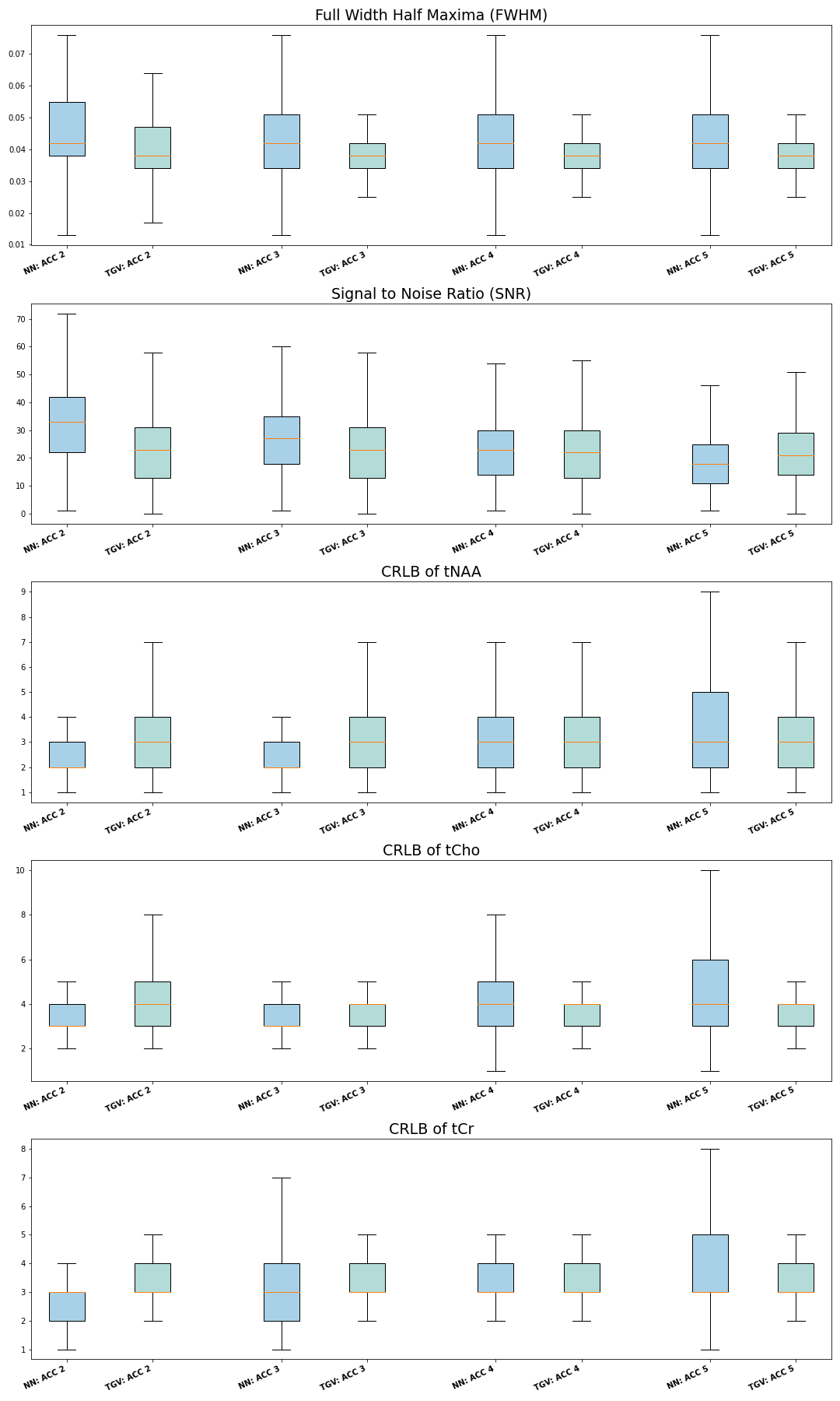
Figure 4 presents full-width-half-maxima (FWHM), signal to noise ratio (SNR) and Cramer-Rao lower bounds (CRLB). DL recon provides slight increase of SNR and CRLB for accelerations 2 & 3.
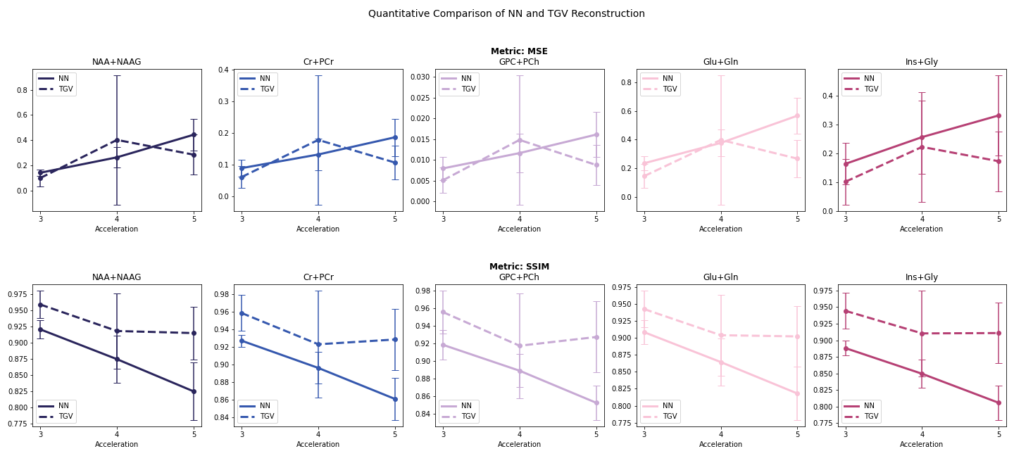
The reconstruction consistency across different accelerations evaluated by MSE and structural similarity index (SSIM) is presented in Figure 5. MSE shows similar results for Deep-ECCENTRIC and conventional TGV reconstruction. The TGV reconstruction provides slightly larger SSIM due to spatial smoothing effect of TGV regularization, while DL provides sharper anatomical boundaries.
CONCLUSION
We present an end-to-end pipeline for rapid DL-based MRSI reconstruction, which demonstrate robust performance to map metabolism of the human brain, including glioma tumors. The architecture of our neural network maintains high temporal consistency that reduces spectral noise. This approach takes a fraction of the time needed by conventional methods, paving the way for clinical use of high-resolution whole-brain MRSI.Acknowledgements
No acknowledgement found.References
[1]. Maudsley, A. A. et al (2020). "Advanced magnetic resonance spectroscopic neuroimaging: Experts' consensus recommendations." NMR Biomed Special Issue: Advanced methodology for in vivo magnetic resonance spectroscopy): e4309.
[2]. Oz, G. et al (2014). "Clinical proton MR spectroscopy in central nervous system disorders." Radiology. 270(3): 658-679.
[3]. Klauser, A. et al (2023). "ECCENTRIC: a fast and unrestrained approach for high-resolution in vivo metabolic imaging at ultra-high field M." Arxiv & Radiology https://arxiv.org/abs/2305.13822.
[4]. Singh, N. M. et al. "Joint frequency and image space learning for MRI reconstruction and analysis." The journal of machine learning for biomedical imaging 2022 (2022).
[5]: Mohammed, A. et al. "Y-net: A deep convolutional neural network for polyp detection." arXiv preprint arXiv:1806.01907 (2018).
[6]: Kingma, Diederik P., and Jimmy Ba. "Adam: A method for stochastic optimization." arXiv preprint arXiv:1412.6980 (2014).
Figures