2733
Efficient Standardization of Clinical T2-Weighted Images: Phase-Conjugacy e-CAMP with Projected Gradient Descent1Department of Biomedical Engineering, Yale University, New Haven, CT, United States, 2Department of Radiology and Biomedical Imaging, Yale University, New Haven, CT, United States
Synopsis
Keywords: Image Reconstruction, Quantitative Imaging
Motivation: Routine clinical images are a massive data source for machine learning. The previously introduced e-CAMP method can convert T2-weighted images of clinical TSE acquisition to quantitative T2 maps, but it requires tuning of many parameters, impeding widespread implementation.
Goal(s): To present an algorithm that requires few parameter choices, is robust to those parameter values, and is faster to convergence.
Approach: Projected Gradient Descent ensures efficient enforcement of the T2-decay model constraint and greatly eliminates parameter tuning. e-CAMP is further enhanced by phase conjugacy with Virtual Conjugate Coils.
Results: The efficient and robust implementation of e-CAMP shows accurate T2 map reconstruction.
Impact: Rather than acquire specific yet time-consuming quantitative images, e-CAMP can efficiently standardize the existing qualitative images from routine clinical scans and exploit the enormous amount of images to create dataset for large-scale machine learning.
Introduction
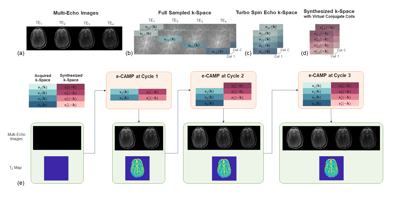
Images from routine clinical MRI scans are a massive data source of machine learning that benefits rapidly evolving fields such as radiomics. Around 30 million MRI studies are conducted annually in the U.S. Yet the potential has not been fully utilized because qualitative images are subject to the scanner and scanning protocol, impeding the direct translation into large-scale datasets.Standardization is required for dataset formation. Turbo Spin Echo (TSE) accounts for a major part of clinical scans, where the band-sampled pattern generates T2-weighted images, as illustrated in Fig. 1(a-c). The state-of-the-art methods for T2 map reconstruction1 rely on accelerated echo images of Cartesian2 or radial trajectories3, and fail on the band-sampling pattern due to the inhomogeneous intensity of echo images4.
e-CAMP was proposed to standardize the T2-weighted images from TSE scans4. Converting the exponential decay to a linear model, e-CAMP forms a biconvex minimization problem of data fidelity and T2-decay model coherence, enabling alternate optimization of multi-echo images and the T2 map. K-space bands are expanded during iterations for careful initialization.
Specifically, the previous version imposed the physics model as a penalty with an increasing penalty coefficient. The parameter tuning causes numerical instability, slow convergence, and limits potential application to highly variable datasets. A more robust and efficient algorithm is desired to avoid the tedious tuning and disseminate the algorithm for broader applications.
Theory
Problem FormulationWe define the underlying multi-echo images $$$\{\rho_p\}_{p=1}^P$$$ of the acquired echoes $$$\{1,\cdots,P\}$$$.The acquired k-space data $$$\{s_p\}_{p=1}^P$$$ is encoded by coil sensitivity $$$C$$$, Fourier transform $$$F$$$, and the sampling mask $$$\{M_p\}_{p=1}^P$$$. Summarizing the encoding matrix as $$$\{E_p\}_{p=1}^P$$$, the forward model is
$$s_p=M_pFC\rho_p+\epsilon_p=E_p\rho_p+\epsilon_p,\quad p\in\{1,\cdots,P\}$$
where $$$\{\epsilon_p\}_{p=1}^P$$$ is the Gaussian noise.
Besides the data fidelity term, we add a constraint to enforce adherence to the T2-decay model. The preliminary constrained minimization problem, including TV regularization, is as follows.
$$\min_{\rho, \alpha} \sum_{p=1}^{P} \left[\left\|s_p - E_p \rho_p \right\|^2 + \lambda TV(\rho_p)\right]$$
$$\text{s.t.} \quad \rho_{p+1} = \alpha \rho_p, \quad p\in\{1,\cdots,P-1\}$$
where $$$\alpha(x)=exp\left(-\frac{\Delta TE}{T_2(x)}\right)$$$.
Phase Conjugacy
Phase-constrained algorithms are used to decrease the number of unknowns by half5. In TSE, the magnetization at each TE has almost the same background phase $$$\Phi$$$ due to refocusing. Extracting $$$\Phi$$$ renders real-valued image series $$$\{\hat{\rho}_p\}_{p=1}^P$$$. However, discarding imaginary components poses too rigorous constraints, as $$$\Phi$$$ varies because of imperfect refocusing. Virtual conjugate coils6 leverages the phase conjugacy by synthesizing conjugate symmetric data shown in Fig. 1(d), and the phase prior is implied in the forward model. The final minimization problem is as follows.
$$\min_{\rho, \alpha} J = \sum_{p=1}^{P} \left[\left\| \begin{pmatrix} s_p - E_p \Phi \hat{\rho}_p s_p^* - E_p^* \Phi^* \hat{\rho}_p \end{pmatrix} \right\|^2 + \lambda TV(\hat{\rho}_p)\right] $$
$$ \text{s.t.} \quad \hat{\rho}_{p+1} = \alpha \hat{\rho}_p, \quad p\in\{1,\cdots,P-1\}$$
Projected Gradient Descent
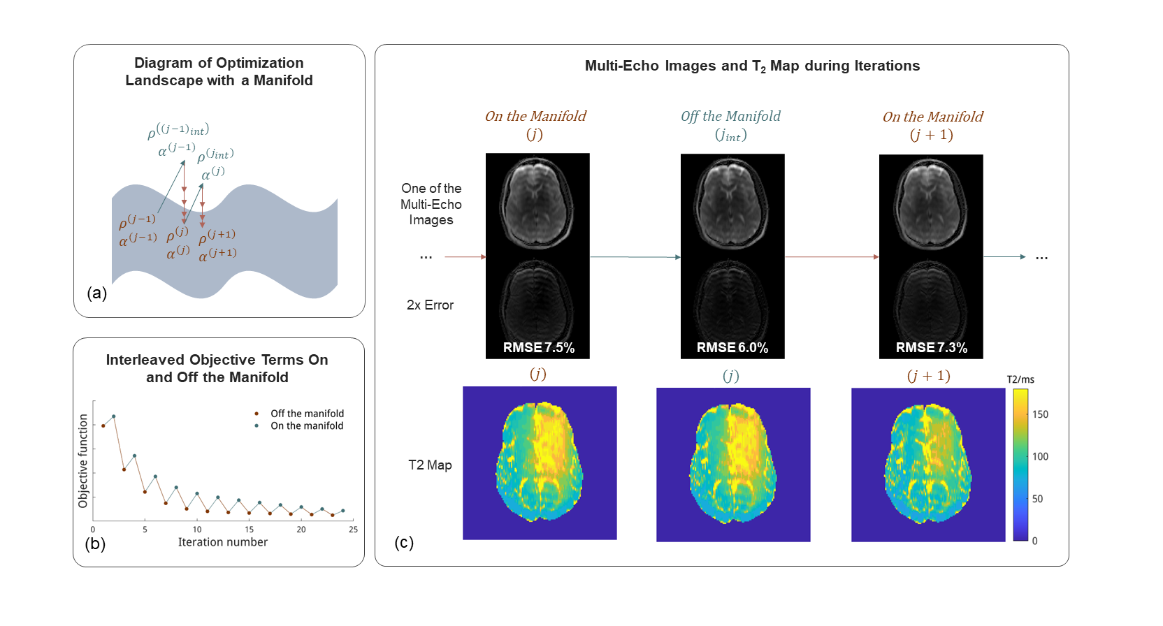
We propose to use the projected gradient descent (PGD) to enforce the model constraint. The projection onto the manifold $$$\phi(\rho_1,\cdots,\rho_P,\alpha)$$$ comprised of the feasible solutions ensures efficient enforcement of the constraint and greatly eliminates parameter tuning. During $$$j$$$-th iteration, $$$\{\hat{\rho}_p^{(j)}\}_{p=1}^P$$$ is first updated to $$$\{\hat{\rho}_p^{(j_{int})}\}_{p=1}^P$$$ regardless of the constraint. The second stage implements the projection by minimizing the Euclidean distance to the manifold.
$$\min_{\hat{\rho}_1^{(j+1)},\alpha^{(j+1)}} \sum_{p=1}^{P} \left\| \left(\hat{\rho}_1^{(j+1)},\alpha^{(j+1)} \right) - \phi\left(\hat{\rho}_1^{(j_{int})},\cdots,\hat{\rho}_P^{(j_{int})},\alpha^{(j)}\right) \right\|^2$$
Then the rest of the images are aligned by $$$\hat{\rho}_1^{(j+1)}$$$ and $$$\alpha^{(j+1)}$$$. A diagram is shown in Fig. (2).
Methods
For retrospective undersampling, fully sampled 8-echo SE brain images were acquired on a 3T MRI scanner with a base resolution of 128 and TE-s spanning from 20ms to 160ms. For prospective undersampling, 9-echo TSE data were acquired with the same base resolution and TE-s from 20ms to 200ms. For a more realistic acquisition in clinical settings, 19-echo TSE data were acquired with a higher resolution of 256 and TE-s from 12ms to 240ms. The first echo was left out in prospective undersampling.Subspace-constrained and model-based algorithms were implemented in BART7.
Results and Discussion
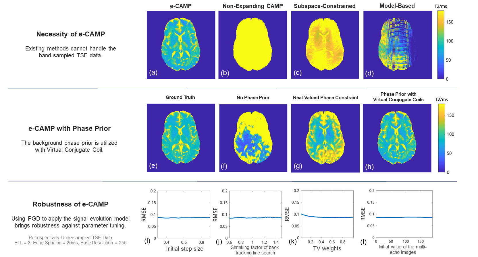
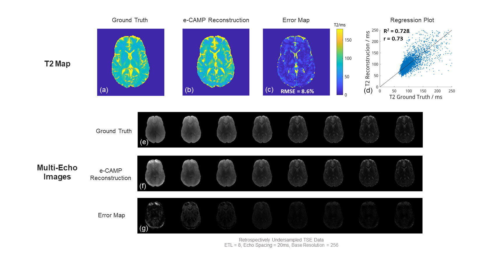
The first row of Fig. 3 shows the necessity of e-CAMP, while non-expanding CAMP (b) and existing state-of-the-art methods (c-d) fail. The second row shows the case without a phase prior (f), or with rigorous phase constraints (g), but virtual conjugate coils (h) handle the phase prior well. The algorithm is robust against hyper-parameter tuning (i-h).Fig. 4 shows a reconstruction of the T2 map and multi-echo images of a retrospectively undersampled dataset. It achieves 8.6% RMSE and accurately recovers the white and grey matter.
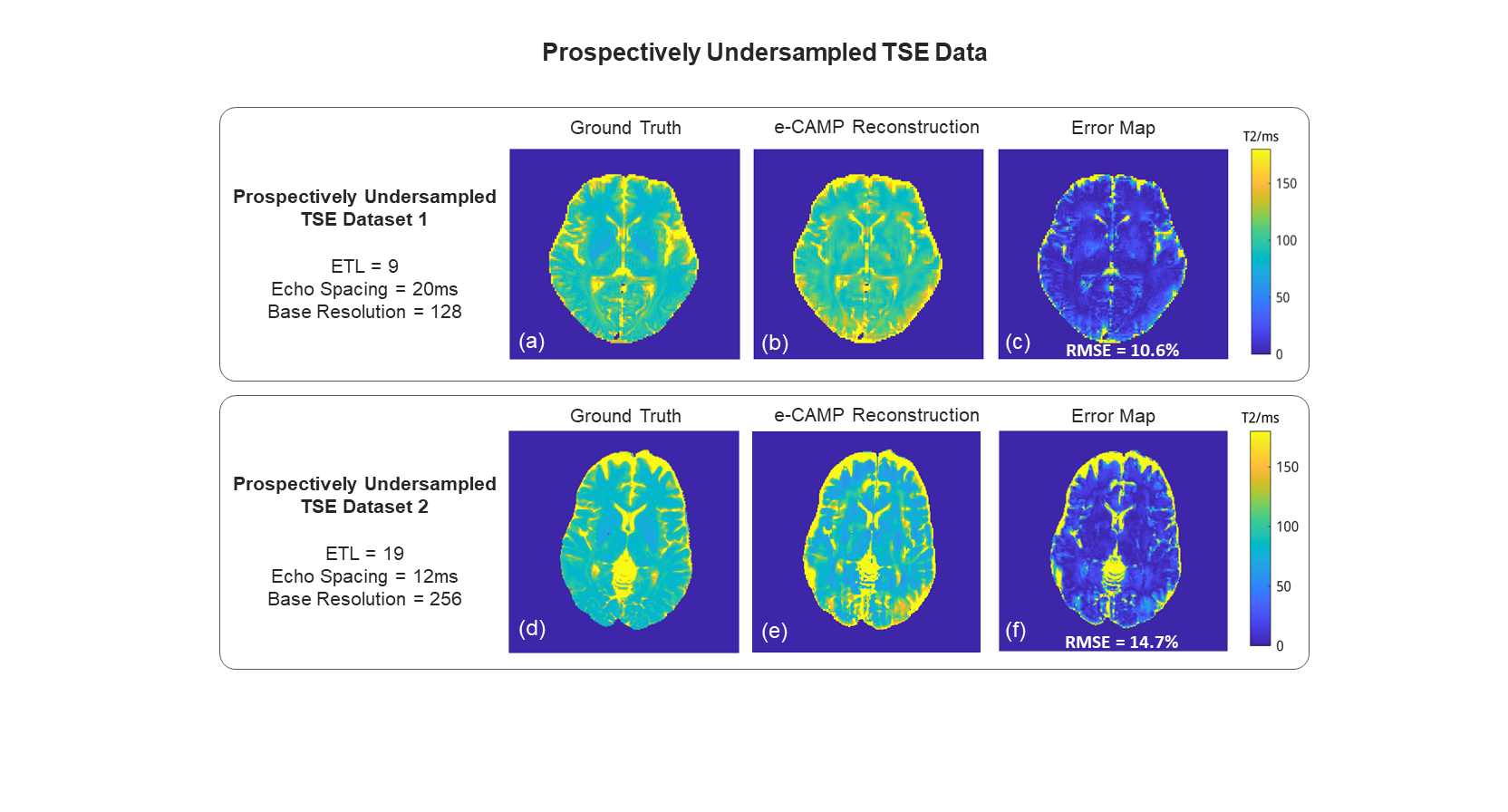
Fig. 5 displays the result of prospectively undersampled datasets. The first row has a protocol close to those processed by earlier versions of e-CAMP, while the latter has a more clinically realistic T2w protocol. Both have similar results to the ground truth.
Acknowledgements
No acknowledgement found.References
1. Wang X, Tan Z, Scholand N, Roeloffs V, Uecker M. Physics-based reconstruction methods for magnetic resonance imaging. Philosophical Transactions of the Royal Society A: Mathematical, Physical and Engineering Sciences. 2021;379(2200):20200196. doi:10.1098/rsta.2020.0196
2. Huang C, Graff CG, Clarkson EW, Bilgin A, Altbach MI. T2 mapping from highly undersampled data by reconstruction of principal component coefficient maps using compressed sensing. Magnetic Resonance in Medicine. 2012;67(5):1355-1366. doi:10.1002/mrm.23128
3. Block KT, Uecker M, Frahm J. Model-based iterative reconstruction for radial fast spin-echo MRI. IEEE Transactions on Medical Imaging. 2009;28(11):1759-1769. doi:10.1109/TMI.2009.2023119
4. Elsaid NMH, Tagare HD, Galiana G. A Physics-Based Algorithm to Universally Standardize Routinely Obtained Clinical T2-Weighted Images. Academic Radiology. July 2023. doi:10.1016/j.acra.2023.05.036
5. Lew C, Pineda AR, Clayton D, Spielman D, Chan F, Bammer R. SENSE phase-constrained magnitude reconstruction with iterative phase refinement. Magnetic Resonance in Medicine. 2007;58(5):910-921. doi:10.1002/mrm.21284
6. Blaimer M, Gutberlet M, Kellman P, Breuer FA, Köstler H, Griswold MA. Virtual coil concept for improved parallel MRI employing conjugate symmetric signals. Magnetic Resonance in Medicine. 2009;61(1):93-102. doi:10.1002/mrm.21652
7. Uecker M, Ong F, Tamir JI, et al. Berkeley Advanced Reconstruction Toolbox. In: Proc. Intl. Soc. Mag. Reson. Med. 23. Toronto; 2015:2486.
Figures