2724
Time-Frequency Multiplexed Wideband Array Beam-Forming Enhances Thermal Magnetic Resonance Theranostics of Brain Tumors1Berlin Ultra High Field Facility, Max-Delbrück-Centrum für Molekulare Medizin, Berlin, Germany, 2Experimental and Clinical Research Center (ECRC), a joint cooperation between the Charité Medical Faculty and the Max-Delbrück Center for Molecular Medicine in the Helmholtz Association, Berlin, Germany, 3Brightmind.AI GmbH, Vienna, Austria, 4MRI.TOOLS GmbH, Berlin, Germany
Synopsis
Keywords: Interventional Devices, MR-Guided Interventions, ThermalMR Brain Tumor
Motivation: Thermal magnetic resonance theranostics combines diagnostic MRI with targeted thermal therapy with an integrated radiofrequency applicator. Precise RF dosimetry is crucial for real-time treatment planning.
Goal(s): Our goal is to evaluate a time-frequency multiplexing wideband RF beamforming method for precise targeting of small and large deep-seated brain tumors for Thermal Magnetic Resonance theranostics.
Approach: We employed a multi-vector field shaping algorithm for optimizing RF channel settings of the RF applicator.
Results: With time-frequency multiplex excitations, we achieved precise SAR10g targeting in the tumor volume while minimizing RF exposure to healthy tissues. Our study advances thermal magnetic resonance theranostics efficacy promising improved outcomes.
Impact: Our approach of utilizing horse-shoe shaped RF applicator comprised of wideband SGBT dipole antenna can be conveniently adapted to individual patient's tumor position and geometry while maintaining the efficiency and quality of RF heating for ThermalMR theranostics of brain tumors.
PURPOSE
Thermal Magnetic Resonance (ThermalMR) is a theranostic approach that combines diagnostic magnetic resonance imaging (MRI) with targeted thermal therapy in the hyperthermia range using an integrated radiofrequency (RF) applicator1-3. ThermalMR’s efficacy encompasses uniform transmission B1+ field for MRI and focal RF heating in the target volume (TV) for thermal therapy. Both criteria govern by RF applicator design and constructive E-field focusing optimization for targeted heating. RF hyperthermia of brain tissue benefits from wideband RF beam-forming6. In this work, we examine the performance of the time-frequency multiplexing beam-forming approach and use this excitation vector optimization approach for targeting small and large deep-seated brain tumors for ThermalMR theranostics at 7.0T, 9.4T and 10.5T.METHODS
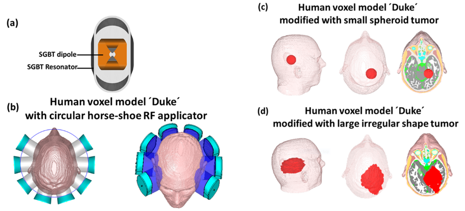
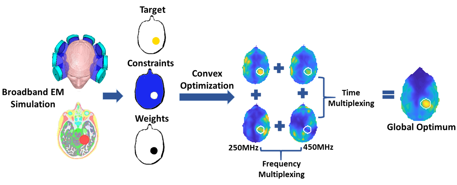
The ThermalMR RF applicator comprises eight compact SGBT4 dipole antennas (size: 42.3x46.3x2.5mm3) arranged in a horse-shoe shaped (arc= 2700) annular array and ensures ample brain coverage while sparing the high conductive regions of the eyes from RF exposure during targeted heating (Fig. 1a,b). The RF applicator supports MRI at 300MHz (B0=7.0T), 400MHz (B0=9.4T) and 450MHz (B0=10.5T) and targeted RF heating at multiple discrete frequencies (f=250, 300, 350, 400, 450MHz). EMF simulations (CST Studio Suite 2020) were performed on the human voxel model ‘Duke' (IT'IS Foundation Zürich). To emulate a real clinical scenario, two patient models were created including a (i) intracranial large irregular shape tumor TVL (volume=500ml, σtumor=1.15S/m, εrtumor=66.5) and (ii) a small spheroid tumor TVS (radius=2cm, volume=33.5ml, σtumor=1.15 S/m, εrtumor=66.5)1 in the right parietal region of Duke’s brain (Fig. 1c,d).A Multiplexed Vector Field Shaping (MVFS)5 (Fig. 2) algorithm was used to provide globally optimal excitation vectors defining phase and amplitude setting for RF channels of RF applicator to focus SAR10g in TV and to reduce RF exposure to the healthy tissues by selecting appropriate intervention frequencies and time-interleaved excitations. For targeted RF heating, time-multiplex (TM) excitation (at 300MHz) and time-frequency multiplex (TFM) excitation of above mentioned multiple discrete frequencies are used to focus SAR10g in the TVL and TVS. The focusing ability of the RF applicator was evaluated using the metrics SAR10g in TV and tumor coverage TCx as local SAR typically used as a representation for tissue heating1,5-6. Tumor coverage TC25, TC50, and TC80 detail the fraction of tumor enclosed in the 25%, 50%, and 80% isolines of peak SAR10g1,5-6. Postprocessing was conducted in MATLAB 2020 to calculate B1+, SAR10g and targeted heating optimization.
RESULTS
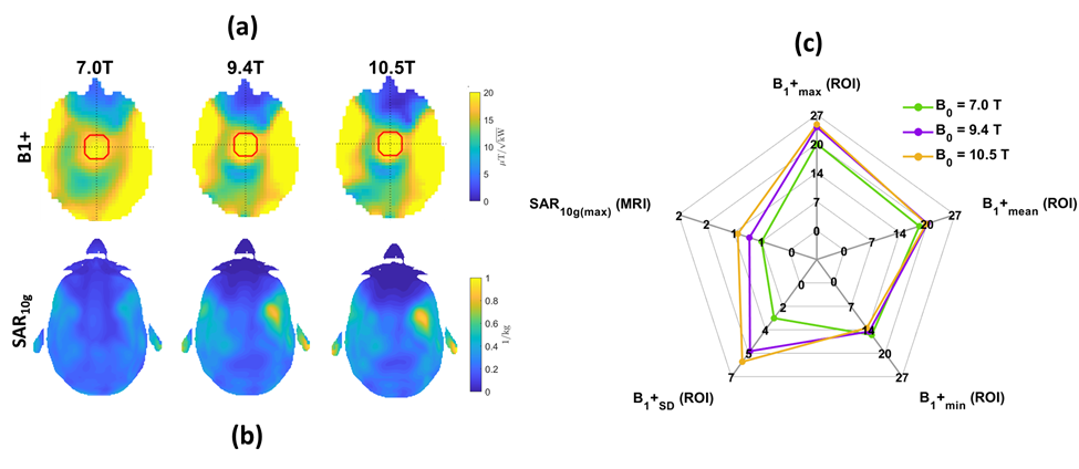
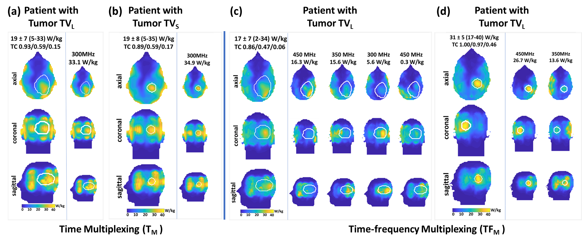
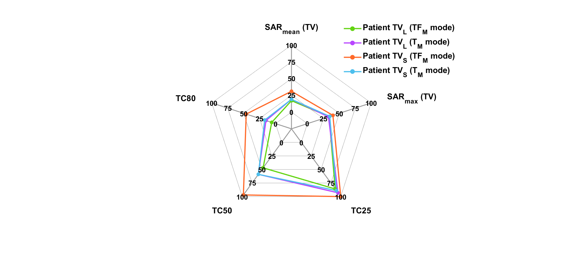
EMF simulations showed that the RF applicator provides B1+ that facilitates brain MRI at 7.0T,9.4T, 10.5T (Fig. 3a,b) with maximum local SAR10g within the IEC limits (Fig. 3a-c). For targeted RF heating, MVFS algorithm derived single excitation in TM mode for both patient models, TVL and TVS, and yielded similar SAR10g(mean~max)=(19~33)W/kg inside the TV (Fig. 4a,b). However, TM mode induced undesired RF power deposition, causing hotspots in peripheral healthy tissues surrounding the head. In TFM mode, MVFS algorithm employed four excitations at frequencies of 300MHz, 350MHz, and 450MHz for TVL patient, and two excitations at 350MHz and 450MHz for TVS patient to focus RF power inside TV (Fig. 4c,d). Patients with TVL demonstrated 8%, 25%, and 15% higher TC25, TC50, and TC80 values in TM mode compared to TFM mode (Fig. 4,5). Conversely, patients with TVS exhibited 11%, 39%, and 64% lower TC25, TC50, and TC80 values in TM mode compared to TFM mode (Fig. 4,5). The TFM mode results showed stronger SAR10g (mean~max)=(31~40)W/kg focus inside TVS, while TVL achieved less TC with SAR10g focus inside full TVL despite the use of four excitation modes. This result is due to the uniformity criterion used for RF power optimization. This constraint limits the maximization of RF power deposition in large volume. The diameter of the spheroid TVS is one-third of the wavelength in brain tissue at ~300MHz which permits surpassing of RF focusing limits and results in excellent tumor coverage. For TVL this problem can be addressed by setting higher target SAR.TFM facilitates significant improvements in protecting healthy tissues from unintended RF power deposition for small and large TV when compared to the TM mode.DISCUSSION & CONCLUSION
This study demonstrates the benefits of time-frequency multiplexing to enhance RF beam-forming for targeted RF heating for ThermalMR of small and large deep-seated brain tumors. The MVFS TFM approach provides numerous degrees of freedom to best deliver RF power at the desired tumor location with an arbitrarily complex target tumor shape. Our simulations provide a framework for ThermalMR treatment planning, allowing for the pre-determination of hotspots for more precise and real-time RF dosimetry. Our work provides a technical foundation for ThermalMR theranostics of brain tumors.Acknowledgements
This project is funded by an advanced ERC grant (EU project Thermal MR: 743077).References
1. Saha, N.; Kuehne, A.; Millward, J.M.; Eigentler, T.W.; Starke, L.; Waiczies, S.; Niendorf, T. Advanced Radio Frequency Applicators for Thermal Magnetic Resonance Theranostics of Brain Tumors. Cancers 2023, 15, 2303. https://doi.org/10.3390/cancers15082303
2. Lee Titsworth W, Murad GJ, Hoh BL, Rahman M. Fighting fire with fire: the revival of thermotherapy for gliomas. Anticancer Res 2014;34(2):565-574.
3. Restivo MC, van den Berg CAT, van Lier ALHMW, et al. Local specific absorption rate in brain tumors at 7 tesla. Magn Reson Med.2016;75(1):381–389.
4. Eigentler TW, Winter L, Han H, Oberacker E, Kuehne A, Waiczies H, Schmitter S, Boehmert L, Prinz C, Trefna HD, Niendorf T. Wideband Self-Grounded Bow-Tie Antenna for Thermal MR. NMR Biomed. 2020 May;33(5):e4274.
5. Kuehne A, Oberacker E, Waiczies H, Niendorf T. Solving the Time- and Frequency-Multiplexed Problem of Constrained Radiofrequency Induced Hyperthermia. Cancers. 2020; 12(5):1072.
6. Zanoli M, Trefná HD. Iterative time-reversal for multi-frequency hyperthermia. Phys Med Biol. 2021 Feb 11;66(4):045027. doi: 10.1088/1361-6560/abd41a. PMID: 33326945.
Figures