2722
Up to 24-channel receiver coil arrays for concurrent TMS-MRI imaging at 3T1Physical Sciences Platform, Sunnybrook Research Institute, Toronto, ON, Canada, 2Department of Medical Biophysics, University of Toronto, Toronto, ON, Canada
Synopsis
Keywords: MR-Guided Interventions, RF Arrays & Systems, Transcranial Magnetic Stimulation, Cocurrent TMS-fMRI
Motivation: Concurrent TMS-fMRI lacks whole-brain coverage or has low SNR.
Goal(s): Develop setups allowing for whole-brain TMS-fMRI with high spatiotemporal resolution and minimizing the interference between MRI and TMS during data collection and neuromodulation.
Approach: Combinations of 8- and 16-channel arrays enabled whole-brain TMS-MRI with 16 or 24 channels.
Results: Structural and functional images of high spatiotemporal resolution (1.5 mm with 2 s/volume or 5 mm with 0.1 s/volume) were demonstrated with an MRI-compatible TMS coil.
Impact: We developed concurrent TMS-MRI setups with whole-brain coverage and high spatiotemporal resolution using combinations of 8- and 16-channel coil arrays. Images of high spatiotemporal resolution were demonstrated.
Introduction
Delivering transcranial magnetic stimulation (TMS) during MRI collection permits 1) monitoring the neuromodulatory effects in real-time with high spatiotemporal resolution using BOLD-contrast fMRI and 2) guiding the neuromodulation at specific cortical areas subserving the information processing evidenced by local hemodynamic changes (for review, see 1). However, a technical challenge is the availability of a receiver coil array to offer high spatiotemporal resolution fMRI with whole-brain coverage. Previously, a body or head volume coils were used at the cost of a reduced signal-to-noise ratio 2–6. A localized coil array immediately around the TMS coil has been developed to enhance the quality of MRI in the vicinity of the stimulation 7. Combinations of such 7-channel arrays were only described briefly 8.Here, we presented the concurrent TMS-MRI setup allowing for whole-head parallel imaging at 3T. Specifically, coil arrays of 8 and 16 channels were constructed to work together with MRI-compatible TMS. The goal of this study is to identify setups allowing for whole-brain TMS-fMRI with high spatiotemporal resolution and minimizing the interference between MRI and TMS during data collection and neuromodulation.
Methods
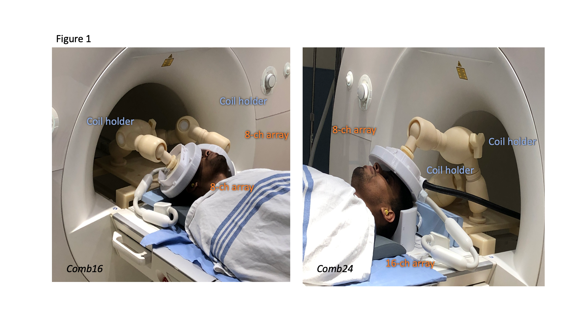
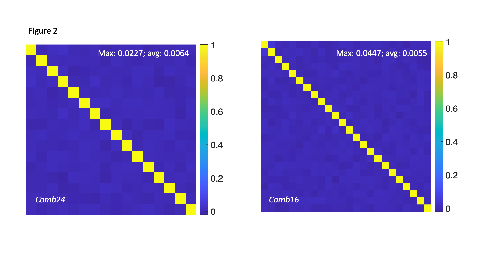
Circular receive-only RF coils with a 5 cm diameter were made from PTFE. Details of RF coil array development was reported7. The mechanical housing of the coil array will be made by a 3D printer using polycarbonate (PC-ISO) plastic (FORTUS, Eden Prairie, MN, USA). Arrays of 8-channel and 16 channels were assembled into a circular disk and a quarter-sphere geometry, respectively.Coupling between channels of a coil array will be quantified by a noise covariance matrix, calculated from the acquired imaging data without any RF transmissioN. We specifically studied two setups for whole-brain TMS-MRI: A 24-channel combination (comb24) with an 8-channel array and a 16-channel array around the frontal and parietal lobes, respectively. There was also a 16-channel combination (comb16) with two 8-channel arrays around the bilateral frontoparietal lobes. Using a novel coil holder allowed for easy positioning of coil arrays in both setups 9.
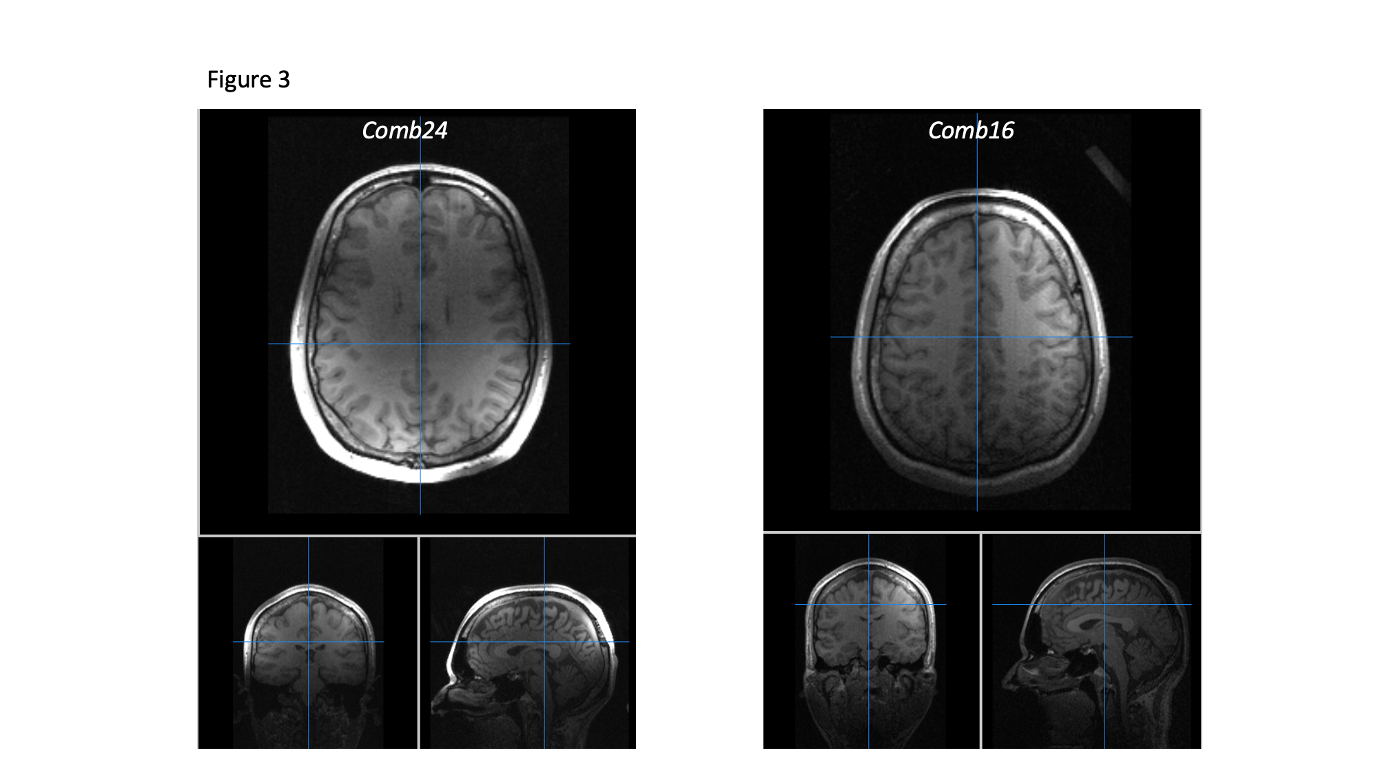
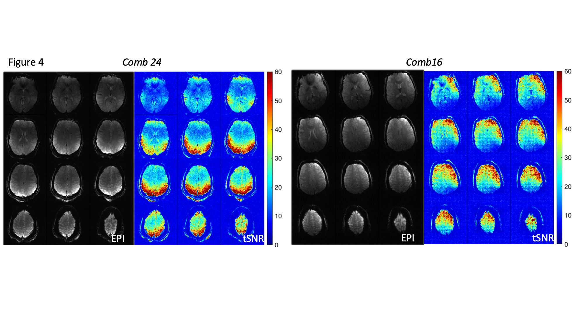
Structural images were taken using an MP-RAGE sequence. High-resolution fMRI was tested on setups using simultaneous mult-slice EPI (comb24: TE/TR=30/2000 ms; flip angle = 60o; 28 slices; 3 mm thickness; in-plane resolution 1.5 mm; FOV=192 mm; 2x slice acceleration; comb16: TE/TR=30/2000 ms; flip angle = 60o; 24 slices; 3 mm thickness; in-plane resolution 2 mm; FOV=192 mm; 2x GRAPPA). We also tested fMRI using highly accelerated fMRI 10, where MRI took 5% of the duty cycle (0.1 s in 2-s repetition time) to allow 95% of time free from the interference between MRI and TMS. The performance of fMRI was quantified by calculating the time-domain signal-to-noise ratio (tSNR) at each image voxel.
Results
Figure 1 illustrates comb16 and comb24 setups. The noise covariance matrices for comb24 and comb16 are shown in Figure 2. The average and maximum were 0.005 and 0.045 in comb24, respectively. The average and maximum were 0.006 and 0.023 in comb16, respectively.Figure 3 shows T1-weighted images using comb24 and comb16, both of which had MRI signals from the whole brain with lower signal strengths at locations away from coils in the vicinity. EPI images and their time-domain SNR (Figure 4) suggested the SNR advantage of comb24 and comb16 setups at frontal and parietal lobes.
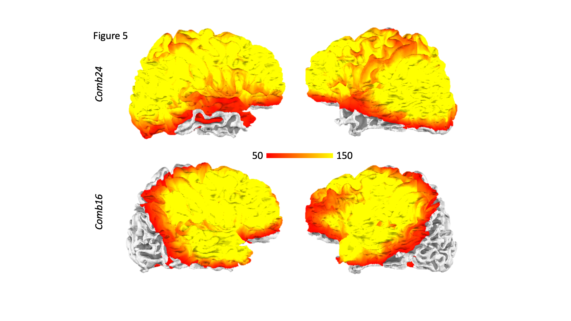
The parallel detection of MRI signals also enabled the fast fMRI, which took only 5% (0.1 s) of the 2-s duty cycle (TR = 2 s). The tSNR distributions at the cortex (Figure 5; comb24: 117 +/- 53; comb16: 91 +/- 51) suggested using flexible TMS pulse schedules, including rTMR, for sensitive fMRI signal detection.
Discussion
The coil array combinations reported here are outcomes of MRI coil arrays of 8 or 16 channels and high degree-of-freedom articulating coil holders 9. Different from the whole-brain TMS fMRI for a specialized instrument 11, the proposed setups can be used by commercially available MRI-compatible TMS systems. The whole-brain MRI enabled parallel imaging applications to trade off the SNR gain with imaging spatiotemporal resolution. The comb24 and comb16 demonstrated the feasibility of whole-brain high-resolution fMRI (1.5 mm image voxel; Figure 4). They also enabled versatile TMS pulse scheduling, such as repetitive TMS, without worrying about the interference between imaging and stimulation and the trade-off of the imaging coverage or spatiotemporal resolutions. Different combinations of coil arrays covering different brain parts, or even with three or more coil arrays, are expected to provide higher freedom and SNR in concurrent TMS-MRI.Acknowledgements
This work was supported by the Natural Sciences and Engineering Research Council of Canada (NSERC; RGPIN-2020-05927), Canada Foundation for Innovation (38913 and 41351), Canadian Institutes of Health Research (PJT 178345 and PJT 185882), MITACS (IT25405 and Global link fellowship).References
- Mizutani-Tiebel, Y. et al. Front. Psychiatry 13, 825205 (2022)
- Bestmann, S. et al. J. Neurosci. 30, 11926–11937 (2010)
- Weiskopf, N. et al. J. Magn. Reson. Imaging 29, 1211–1217 (2009)
- Shitara, H. et al. Neuroimage 56, 1469–1479 (2011)
- Shitara, H. et al. Front. Hum. Neurosci. 7, 554 (2013)
- Blankenburg, F. et al. J. Neurosci. 28, 13202–13208 (2008)
- Navarro de Lara, L. I. et al. Neuroimage 150, 262–269 (2017)
- Windischberger, C. et al. Brain Stimulation: Basic, Translational, and Clinical Research in Neuromodulation 14, 1723 (2021)
- Lee, H.-J. et al. Brain Stimul. 16, 966–968 (2023)
- Hsu, Y.-C. et al. Sci. Rep. 7, 17019 (2017)
- Navarro de Lara, L. I. et al. Brain Stimul. 16, 1021–1031 (2023)
Figures