2721
A radiation-transparent, rotatable receive-only extremity RF coil for flexible MR-guided particle therapy at 0.25 T1Department of Radiation Oncology, Heidelberg University Hospital, Heidelberg, Germany, 2Department of Medical Physics in Radiology, German Cancer Research Center (DKFZ), Heidelberg, Germany, 3Faculty of Physics, Heidelberg University, Heidelberg, Germany, 4Clinical Cooperation Unit Radiation Oncology, German Cancer Research Center (DKFZ), Heidelberg, Germany, 5Division of Medical Physics in Radiation Oncology, German Cancer Research Center (DKFZ), Heidelberg, Germany, 6Heidelberg Institute of Radiation Oncology (HIRO), Heidelberg, Germany, 7National Center for Radiation Research in Oncology (NCRO), Heidelberg, Germany, 8Heidelberg Ion-Beam Therapy Center (HIT), Heidelberg University Hospital, Heidelberg, Germany, 9German Cancer Consortium (DKTK), Heidelberg, Germany, 10National Center for Tumor Diseases (NCT), Heidelberg, Germany, 11Faculty of Medicine, Heidelberg University, Heidelberg, Germany
Synopsis
Keywords: MR-Guided Radiotherapy, MR-Guided Interventions, Particle therapy
Motivation: MR-guided particle therapy (MRgPT) could improve tumor control and reduce healthy tissue dose, thereby increasing quality of therapy compared to conventional CT-guided radiotherapy.
Goal(s): Development of a rotatable, radiation-transparent extremity radiofrequency (RF) coil suitable for MRgPT at a 0.25T C-shaped magnet.
Approach: The receive field homogeneity of the RF coil was evaluated for different rotation angles in electromagnetic field simulations, and the SNR was investigated in a homogeneous phantom in comparison to two commercial coils.
Results: The constructed extremity RF coil including a custom-built preamplifier achieves improved receive field homogeneity and SNR compared to two commercial RF coils and is compatible with MRgPT.
Impact: The rotatable, radiation-transparent RF coil enables MRgPT with through-coil irradiation and multi-angle access to the patient. The developed RF coil and preamplifier design provides better RF field homogeneity and SNR over a larger FOV compared to two commercial RF coils.
Introduction
In radiotherapy, the ability to precisely target tumor tissue while sparing surrounding healthy tissue is critical to treatment success. Due to the excellent soft tissue contrast of MRI and the associated improved differentiation between different tissue types during treatment planning and delivery, MR-guided radiotherapy is gaining popularity as an alternative to conventional CT-guided radiotherapy1. The absence of ionizing radiation allows for more frequent imaging and closer monitoring of the patient's anatomy and physiology. For optimal treatment quality, inter- and intra-fractional modifications to the treatment plan based on MRI are possible, taking into account the patient's position or real-time gating. Further dose reduction in healthy tissue could be achieved by combining precise MRI with particle therapy due to the sharp and spatially limited dose maximum (Bragg peak) of particle beams in water-like tissues2. Lower magnetic field strengths, although associated with a lower signal-to-noise ratio, have the advantage of less beam deflection due to the Lorenz force and may be more practical for MRgPT than standard MR systems. In addition, the particle range and thus the dose distribution in the patient is strongly influenced by the materials in the beam path. Therefore, MR systems with open access to the patient (e.g. double-doughnut or C-shaped configuration)3, are particularly suitable. In previous work, a rotatable, radiation-transparent transmit/receive RF body coil was constructed for a 1.5T MR system, allowing 360° rotation of the patient4. In this work, the concept of a rotatable RF coil made of radiation-transparent materials compatible with flexible multi-angle irradiation is transferred to a low-field MR system.Methods
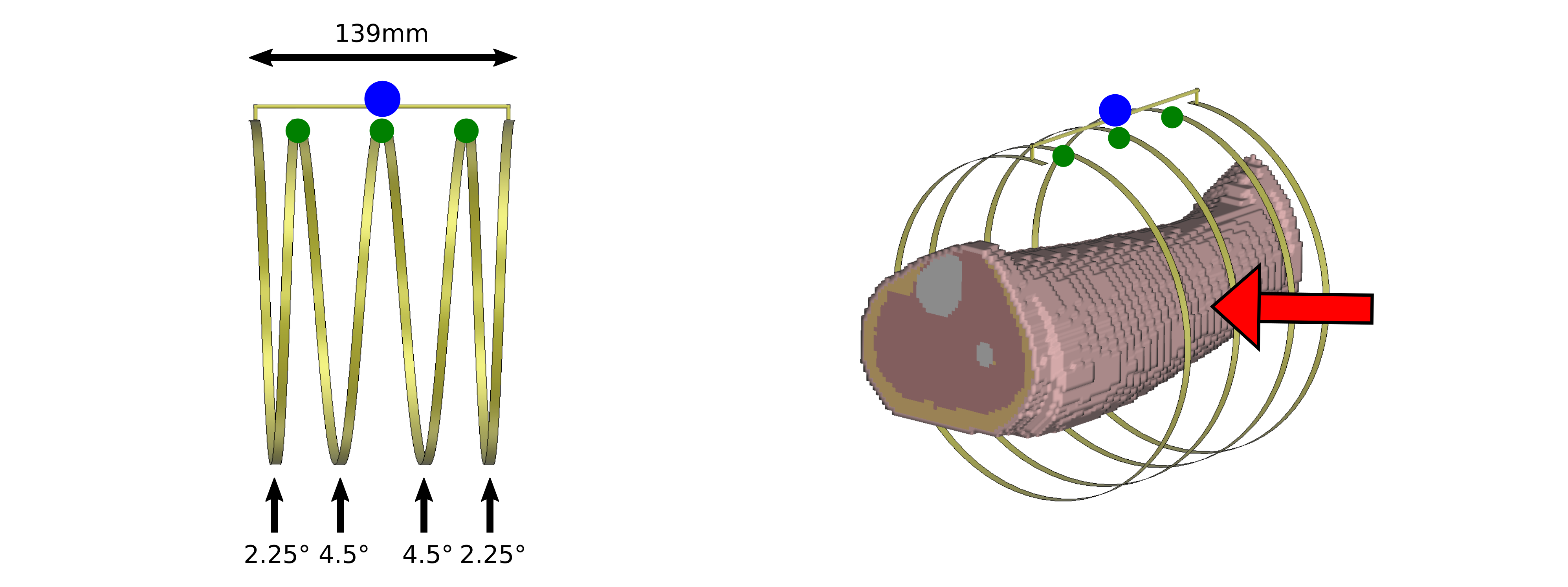
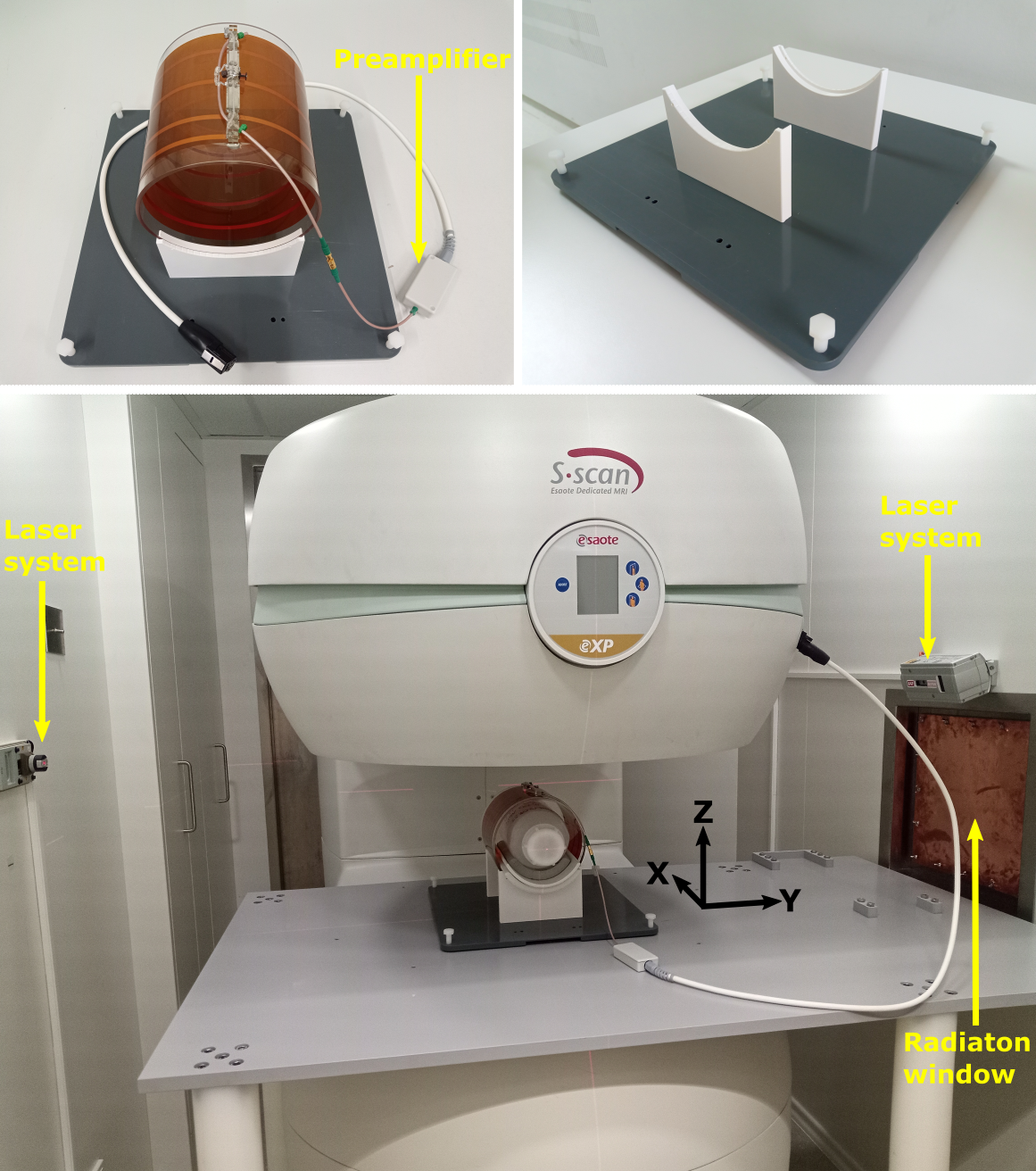
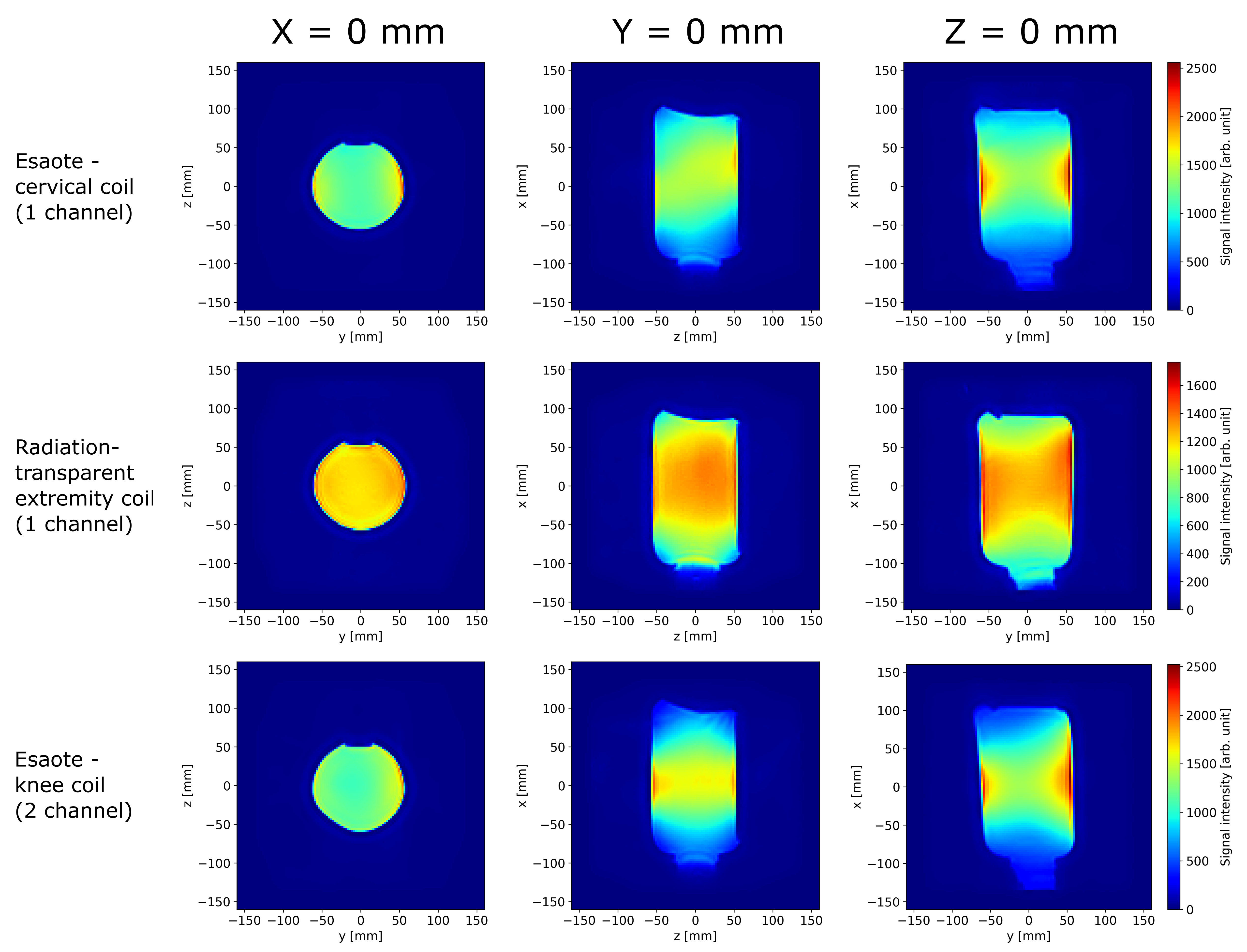
Coil: The developed 1-channel receive-only RF coil (length:14cm) for extremity imaging is based on a four-turn solenoid configuration with increased turn density at the coil ends (Figure 1) to achieve higher RF field homogeneity5, especially in axial (x) direction (cf. Figure 2). The conductive layer (thickness:35µm) is embedded between adhesive and polyimide layers with a negligible water-equivalent thickness and can therefore be considered radiation-transparent4. The RF coil is wrapped around a PMMA cylinder (outer diameter:18cm,thickness:4mm) and fixed on a mounting plate. The coil can be rotated about the x-axis allowing multi-angle access for particle therapy. Furthermore, a custom-designed low-noise preamplifier (gain:23.5dB,noise figure:1.0dB) was developed for signal acquisition with the coil.Measurements: SNR ($$$\frac{signal}{noise}$$$) was determined in a homogeneous phantom and compared with two commercial imaging coils (Figure 4). To calculate the average signal in the phantom and determine noise from the background6 (NEMA,method 5), masks were defined in the three central slices.
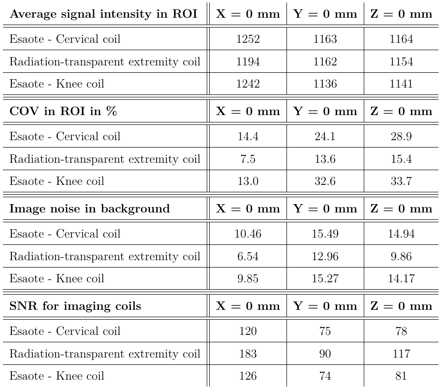
Additionally, COV (coefficient of variation:$$$\frac{standard~deviation}{mean}$$$) values were determined in the MR images (Table 1).
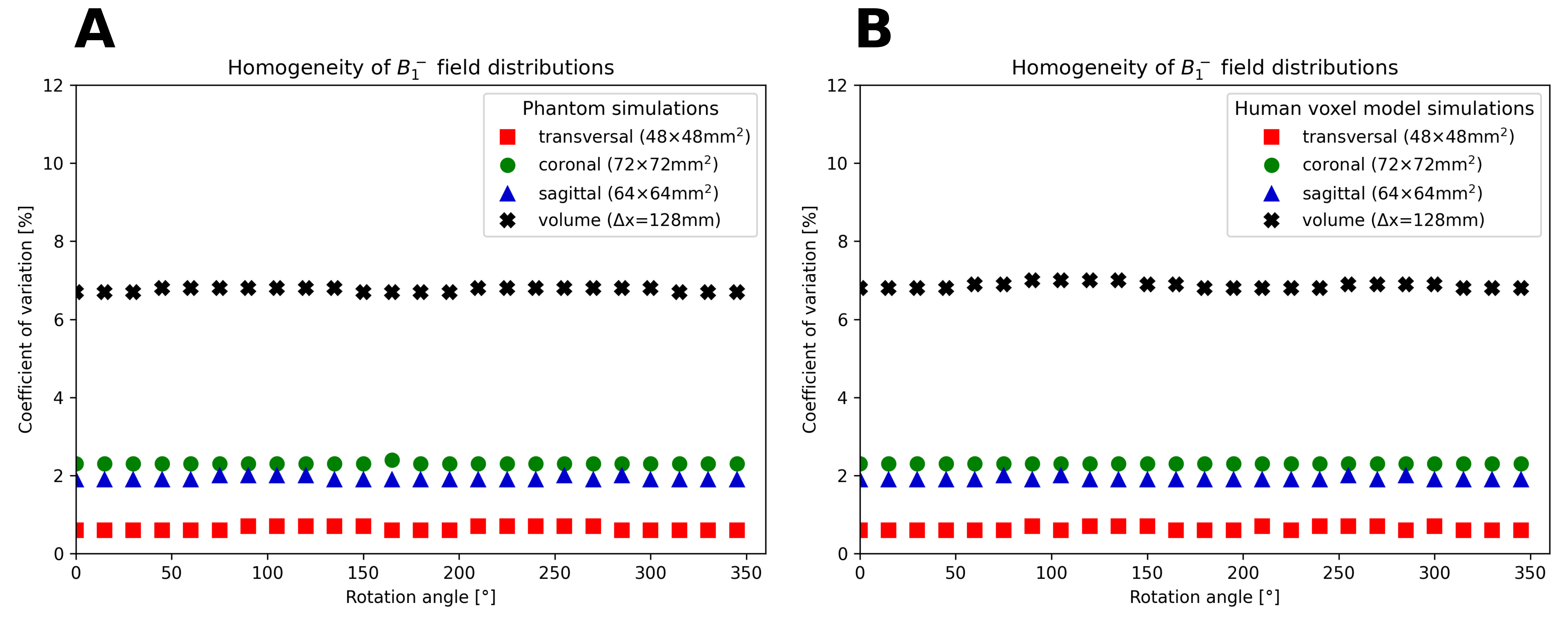
Simulations: A simplified 3D model, including the C-shaped magnet (0.25T) and the RF cabin as boundary conditions, was implemented in electromagnetic field simulations7 at 10.25MHz and the receive field (B1-) of the extremity coil was calculated using the Finite-integration method8. To evaluate the simulated field homogeneity, the COV was calculated in a ROI in the central transverse, coronal, and sagittal slices (slice thickness:2.5mm) as well as a volume (-64mm≤x≤+64mm,complete y/z range) of a homogeneous phantom and a human voxel model for a complete rotation (0-360°) in steps of 15° (Figure 3).
Phantoms: A voxel model of a human calf (Gustav7) and a cylindrical, homogeneous phantom of similar size
(diameter:11cm,height (x-direction):20cm,volume:2L) and with average permittivity and conductivity of the calf model (εr=150.5,σ=0.45$$$\frac{S}{m}$$$) was used for simulations. Measurements were performed in a homogeneous phantom filled with water and sodium chloride ($$$\frac{m_{NaCl}}{m_{total}}$$$=0.23%) with comparable conductivity (σ=0.47$$$\frac{S}{m}$$$) and lower permittivity (εr=80).
Results
COV values (Figure 3) demonstrate good receive field homogeneity for both phantoms in all slices and only slightly increased inhomogeneity in the 3D volumes. Furthermore, the receive field homogeneity is almost constant and shows no significant dependence on the rotation angle. High field homogeneity can also be observed in the center of the MR images of the rotatable, radiation-transparent extremity coil (Figure 4). Table 1 shows better image homogeneity, similar signal intensity, and lower image noise for the developed extremity coil (including the custom-built preamplifier), resulting in a 20-50% improvement in SNR (depending on the image slice) compared to the commercial coils.Discussion & Conclusion
A radiation-transparent imaging coil compatible with rotation for multi-angle irradiation was developed for a low-field MR system connected to a particle beam with fixed position. Simulated receive field distributions and acquired MR images show good homogeneity of the receive sensitivity profile, and the determined SNR exceeds that of two commercial coils. Therefore, the developed RF coil is suitable for extremity imaging and flexible MRgPT including coil and patient rotation. Further studies should confirm the presented results in in vivo measurements.Acknowledgements
This work received financial support from the German Federal Ministry of Education and Research (BMBF, ARTEMIS project WP8, funding reference 13GW0436)References
1Pollard, J. M. et al., "The
future of image-guided radiotherapy will be MR guided", The British Institute of
Radiology (2017)
2Hoffmann A., et al., "MR-guided proton
therapy: a review and a preview", Radiation Oncology 15 (2020); 1-13
3Mislow J. M. K., et al.,
"Origins of Intraoperative MRI", Magnetic Resonance Imaging Clinics
of North America (2010); 1-10
4Dietrich K. A. et al.,
"Construction of a Tx/Rx body coil on a rotatable patient capsule for
MR-guided particle therapy", Proceedings Intl. Soc. Mag. Reson. Med. 30
(2022)
5Leifer M. C., "RF
Solenoid with Extended Equiripple Field Profile", Magnetic Resonance,
Series A (1993); Vol. 105, No. 1, 1-6
6NEMA,
"Determination of Signal-to-Noise Ratio (SNR) in Diagnostic Magnetic
Resonance Imaging", NEMA Standards Publication MS 1 (2008)
7CST Studio Suite 2021, Dassault Systèmes, Vélizy-Villacoublay,
France
8Weiland T., "A
discretization method for the solution of Maxwell’s equations for six-component
fields", Electronics and Communications AEU (1977); Vol. 31, No. 3,
116-120
Figures