2705
MR Thermometry with a Deep-Learning Reconstruction1GE HealthCare, Royal Oak, MI, United States, 2GE HealthCare, Houston, TX, United States, 3GE HealthCare, Calgary, AB, Canada, 4Technology and Innovation Center, GE HealthCare, Niskayuna, NY, United States, 5GE HealthCare, Waukesha, WI, United States, 6Department of Radiology, University of Iowa, Iowa City, IA, United States
Synopsis
Keywords: Thermometry/Thermotherapy, Thermometry
Motivation: Improved MR thermometry is needed for thermal-based therapies.
Goal(s): Improve precision while maintaining accuracy by denoising proton resonance frequency shift (PRFS)-based thermometry images using a deep learning-based reconstruction (DLR).
Approach: 2D fast spoiled gradient-echo (FSPGR) images were acquired on a variety of phantoms with and without heating. Complex images were reconstructed with and without DLR to calculate temperature change maps. The mean and standard deviation of ROIs were analyzed to demonstrate any changes in accuracy and precision.
Results: DLR improves precision and maintains accuracy in PRFS temperature change maps in phantoms.
Impact: The improvements indicate an opportunity to increase spatial and/or temporal resolution in MRI thermometry. It may be possible to improve MRI-based heating optimization during clinical thermal therapies.
Introduction:
In recent years, significant research into deep learning based reconstruction (DLR) has produced improved image quality in several clinical applications1–4. However, these improvements have primarily been applied to magnitude images. This work aims to extend the reconstruction pipeline of GE HealthCare’s AIR Recon DL (ARDL) (GE HealthCare, Waukesha, WI)5, to denoise complex images in Magnetic Resonance Thermometry imaging (MRTI).MRTI is currently used to guide minimally-invasively thermal therapies, such as laser6, radiofrequency (RF)7, and ultrasound8. Most MR parameters experience a temperature dependence, and thus can be used as a surrogate for temperature change. Because of its simplicity and linear response with temperature change, proton resonance frequency shift (PRFS) MRTI has emerged as a dominant MRTI approach for aqueous tissues, where temperature changes are computed via the phase difference in complex MRI images9. In this preliminary study, we propose to analyze the precision and accuracy of a DL based complex image reconstruction technique for PRFS temperature change calculation.
Methods:
Acquisition:2D fast spoiled gradient-echo (FSPGR) images were acquired on a 3T MR750 (GE HealthCare, USA) on an ACR phantom, with no temperature modulation, for image quality analysis and baseline PRFS calculation with the following scan parameters:
Matrix size: 128x128, TR: 140.05ms, TE: 2.96ms, 1Tx32Rx Nova Coil (Nova Medical, USA)
2D FSPGR images were acquired on a 3T SIGNA Premier (GE HealthCare, USA) with the following parameters were used for temperature experiments described below.
Matrix size: 256x256, TR: 6.9ms, TE: 3.14ms, 60-Channel Posterior Array (GE HealthCare, USA)
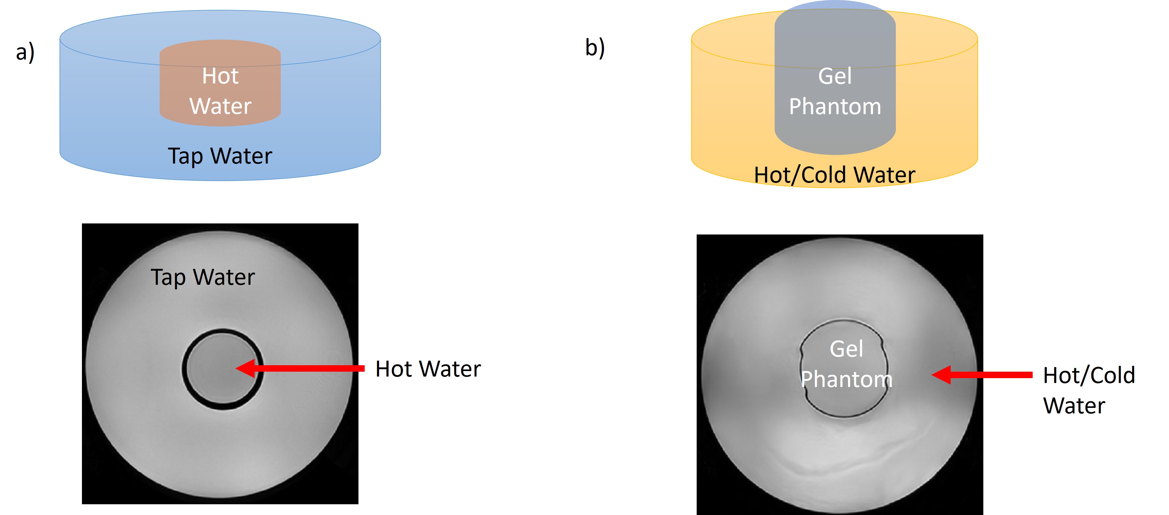
First, a cooling experiment was performed where a beaker containing boiling water was placed in the center of a basin of tap water (Figure1a). Baseline image was acquired when the beaker was placed in the basin, and subsequent acquisitions tracked the temperature change in the two water volumes.
Another heating and cooling experiment was performed using a custom-made 2% agarose phantom (Figure1b). The basin was filled with warm water to heat up the phantom, then ice was added to the water bath to introduce cooling after 15 minutes. Images were acquired at designated time points to track the temperature change.
Reconstruction & Analysis: Images were reconstructed two ways to produce two sets of complex images from each raw data: i) conventional reconstruction and ii) ARDL DL 2D reconstruction5. The latter has preset denoising level settings (0% (no noise removal), 25%, 50%, 75%, and 100%). The different denoising settings are noted as DL0, DL25, DL50, DL75, and DL100.The complex images were outputted from the reconstruction pipeline before taking the absolute value. MATLAB (2022a; MathWorks, Natick, MA) was used for analysis.
PRFS-MRTI maps were calculated using:
$$ΔT=T(t_2)-T(t_1)=(ϕ(t_2 )-ϕ(t_1 ))/(αγB_0 TE)$$
Where ϕ is the phase of the complex image at specified time points, α ≈ -0.01 ppm/C, γ is the gyromagnetic ratio, and TE is the echo time.
Mean and standard deviation were measured from select ROIs in temperature difference maps calculated using DL and non-DL reconstruction.
Results:
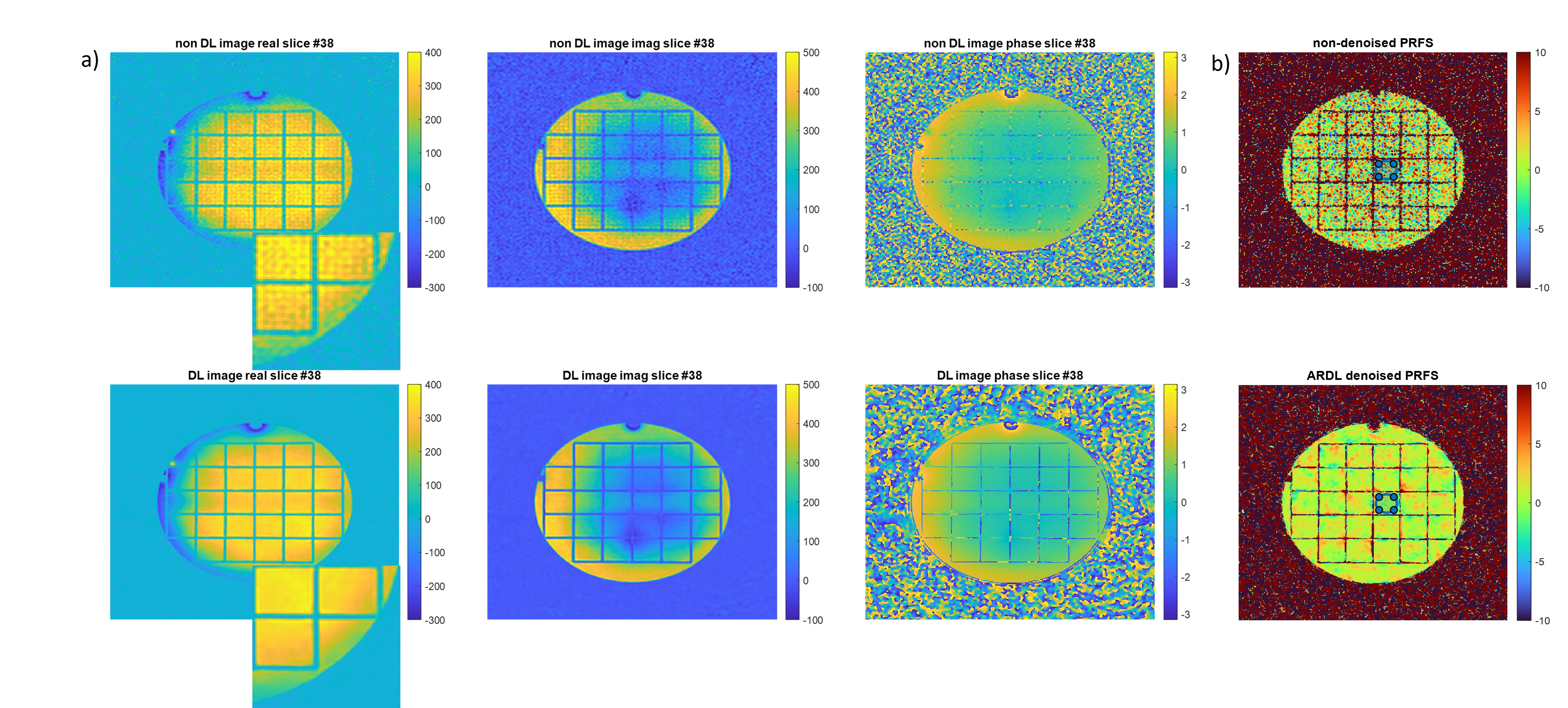
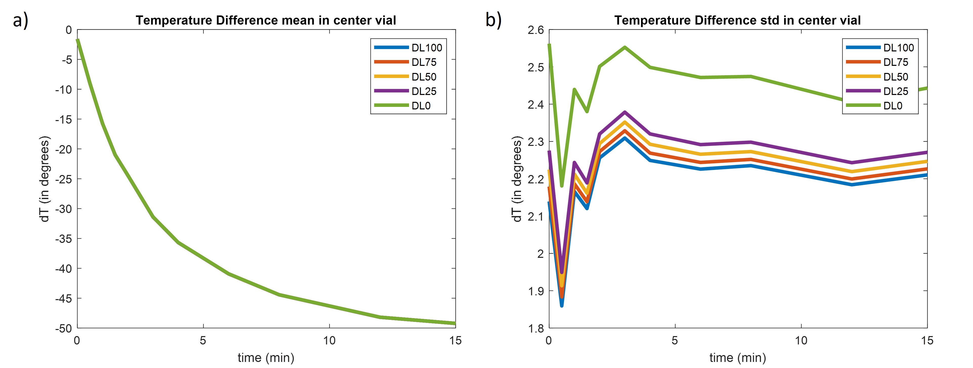
Figure 2 shows the performance of DL75 vs non-DL reconstruction in the ACR phantom. The non-DL denoised complex images are visually noisier with ringing artifacts along the grids of the phantom. The temperature difference map of the phantom indicates DL reconstruction does not introduce a temperature bias in the non-heated phantom experiment but does reduce the standard deviation.Figure 3 shows the results from the cooling experiment. The data is reconstructed with preset denoising levels. DL reconstruction did not introduce a temperature bias in this heating/cooling phantom experiment, and standard deviation reduction correlates with the level of denoising.
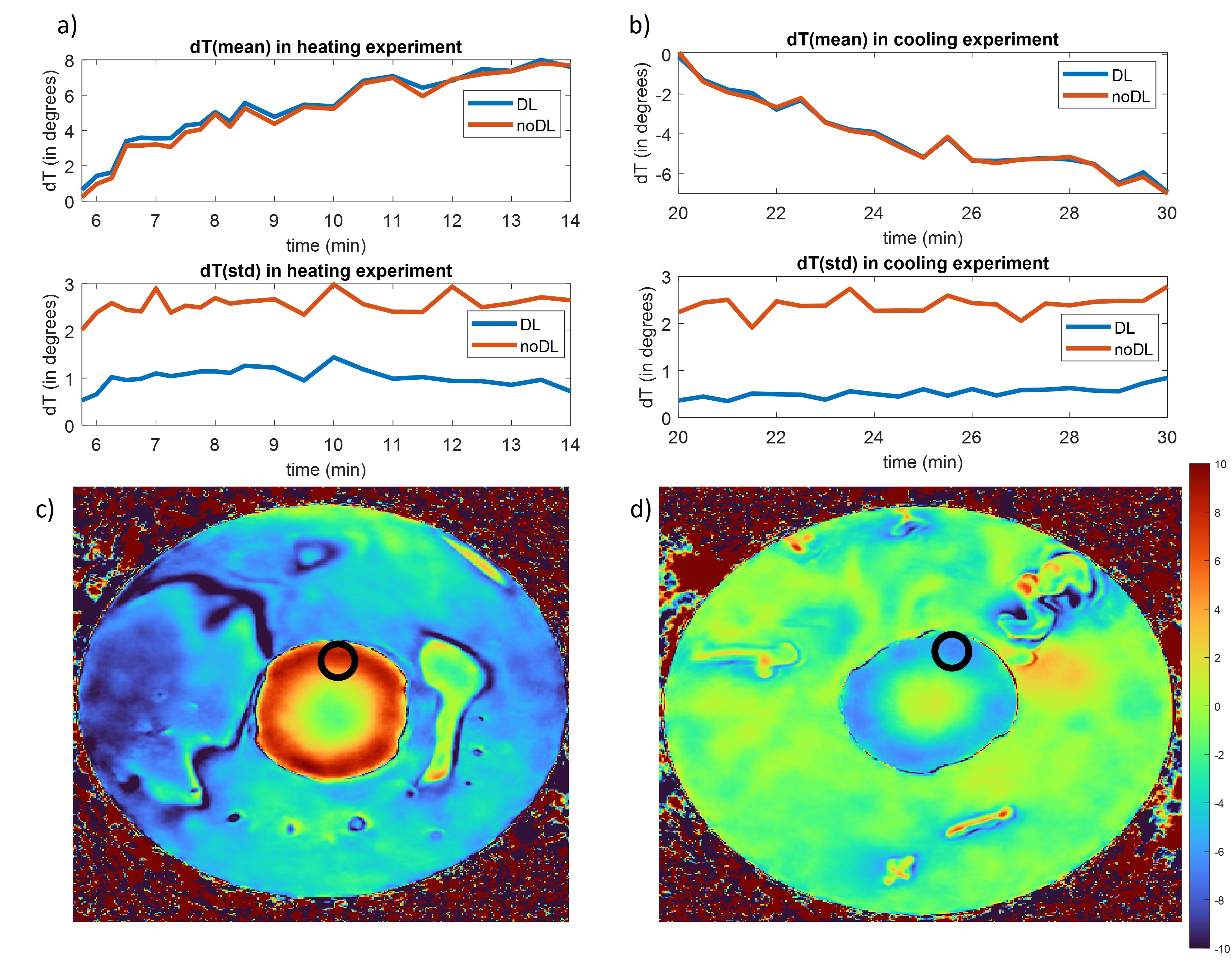
Figure 4 shows the results from the heating and cooling agarose gel experiment. Comparison of the DL-reconstructed images to the non-DL images shows there is no temperature bias and accuracy is improved. The image derived temperature change correlates nicely with the expected temperature change.
Discussion and Conclusion:
This preliminary work shows the proposed modification to ARDL can enable denoising of complex images thereby reducing the standard deviation of the ROI while maintaining the average value of the temperature change map, in comparison to conventional reconstruction.MR thermometry experiments are typically limited by poor SNR leading to low spatial resolution acquisitions. However, these results suggest the proposed modified ARDL prototype can substantially improve the SNR for MR thermometry that may enable higher spatial resolution or faster acquisitions. These could in turn minimize over-heating of healthy tissues or avoiding not fully treating tumor margins and failing to achieve the desired treatment effect.
ARDL has demonstrated the ability to retain SNR while reducing scan time5,10–13. Future work will include protocol optimization, SNR analysis, and developing an integrated postprocessing pipeline.
Acknowledgements
This work was supported from equipment funded by NIH R01CA266879 and NIH S10OD030220References
1. Aggarwal HK, Mani MP, Jacob M. MoDL: Model-Based Deep Learning Architecture for Inverse Problems. IEEE Transactions on Medical Imaging. 2019;38(2):394-405. doi:10.1109/TMI.2018.2865356
2. Herrmann J, Koerzdoerfer G, Nickel D, et al. Feasibility and Implementation of a Deep Learning MR Reconstruction for TSE Sequences in Musculoskeletal Imaging. Diagnostics. 2021;11(8):1484. doi:10.3390/diagnostics11081484
3. Chen F, Taviani V, Malkiel I, et al. Variable-Density Single-Shot Fast Spin-Echo MRI with Deep Learning Reconstruction by Using Variational Networks. Radiology. 2018;289(2):366-373. doi:10.1148/radiol.2018180445
4. Fuin N, Bustin A, Küstner T, et al. A multi-scale variational neural network for accelerating motion-compensated whole-heart 3D coronary MR angiography. Magnetic Resonance Imaging. 2020;70:155-167. doi:10.1016/j.mri.2020.04.007
5. Lebel RM. Performance characterization of a novel deep learning-based MR image reconstruction pipeline. Published online August 14, 2020. doi:10.48550/arXiv.2008.06559
6. Carpentier A, Itzcovitz J, Payen D, et al. REAL-TIME MAGNETIC RESONANCE-GUIDED LASER THERMAL THERAPY FOR FOCAL METASTATIC BRAIN TUMORS. Operative Neurosurgery. 2008;63(1):ONS21. doi:10.1227/01.NEU.0000311254.63848.72
7. Anzai Y, Lufkin R, DeSalles A, Hamilton DR, Farahani K, Black KL. Preliminary experience with MR-guided thermal ablation of brain tumors. AJNR Am J Neuroradiol. 1995;16(1):39-48; discussion 49-52.
8. Ram Z, Cohen ZR, Harnof S, et al. MAGNETIC RESONANCE IMAGING-GUIDED, HIGH-INTENSITY FOCUSED ULTRASOUND FOR BRAIN TUMOR THERAPY. Neurosurgery. 2006;59(5):949. doi:10.1227/01.NEU.0000254439.02736.D8
9. Odéen H, Parker DL. Magnetic resonance thermometry and its biological applications – Physical principles and practical considerations. Prog Nucl Magn Reson Spectrosc. 2019;110:34-61. doi:10.1016/j.pnmrs.2019.01.003
10. Hahn S, Yi J, Lee HJ, et al. Image Quality and Diagnostic Performance of Accelerated Shoulder MRI With Deep Learning-Based Reconstruction. AJR Am J Roentgenol. 2022;218(3):506-516. doi:10.2214/AJR.21.26577
11. Chazen JL, Tan ET, Fiore J, Nguyen JT, Sun S, Sneag DB. Rapid lumbar MRI protocol using 3D imaging and deep learning reconstruction. Skeletal Radiol. 2023;52(7):1331-1338. doi:10.1007/s00256-022-04268-2
12. Kim E, Cho HH, Cho SH, et al. Accelerated Synthetic MRI with Deep Learning–Based Reconstruction for Pediatric Neuroimaging. American Journal of Neuroradiology. 2022;43(11):1653-1659. doi:10.3174/ajnr.A7664
13. Allen TJ, Henze Bancroft LC, Unal O, et al. Evaluation of a Deep Learning Reconstruction for High-Quality T2-Weighted Breast Magnetic Resonance Imaging. Tomography. 2023;9(5):1949-1964. doi:10.3390/tomography9050152
Figures