2701
Extrapolated-B1-corrected Variable Flip Angle T1 Mapping in Cortical Bone with a Phase-Sensitive Method1Biomedical Engineering, University of Virginia, Charlottesville, VA, United States, 2Netflix, Los Gatos, CA, United States, 3U.S. Food and Drug Administration, Silver Spring, MD, United States, 4Electrical & Computer Engineering, Brigham Young University, Provo, UT, United States, 5Radiology and Medical Imaging, University of Virginia, Charlottesville, VA, United States
Synopsis
Keywords: MR-Guided Focused Ultrasound, Bone, Skull, UTE, B1, T1, VFA
Motivation: Unintended heating of the skull and nearby soft tissue during transcranial MR-guided Focused Ultrasound surgery needs to be monitored. However, T1-based thermometry in cortical bone may be biased with variable flip angles.
Goal(s): Our goal was to correct flip angles to improve the T1 mapping accuracy in cortical bone.
Approach: In phantom and human studies, B1 maps of soft tissue were measured with a phase-sensitive method, extrapolating from which B1 maps of bone were generated and were used to correct flip angles for T1 mapping.
Results: Improved accuracy and homogeneity of T1 maps were demonstrated as validation.
Impact: Extrapolated-B1 correction with a phase-sensitive method should improve accuracy and homogeneity of T1 maps in cortical bone and is promising in monitoring unintended heating in the skull and nearby brain tissue during transcranial MR-guided Focused Ultrasound surgery.
Introduction
Monitoring the temperature of unintended heating in the skull and adjacent soft tissue during transcranial MR-guided Focused Ultrasound (MRgFUS) surgery presents a major challenge, demanding extensive spatial coverage and frequent measurements. A method using a dual-echo 3D spiral ultra-short echo-time (UTE) sequence has been introduced to tackle this issue, which combines the variable flip angle (VFA) T1 mapping technique for MRI thermometry in cortical bone and the proton resonance frequency (PRF) shift method for soft tissue1.The VFA method requires accurate flip angles. Therefore, it may be biased by the spatial inhomogeneity of RF transmit field2. B1 mapping methods have been proposed including the dual-angle method3, the actual-flip-angle method4, and the phase-sensitive method5. Multiple studies comparing these methods have been published5,6,7 suggesting that the phase-sensitive method demonstrates excellent accuracy and precision particularly in a low signal-to-noise ratio (SNR) environment. However, its efficacy is constrained by the assumption of minimal relaxation during RF pulses, rendering it less suitable for direct use in short-T2 tissues such as cortical bone.
In this study, we propose a method aimed at extrapolating the phase-sensitive B1 map of adjacent soft tissue into cortical bone, thereby facilitating the correction of its T1 map. Phantom and volunteer experiments were conducted for validation.
Methods
The phase-sensitive method uses a non-selective RF pulse that is composed of a pulse about the x axis with flip angle 2α followed immediately by a pulse about the y axis with flip angle α5. It was implemented based on the same 3D spiral UTE sequence used for VFA T1 mapping and PRF temperature mapping. A lookup table for flip angles, off-resonance phases accrued during RF, and signal phases was created via simulation.A phantom was constructed composed of bovine femur cortical bone immersed in 1% agar and 7.8mM CuSO4 solution. It was pre-heated by ~35°C water bathing for 40min before being imaged transaxially in a cooling experiment with ~10°C temperature change. The bone’s long axis was aligned with the B0 field to minimize susceptibility effects. Two cylinders of unheated agar gel phantom were placed next to the bone-gel phantom to detect potential field drift. MRI data was collected on a 1.5T scanner (MAGNETOM Avanto, Siemens Healthcare, Erlangen, Germany) using the standard head coil. The UTE sequence parameters were: TR 20ms, TE 0.05/10ms, FAs 15°/25°, matrix 128x128x10, volume 200x200x80mm3, 64 spiral interleaves, RF duration 60µs, 7/8 Kz partial Fourier sampling, and three temporal frames with ~20min intervals. Scan time for each FA was 16s and each frame consisted of two FAs. In a human study, the sequence volume was changed to 256x256x140mm3 and sagittal images were scanned without heating. In both studies, the phase-sensitive sequence used FA 25°, TE/TR=0.29/30ms, full Kz sampling, and the other parameters were identical to those for the UTE sequence. 3D field maps were acquired.
VFA and PRF were applied on the first and second echoes respectively. The bone regions were segmented via scaled subtraction8. B1 maps were generated with the phase sensitive method and extrapolated into the bone regions using nearest neighbors, which were then used to correct the flip angles applied for VFA acquisition. With the short RF duration in the UTE sequences, the transverse relaxation of the bone during RF was considered minimal such that B1 maps alone could be used for flip angle correction.
Results/Discussion
In the cooling experiment, three frames of uncorrected and corrected T1 maps of bovine cortical bone were calculated and shown in Figure 1. The mean bone T1 and gel delta temperature are plotted in Figure 2. The corrected bone T1 values are ~15ms higher than the uncorrected. The delta temperature maps are plotted in Figure 3. Downward trends of T1 and temperature match in Figures 2 and 3.In the human scan, the skull was segmented in Figure 4. In Figure 5, the corrected T1 map on the right is more homogenous than the uncorrected on the left, particularly in the back portion of the skull.
The calculated T1 values of the bovine cortical bone and the human skull are significantly different, which is similar to the variance in the reported values. Katsiri et al explained this as the result of percentage of bound and pore water, field strength, etc.9
Conclusion
A method using extrapolated B1 maps to correct VFA T1 mapping in cortical bone was proposed, implemented, and tested in both phantom and human experiments. The T1 maps matched the PRF temperature in the phantom experiment and rendered higher homogeneity in the human skull scan. Future work will include increasing SNR and image acceleration.Acknowledgements
This research was partly supported by NIH R01 EB028773, the Focused Ultrasound Foundation, and Siemens Medical Solutions. The authors acknowledge Josef Pfeuffer, Thomas Benkert, and Berthold Kiefer of Siemens for their help with this project.References
[1] Chen S, Gilbo YK, Sporkin HL, Fielden SW, Allen SP, Mugler JP, Miller GW, Meyer CH. Combining Proton Resonance Frequency Shift and T1-mapping Thermometry with a 3D Spiral Ultra-Short Echo Time Sequence. Proc. Intl. Soc. Mag. Reson. Med. 2022;30:2151.
[2] Lee Y, Callaghan MF, Nagy Z. Analysis of the Precision of Variable Flip Angle T1 Mapping with Emphasis on the Noise Propagated from RF Transmit Field Maps. Front Neurosci. 2017 Mar 9;11:106.
[3] Cunningham CH, Pauly JM, Nayak KS. Saturated double-angle method for rapid B1+ mapping. Magn Reson Med. 2006 Jun;55(6):1326-33.
[4] Yarnykh VL. Actual flip-angle imaging in the pulsed steady state: a method for rapid three-dimensional mapping of the transmitted radiofrequency field. Magn Reson Med. 2007 Jan;57(1):192-200.
[5] Morrell GR. A phase-sensitive method of flip angle mapping. Magn Reson Med. 2008 Oct;60(4):889-94.
[6] Morrell GR, Schabel MC. An analysis of the accuracy of magnetic resonance flip angle measurement methods. Phys Med Biol. 2010 Oct 21;55(20):6157-74.
[7] Pohmann R, Scheffler K. A theoretical and experimental comparison of different techniques for B₁ mapping at very high fields. NMR Biomed. 2013 Mar;26(3):265-75.
[8] Fielden S, Mugler J, Miller W, Stemmer A, Pfeuffer J, Kiefer B, Meyer C. A variable-TE stack-of-spiral sequence for 3D UTE imaging. Proc. Intl. Soc. Mag. Reson. Med. 2016;24:1062
[9] Ketsiri T, Uppuganti S, Harkins KD, Gochberg DF, Nyman JS, Does MD. T1 relaxation of bound and pore water in cortical bone. NMR Biomed. 2023 May;36(5):e4878.
Figures
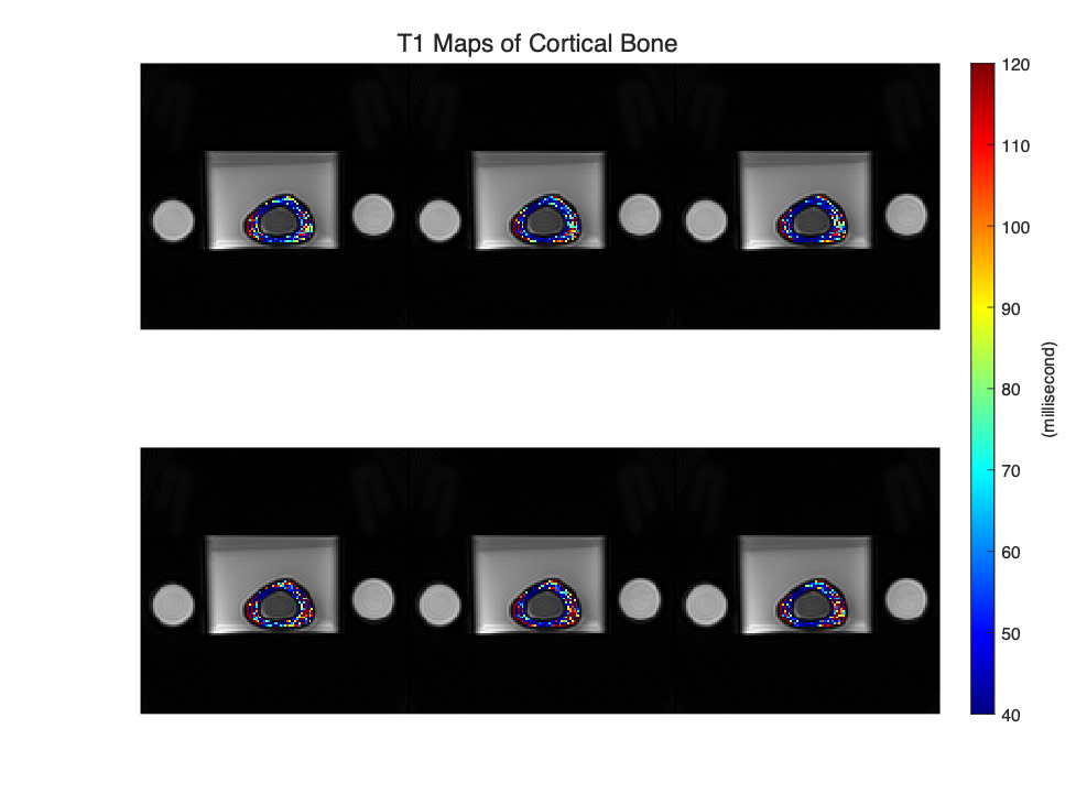
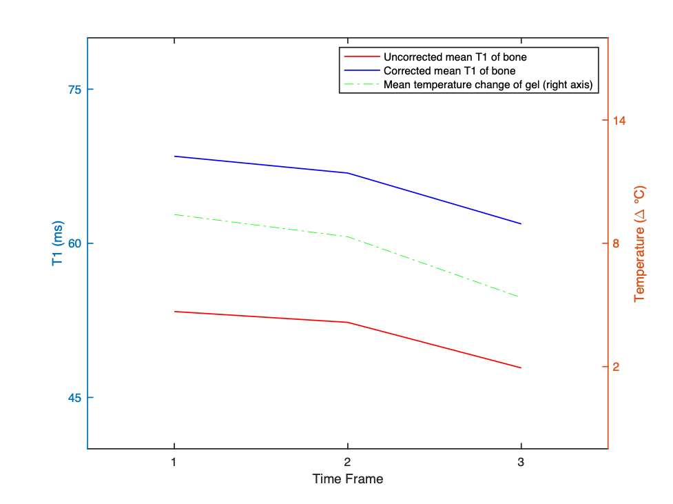
Figure 2. Three frames of mean T1 values of cortical bone and mean temperatures of agar gel. The corrected T1 values are ~15ms higher than the uncorrected values. Downward trends of T1 and temperature match. The frames correspond to those in Figures 1 & 3.
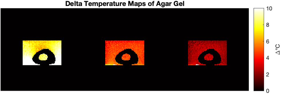
Figure 3. Three frames of delta temperature maps of agar gel by PRF. The frames correspond to those in Figures 1 & 2.
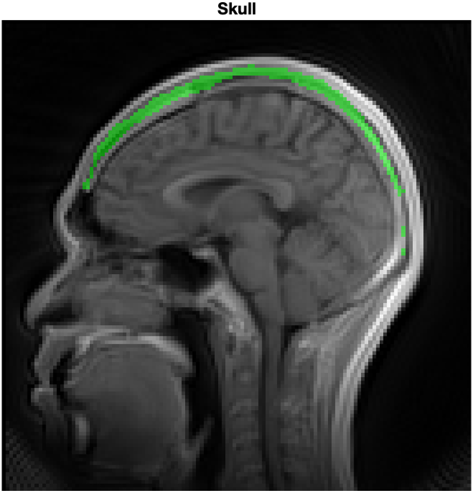
Figure 4. The skull is highlighted green by scaled subtraction.
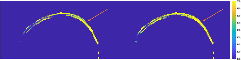
Figure 5. The uncorrected (left) and corrected (right) T1 maps of the skull. Arrows show improved homogeneity.