2699
Improved Tx Efficiency in Mock Transcranial MRgFUS with Modified Transfer Medium & Transducer Ground at 3T: Improved Results with Updated Phantom1Bernard and Irene Schwartz Center for Biomedical Imaging, Department of Radiology, New York University Grossman School of Medicine, New York, NY, United States, 2Center for Advanced Imaging Innovation and Research (CAI2R), Department of Radiology, New York University Grossman School of Medicine, New York, NY, United States
Synopsis
Keywords: MR-Guided Focused Ultrasound, MR-Guided Interventions
Motivation: In MRI at 3T, the typical configuration of MR-guided focused Ultrasound (MRgFUS) results in a dark band through the brain in MRI.
Goal(s): We introduce an approach to significantly improving MRI in the whole brain in addition to removing the dark band artifact, demonstrating here with an improved phantom for better results.
Approach: An improved phantom for experimental validation of our method was developed by using a spheroidal compartment rather than a hemispherical compartment to represent the head.
Results: Transmit Efficiency was improved by more than a factor of 4 on average in the ROI using our proposed method.
Impact: By strategically adjusting electric permittivity of the transfer medium and slotting the transducer ground plane, transmit efficiency of MRI in transcranial MRgFUS can be improved by a factor of up to 4 while also eliminating a well-known dark band.
Introduction
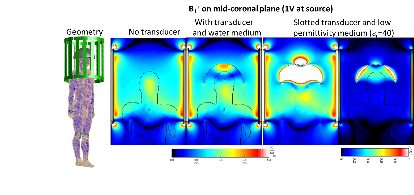
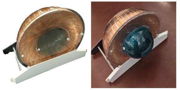
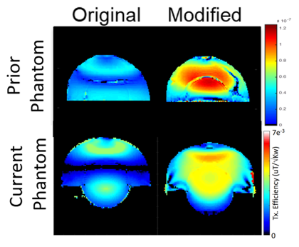
Transcranial MRI-guided focused ultrasound (TMRgFUS) is used for treatment for essential tremor (1). It requires both an array of ultrasound transducers with a common conductive ground and a large bath of fluid (transfer medium). These systems adversely affect the distribution of radiofrequency magnetic fields in 3T MRI. Previously we used simulations and experiment to show potential artifact reduction and significant increase in SNR and transmit efficiency with modification of the relative electric permittivity in the transfer medium and slotting the ground plane in the transducer array (2, 3)(Figure 1), but the phantom produced an artifactual low-B1 region near the boundary of the phantom, which was unfortunately within the ROI (3). Here we present results with a new phantom design (Figure 2, right) that does not have the non-anatomical boundary.Methods
Three hemispherical acrylic shells (one with diameter 35cm and two with diameter 18cm) were mounted on a circular acrylic plate with nylon screws and silicone sealant to approximate the geometry of a TMRgFUS transducer containing the upper portion of the human head (Figure 2, Figure 3). The inner region was filled with saline solution (1g NaCl/1L H20 for conductivity 0.21S/m). The outer compartment was filled alternately with distilled water (electric permittivity of 78) or a mix of 55% isopropyl alcohol and 45% water with a permittivity near 40. The outer surface of the phantom was covered with copper tape except for longitudinal slots producing 8 segments. Slots were bridged with multiple large (390pF) capacitors when outer compartment was filled with water (“Original” configuration) and were left open when outer compartment was filled with low-permittivity mixture (“Modified” configuration). Transmit efficiency (TxEff) was characterized at 3T with the system body coil used in excitation and reception. B1+ maps were acquired using TurboFLASH(4) (TE/TR-1.9/10000ms) and GRE acquisitions.Results
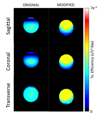
Figure 4 shows experimentally-measured maps of TxEff on three orthogonal planes passing through the middle of the smaller compartment in both configurations. By reciprocity, SNR for the body coil should see similar improvement. In the Original configuration, a band of near-zero signal is seen to pass through the upper portion of the inner compartment, similar to a dark region seen in images of the head in the ExAblate system at 3T. In Modified configuration the signal is many times higher in this region, but lower towards the inferior surface of the phantom. In Modified configuration, TxEff is lower in the outer compartment than the inner compartment even in neighboring regions with similar flip angles due to the alcohol/water mixture in the outer compartment.Discussion
The modified configuration results in improved TxEff by a factor of 2.5-5.44 in the ROI on the three orthogonal planes, and by a factor of 4.06 when averaging the planes together. This is notably better than for the Prior phantom (3), and much closer to improvements in simulation (2). By reciprocity, similar patterns are expected for both transmit efficiency and normalized SNR. Other approaches including introduction of conductors to the transfer medium (5-7), use of receive coils (8), or addition of salt to the transfer medium (9) do not appear to show this level of improvement.Acknowledgements
This work was performed under the rubric of the Center for Advanced Imaging Innovation and Research (CAI2R, www.cai2r.net), an NIBIB National Center for Biomedical Imaging and Bioengineering (NIH P41 EB017183).References
1. V Krishna et al., JAMA Neurology 2018;75(2):246-254
2. CM Collins et al., 2020 ISMRM, p. 1279
3. K Lakshmanan, CM Collins, 2023 ISMRM, p. 0745
4. H-P Fautz et al., 2008 ISMRM, p.1247
5. X Yan et al., 2020 ISMRM, p. 0113
6. X Yan and W Grissom, US Patent application 2020/0360733 A1
7. R Hadley et al., 2020 ISMRM, p. 1268
8. J Corea et al., Scientific Reports 2018;8:3392
9. S Leung et al., J Therapeutic Ultrasound 2015;3:P27
Figures