2694
Low-rank reconstruction of variable-density random undersampled data for low-field MRI: accelerated T1 mapping at 46 mT1C.J. Gorter MRI Center, Department of Radiology, LUMC, Leiden, Netherlands, 2Philips Research Hamburg, Hamburg, Germany
Synopsis
Keywords: Low-Field MRI, Low-Field MRI
Motivation: Limited access to medical equipment makes low-field MRI an interesting option in many settings. One challenge is the prolonged scan time, especially for quantitative imaging. However, parallel imaging is typically not used due to the very high sensitivity of a single solenoid receive coil.
Goal(s): This study aims to accelerate T1 mapping (used for estimating brain myelination) on a Halbach low-field system.
Approach: A locally low-rank reconstruction was applied to diminish undersampling artifacts from the variable-density random sampling trajectory.
Results: The study highlights the potential for both, fast lower-resolution (2.5mm2 in-plane) and higher-resolution (1.5mm2 in-plane) T1 mapping with an acceleration factor of R=4.
Impact: Our study's advanced low-rank reconstruction approach for low-field MRI could transform imaging methods in regions where high-field MRI is inaccessible, enabling precise and fast T1 brain mapping, which is critical for assessing myelination-related diseases with newfound speed and reliability.
Introduction
Access to high-end medical equipment remains limited in many settings, making low-field MRI a potentially valuable diagnostic tool1. While cost-effective and suitable for environments with limited infrastructure, the primary limitation of low-field MRI is its lengthy scan time2,3. T1 mapping is an important tool for future neuroscience studies into brain development, disease progression and brain myelination. However, the scan time for T1 mapping at 50mT is usually more than 30 minutes3 due to the limited gradient strengths. Acceleration approaches such as parallel imaging4 are not typically used since the solenoid coil is much more sensitive than multiple loop receivers. In this work, we accelerated T1 mapping using locally low-rank reconstruction5 with variable-density random sampling on a 46 mT Halbach scanner. The method was tested both on phantoms and in-vivo.Methods
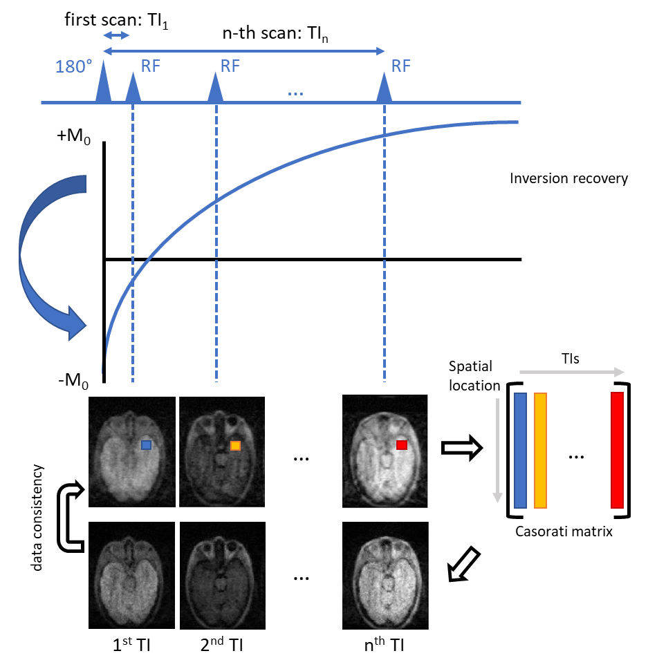
Despite using only a single receive coil, data redundancy can be assumed along the parameter mapping dimension5. The corresponding optimization problem can be expressed as:$$\left\{\hat{x}_{1,\ldots,N}\right\}=\underset{x_{1,\ldots,n}}{\operatorname{argmin}}\sum_{n=1}^N\left\|D_nFx_n-y_n\right\|_2^2+\lambda\sum_{b\in\Omega}\left\|R_b\left\{x_{1,\ldots,N}\right\}\right\|_{*},\quad(1)$$Where $$$D_n$$$ represents the sampling mask for each $$$TI_n(n=1,2,\ldots,N)$$$, $$$F$$$ the 3D FFT, $$$x_n$$$ the unknown images to be reconstructed, and $$$y_n$$$ the undersampled k-space data. The operator $$$R_b$$$ extracts a small spatial 3D block around pixel-index $$$b$$$ in image space, vectorizes each one and concatenates them along all vectorized arrays from all different TIs to form a Casorati matrix. By minimizing its nuclear norm, one can enforce the low-rankness property along the TI dimension, using the redundant information to guide the reconstruction of each individual TI sample.All experiments were performed on a 46 mT Halbach low-field MRI system3. TSE-data with 6 echoes were acquired using TE/TR=20/1200ms at 6 TIs of 50,91,166,302,549, and 900ms. Fully sampled (R=1) data at low-resolution (2.5×2.5×5mm3), and variable-density undersampled data (R=4) at both low (2.5×2.5×5mm3) and high-resolution (1.5×1.5×5mm3) were collected in a morphometric brain-like phantom7 and in-vivo (one subject). Data with an acceleration factor of R=8 were also acquired in the phantom. The acquisition time was 7.5/1.9/1.0 minutes per TI for the low-resolution R=1/4/8 and 3.1/1.6 minutes for the high-resolution R=4/8 scans. 6 different variable density Poisson disk sampling patterns were generated for each undersampled TI scan, along the two phase-encoding directions, with each dimension covering approximately 12.5% of the k-space in the center.
To solve Eq.1, 3D non-overlapping blocks (10×10×4) from each of the TIs were used to form the Casorati matrix and a proximal gradient descent solver was used with associated single value decomposition (SVD) for each Casorati matrix + soft-thresholding to minimize its nuclear norm. The reconstruction pipeline (2D example) is shown in Fig.1. Prior to T1 fitting, a simple phase correction8 was applied using the phase of the last TI to obtain real-valued images. In the phantom study, paired t-tests were performed between R=1 low-resolution data and all four individually undersampled data sets (R=4/8, low/high resolution), with two thresholding masks created independently on each T1 map to report the T1 values of two target tissues.
Results
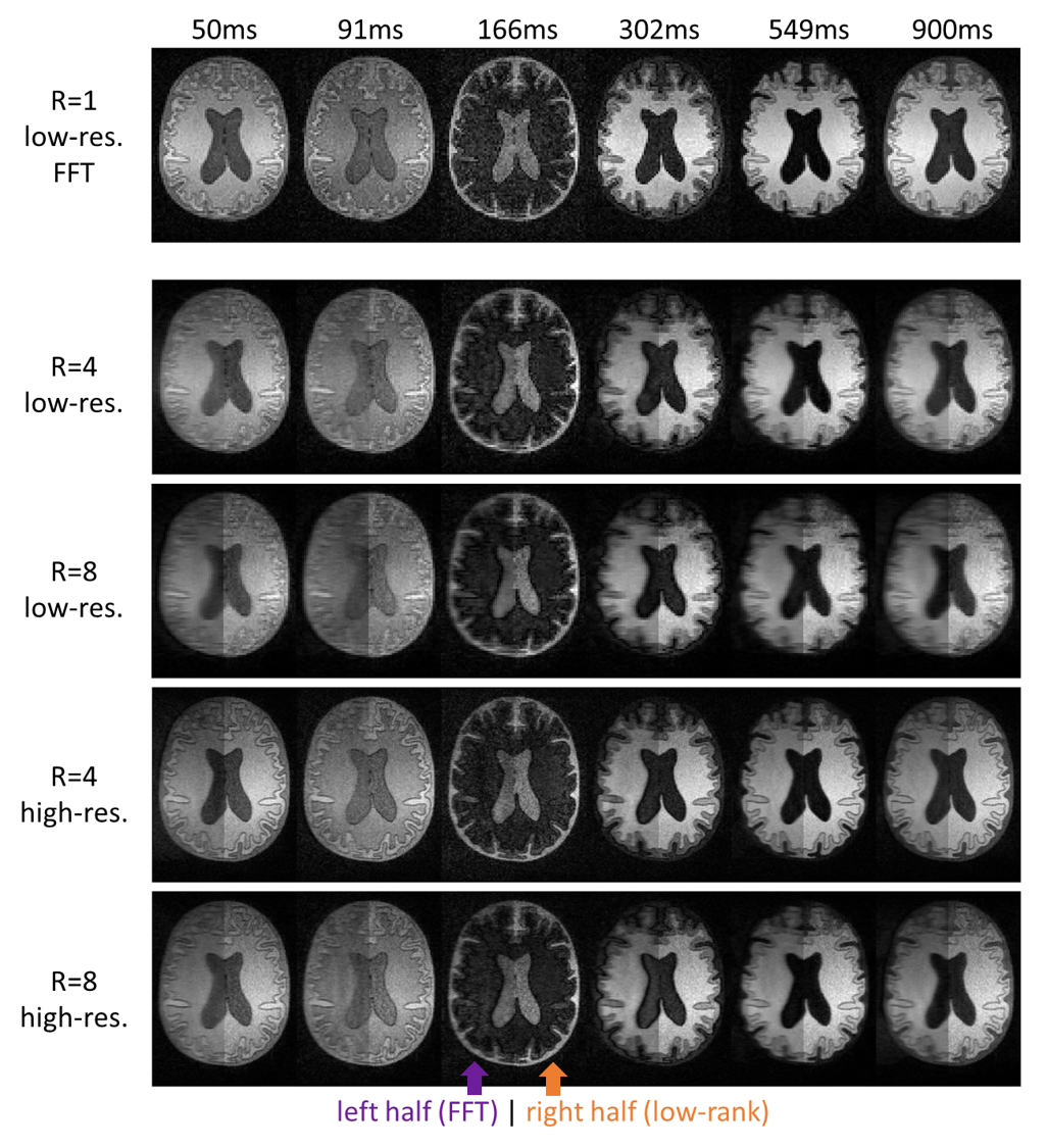
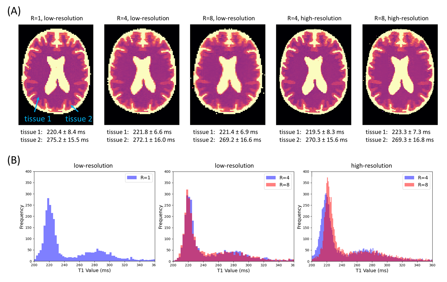
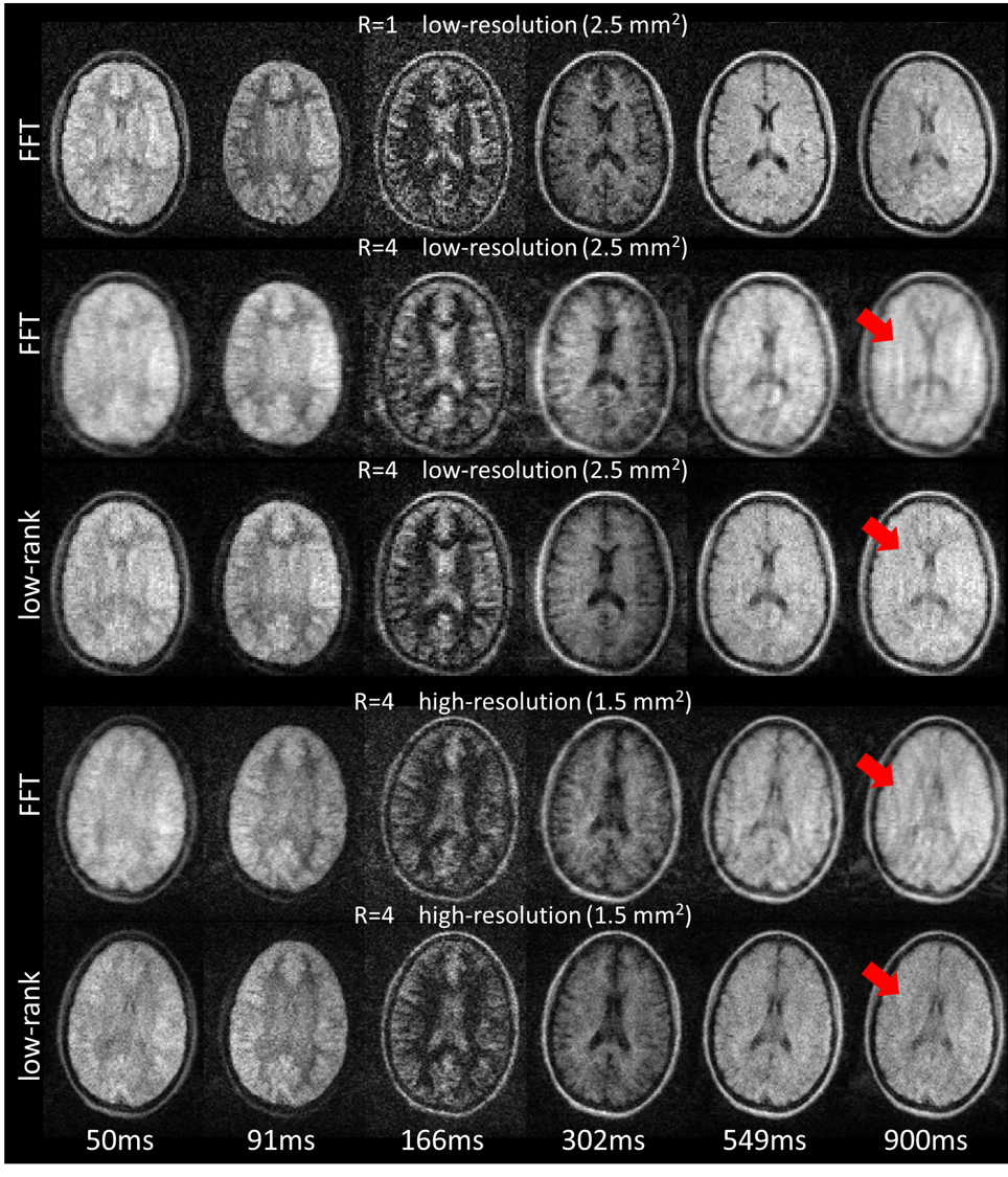
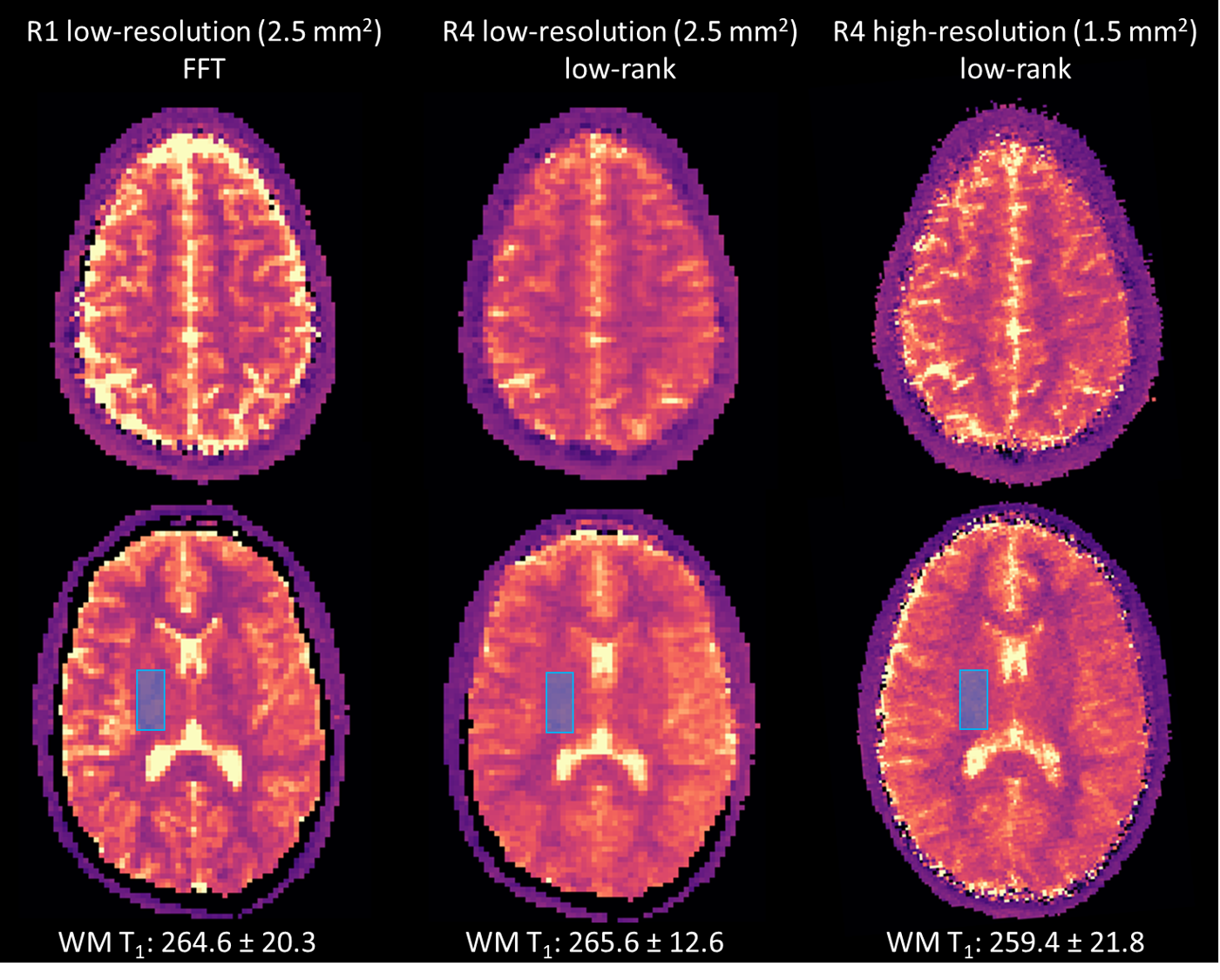
Figure 2 shows the reconstructed results of the low-resolution phantom study, with R=1 using direct FFT and both R=4 and R=8 FFT/low-rank reconstruction, respectively. Results show that the locally low-rank reconstruction can effectively mitigate the undersampling artifacts in all cases compared to FFT. Figure 3 shows low-resolution T1 maps (phantom data) obtained with fully-sampled k-space and at different undersampling factors (R=4 and 8), and high-resolution (R=4 and 8) low-rank results of the phantom. The histogram of the T1 distribution also shows a good match among different scans in both tissues. Figure 4 shows in-vivo images of R=1 (FFT reconstructed), R=4 low and high-resolution images (FFT and low-rank reconstructed). Figure 5 shows the corresponding T1 maps for each in-vivo data set, illustrating only the FFT result for R=1 and low-rank results of undersampled data. Differences in CSF T1 maps (which are not diagnostically relevant) between R=1 and R=4 are mainly due to the very long T1 value with respect to the TR, making the fit more susceptible to undersampling-induced blurring and partial-volume effects.Discussion and conclusion
In this study, we have demonstrated the benefits of using locally low-rank reconstruction to accelerate low-field T1 mapping. For a protocol using six different inversion times, we achieved R=4 acceleration for the in-vivo scans, reducing total scan time from 45 minutes to 11 minutes (2.5×2.5×5 mm3). This study also presented the potential for high-resolution T1 mapping in-vivo in 18 mins (1.5×1.5×5mm3 with R=4). Future improvements in T1 mapping quality could be achieved by integrating AI-based denoising9,10 with low-rank reconstruction, potentially helping to further reduce the number of TIs required.Acknowledgements
This work was partly funded by the Dutch Science Foundation Open Technology 18981.References
1. Kimberly WT, Sorby-Adams AJ, Webb AG, et al. Brain imaging with portable low-field MRI. Nature Reviews Bioengineering. 2023;1(9).
2. O’Reilly T, Webb AG. In vivo T1 and T2 relaxation time maps of brain tissue, skeletal muscle, and lipid measured in healthy volunteers at 50 mT. Magn Reson Med. 2022;87(2).
3. O’Reilly T, Teeuwisse WM, de Gans D, Koolstra K, Webb AG. In vivo 3D brain and extremity MRI at 50 mT using a permanent magnet Halbach array. Magn Reson Med. 2021;85(1).
4. Pruessmann KP, Weiger M, Scheidegger MB, Boesiger P. SENSE: Sensitivity encoding for fast MRI. Magn Reson Med. 1999;42(5):952-962.
5. Zhang T, Pauly JM, Levesque IR. Accelerating parameter mapping with a locally low rank constraint. Magn Reson Med. 2015;73(2):655-661.
6. Hu Y, Levine EG, Tian Q, et al. Motion-robust reconstruction of multishot diffusion-weighted images without phase estimation through locally low-rank regularization. Magn Reson Med. 2019;81(2):1181-1190.
7. Najac C, Koolstra K, O’Reilly T, Webb A. Reducing scan-time for 3D imaging with undersampled cartesian-radial phase encoding on a point-of-care 46 mT Halbach MRI scanner. Published online 2023: In: Proceedings of 33rd Annual Meeting of ISMRM.
8. Prah DE, Paulson ES, Nencka AS, Schmainda KM. A simple method for rectified noise floor suppression: Phase-corrected real data reconstruction with application to diffusion-weighted imaging. Magn Reson Med. 2010;64(2).
9. Koonjoo N, Zhu B, Bagnall GC, Bhutto D, Rosen MS. Boosting the signal-to-noise of low-field MRI with deep learning image reconstruction. Scientific Reports 2021 11:1. 2021;11(1):1-16.
10. Le DBT, Sadinski M, Nacev A, Narayanan R, Kumar D. Deep Learning-based Method for Denoising and Image Enhancement in Low-Field MRI. IST 2021 - IEEE International Conference on Imaging Systems and Techniques, Proceedings. Published online 2021.
Figures