2693
Methods for portable magnetic resonance elastometry1Physics, University of New Brunswick, Fredericton, NB, Canada, 2Mechanical Engineering, University of New Brunswick, Fredericton, NB, Canada
Synopsis
Keywords: Low-Field MRI, Elastography
Motivation: Magnetic resonance elastography can provided quantitative assessment of tissue mechanical properties, but requires large, expensive, in demand clinical MRI scanners, this motivates a more accessible portable MR technique.
Goal(s): To develop robust experimental protocols for bulk assessment of tissue mechanical properties using portable magnetic resonance and explore clinical applications.
Approach: Effects of phase interference on the MR signal magnitude and phase are investigated to characterize the shear wave velocity for samples with differing mechanical properties under harmonic and transient excitation.
Results: Preliminary measurements show that a portable MR instrument can be used to quantify the shear wave velocity in homogeneous samples.
Impact: These results show the potential of a portable magnetic resonance instrument for quantification of shear mechanical properties in biological tissues. This merits further investigation of applications to the diagnosis of specific diseases which could potentially improve patient outcomes.
Introduction
Magnetic resonance elastography (MRE) uses phase-contrast magnetic resonance imaging (MRI) sequences, synchronized with harmonic sample displacements, to assess the mechanical properties of tissues [1]. Our approach utilizes compact and cost-effective portable MR sensors that provide a bulk characterization of mechanical properties in homogeneous regions, close to the sample surface [2]. We aim to achieve direct measurements of mechanical properties by observing signal modulations due to phase interference caused by a non-uniform velocity distribution [3].Methods
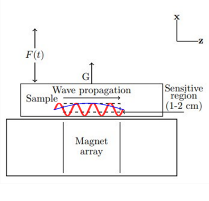
Portable MRE techniques can be either phase-interference-based or net-phase-based. Net-phase techniques are applicable when the wavelength is much larger than the size of the sensor, typically this is the case for longitudinal waves. We have used the net-phase approach to obtain relative viscoelasticity measurements in samples under bulk longitudinal excitation [2].Phase-interference techniques [3] are applicable when the wavelength is comparable to the sensor size (as shown in Figure 1), which is typically the case for shear waves in biological tissues at frequencies typical of conventional MRE (50-100 Hz). We have developed two procedures based on the phase interference of the MR signal that can be used to characterize the shear wave velocity.
The harmonic procedure uses sinusoidal oscillations at varying frequencies to change the wavelength and observe the signal magnitude and phase vs amplitudes and echo times. The integer number of wavelengths over the sensitive region causes the complete signal decay (at certain amplitudes and echo times), enabling measurement of the shear wave velocity.
The transient procedure is a time-of-flight-based approach and involves pulsed excitation of samples synchronized with MR acquisition. The delay between excitation and echo acquisition is varied until the pulse reaches the sensitive region, indicated by a phase-interference-induced change in the MR signal. This time can be used to determine the shear wave velocity provided the distance between the vibration source and the MR sensor is known.
The sole requirement of the MR instrumentation is a well-defined region of constant gradient in one direction. We employed a unilateral three-magnet array with a constant gradient of 254 G/cm perpendicular to its surface. In addition, reliable acoustic excitation is required. For this, we have used an electromagnetic vibration shaker and an excitation stinger to transfer vibrations into the samples.
Results and Discussion
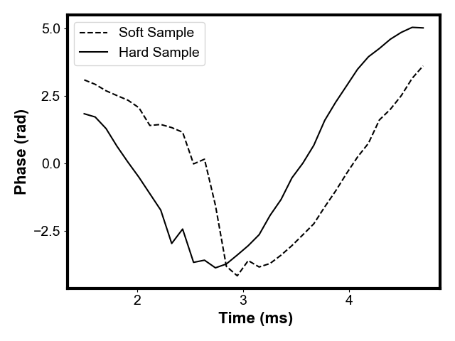
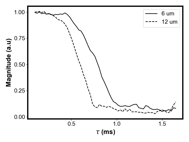
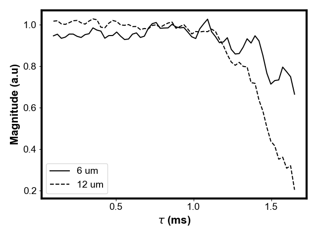
We carried out preliminary measurements on two elastomers with differing stiffness (different shear wave velocities). In the transient approach, we excited the samples with a 100-microsecond pulse. Echoes were recorded with an echo time of 1.5 milliseconds at various delays. Figure 2 shows that the phase for the stiffer sample precedes that of the softer sample due to its higher shear wave velocity.In the harmonic approach, the samples were excited sinusoidally at 125 Hz at two different amplitudes. We adjusted the echo time to investigate the impact of phase interference on signal magnitude (Figures 3 and 4). A three-point moving average filter was used to smooth out noise, and better represent the general trend. Notably, we observed that higher amplitude excitation resulted in a more pronounced and rapid decline in signal magnitude. Furthermore, there is a clear dependence on the shear wavelength and the decay in signal magnitude. In future measurements, we will investigate using this approach to directly extract the shear wave velocity.
Conclusion
Our measurements demonstrate the potential of a simple measurement capable of directly measuring the shear wavelength in biological tissues. It can already achieve a rough estimate that aligns with expectations. In future measurements, we will investigate combining the two techniques: first, using the transient approach for a rough estimate of the shear wave velocity, which can be used to select frequencies used in the harmonic approach for a more accurate measurement. Once we have established robust procedures for characterizing shear properties, we can aim to combine them with conventional MR measurements of relaxation and diffusion, providing an additional layer of information that could potentially be useful in clinical applications.Acknowledgements
Funding from the Natural Sciences and Engineering Council of Canada (NSERC) and the New Brunswick Innovation Foundation (NBIF) is gratefully acknowledged.References
[1] Muthupillai, R. et al., "Magnetic resonance elastography by direct visualization of propagating acoustic strain waves", Science 269 (1995)
[2] Selby, W., Garland, P., Mastikhin, I., "Dynamic Mechanical Analysis with Portable NMR", JMR 339 (2022).
[3] Selby W., Balcom, B.J., Newling, B., Mastikhin, I., "Using Phase Interference to Characterize Dynamic Properties-A Review of Constant Gradient, Portable Magnetic Resonance Methods", Frontiers in Physics (2023)
Figures