2692
Fast and pseudo-random: optimization of settings for rapid quantification of T1 in white and grey matter at 64 mT1C.J. Gorter MRI Center, Radiology Department, Leids Universitair Medisch Centrum, Leiden, Netherlands, 2Hyperfine Inc., Guilford, CT, United States, 3Section of Diagnostic Radiology,Department of Clinical Sciences Lund, Lund University, Lund, Sweden, 4Lund BioImaging Center, Lund University, Lund, Sweden, 5Department of Medical Radiation Physics,, Lund University, Lund, Sweden, 6Department of Neuroimaging, King’s College London, London, United Kingdom
Synopsis
Keywords: Low-Field MRI, Low-Field MRI, Relaxometry
Motivation: Low-field MRI holds promise for efficient diagnostics. T1 mapping is valuable in neuroscience for studying myelination and brain development. To reduce scan time, incoherent, variable density trajectories are often used.
Goal(s): to reach high image quality and T1 accuracy for fast T1 mapping at 64 mT.
Approach: Using the 64-mT, Hyperfine Swoop scanner, we compared T1 maps acquired with fully sampled and undersampled trajectories (with and without incoherence) and reconstructed them with locally low rank regularization at different regularization factors λ.
Results: Our findings showed that the most effective approach involves the use of a customized trajectory with λ around 0.004.
Impact: Since fast and accurate T1 mapping in the context of low-field MRI is achievable, it would now be interesting to study brain developement in children, that present a unique challenge due to movement.
Introduction
Low-field, point-of-care (POC) MRI is a promising technology with the potential to increase access to diagnostic imaging[1]. Quantitative MR parameter mapping (qMRI) aids in characterizing tissue properties for medical diagnosis, treatment monitoring, and research into tissue physiology[2]. T1 mapping, a key component of qMRI, is particularly valuable in neuroscience for studying brain myelination, assessing brain development, and identifying myelin-related conditions and diseases[3, 4]. However, T1 mapping with low-field MRI currently suffers from long scan times[2, 5]. In the context of POC MRI, studying children presents a unique challenge due to movement[6]. To tackle this, one approach is to accelerate POC T1 mapping, with a specific focus on employing Locally Low Rank (LLR) regularization as a potential solution [7]. The current study was conducted on a 64 mT Hyperfine Swoop System, aiming to obtain an efficient T1 mapping strategy for pediatric imaging. We focused on refining both data acquisition and reconstruction strategies. This involves the implementation of incoherent variable density sampling trajectories and sparse reconstruction techniques with a locally low rank constraint. These approaches are evaluated by comparing the results with cases where acceleration and incoherence were not employed. Furthermore, we explored the impact of different regularization factors on the quantitative aspects of the reconstructed data.Methods
AcquisitionA Hyperfine Swoop (Hyperfine Inc., CT) operating at 64 mT with a single coil transmit and 8-channel receive head coil was used for all experiments. T1 maps were acquired using a 3D inversion recovery turbo spin-echo (TSE) sequence, with acquisition parameters: TR=1500 ms, effective TE=5 ms, echo train length 32, 6 inversion times (TI) logarithmically spaced between 50 and 1000 ms, FOV 220x180x200 mm3, resolution 1.6x1.6x5 mm3. k-space encoding in the phase-slice direction followed a pseudo random radial sampling, customized for the Hyperfine scanner (called here “Default”)[8]. A variable density Poisson-disk mask was also implemented, as suitable trajectory known to provide the best results for compressed sensing and LR reconstruction[7, 9]. Cartesian encoding was used along the readout direction for all trajectories. The scans consisted of four T1 mapping protocols: (A) fully-sampled Default without incoherence between TIs; undersampled Default with (B) and without (C) incoherence; (D) undersampled Poisson-disk mask, with incoherence. The total scan time for the fully sampled data set was 42.3 min. With undersampling, the total scan time was reduced to 10.8 min for four-times undersampling (R4).
Reconstructions and Fitting
Image reconstruction was performed with the pics -e -S -N -d 4 -R L:7:7:λ -i 20 -b 4 -U command of the BART toolbox [10], using a local low rank regularization constraint (ten λ's between 0.0-0.01 and block size 4 × 4 × 4)[7, 11]. An ADMM solver was used to reconstruct all TIs jointly with 20 iterations. Coil sensitivity maps were calculated with ESPIRIT [12] using the longest TI scan. To obtain real-valued data for T1-fitting, phase correction was performed using the longest TI image (8). The real data were then fitted to the signal equation presented by Padormo et al. (4).
Experiments
The performance of the different accelerated T1 protocols were tested against the fully sampled case (R1) in a calibrated quantitative phantom (Caliber MRI, Boulder, CA, model 137) containing T1-mimics with known concentrations of NiCl2. To demonstrate the method in vivo, the same protocols were acquired in two healthy volunteers.
Results
In Figure 1, increasing λ reduces noise but decreases T1 accuracy, with up to a 50 ms difference for λ=0.01. Poisson-disk trajectory T1 maps deviate the most from R1, while Default R4 trajectories yield similar estimates, with slightly higher standard deviation for coherent Default. Phantom image quality (Figure 2) remained consistent regardless of λ or trajectory. Both Figure 1 and 2 suggest that the lower the λ the higher the accuracy. In contrast, in-vivo T1 maps (Figure 3-4) show clear over-regularization at high λ, especially with the Poisson-disk trajectory, and under-regularization at λ=0 for the undersampled trajectories. The best image quality was achieved with Default R4 and λ=0.04. Quantitatively (Figure 5), Default R4 with incoherence provides T1 values closest to the reference in white and grey matter.Discussions and Conclusions
A rapid T1 mapping method was developed on a portable 64 mT MRI system, achieving accurate T1 quantification in white (WM) and grey matter (GM) with minimal image quality compromise. The customized Hyperfine trajectory performs best for this purpose, possibly due to explicit reconstruction using known hardware/artefact characteristics. In our current settings, an optimal λ around 0.004 reduced noise while providing T1 accuracy. However, our sample size was small, so future research will assess the method's reproducibility in a larger volunteer cohort.Acknowledgements
This work was part funded by the Crafoord Foundation (ref 20220898) and by the Dutch Science Foundation Open Technology (ref 18981)References
1. Arnold TC, Freeman CW, Litt B, Stein JM (2023) Low-field MRI: Clinical promise and challenges. Journal of Magnetic Resonance Imaging 57
2. Jordanova K V., Martin MN, Ogier SE, et al (2023) In vivo quantitative MRI: T1 and T2 measurements of the human brain at 0.064 T. Magnetic Resonance Materials in Physics, Biology and Medicine. https://doi.org/10.1007/s10334-023-01095-x
3. Padormo F, Cawley P, Dillon L, et al (2023) In vivo T1 mapping of neonatal brain tissue at 64 mT. Magn Reson Med 89:. https://doi.org/10.1002/mrm.29509
4. Eminian S, Hajdu SD, Meuli RA, et al (2018) Rapid high resolution T1 mapping as a marker of brain development: Normative ranges in key regions of interest. PLoS One 13:. https://doi.org/10.1371/journal.pone.0198250
5. O’Reilly T, Webb AG (2022) In vivo T1 and T2 relaxation time maps of brain tissue, skeletal muscle, and lipid measured in healthy volunteers at 50 mT. Magn Reson Med 87:. https://doi.org/10.1002/mrm.29009
6. Eichhorn H, Vascan A-V, Nørgaard M, et al (2021) Characterisation of Children’s Head Motion for Magnetic Resonance Imaging With and Without General Anaesthesia. Frontiers in Radiology 1:. https://doi.org/10.3389/fradi.2021.789632
7. Zhang T, Pauly JM, Levesque IR (2015) Accelerating parameter mapping with a locally low rank constraint. Magn Reson Med 73:655–661. https://doi.org/10.1002/mrm.25161
8. O’Halloran R, Dyvorne H, Sacolick L, et al (2022) Diffusion-Weighted Imaging at 0.064 T. Joint Annual Meeting ISMRM-ESMRMB ISMRT 31st Annual Meeting. https://doi.org/10.58530/2022/0043
9. Doneva M, Börnert P, Eggers H, et al (2010) Compressed sensing reconstruction for magnetic resonance parameter mapping. Magn Reson Med 64:. https://doi.org/10.1002/mrm.22483
10. Uecker M, Tamir JI, Ong F, Lustig M (2016) The BART Toolbox for Computational Magnetic Resonance Imaging. Proceedings of the 23rd Annual Meeting of ISMRM, Toronto, Canada
11. Tamir JI, Uecker M, Chen W, et al (2017) T2 shuffling: Sharp, multicontrast, volumetric fast spin-echo imaging. Magn Reson Med 77:. https://doi.org/10.1002/mrm.26102
Figures
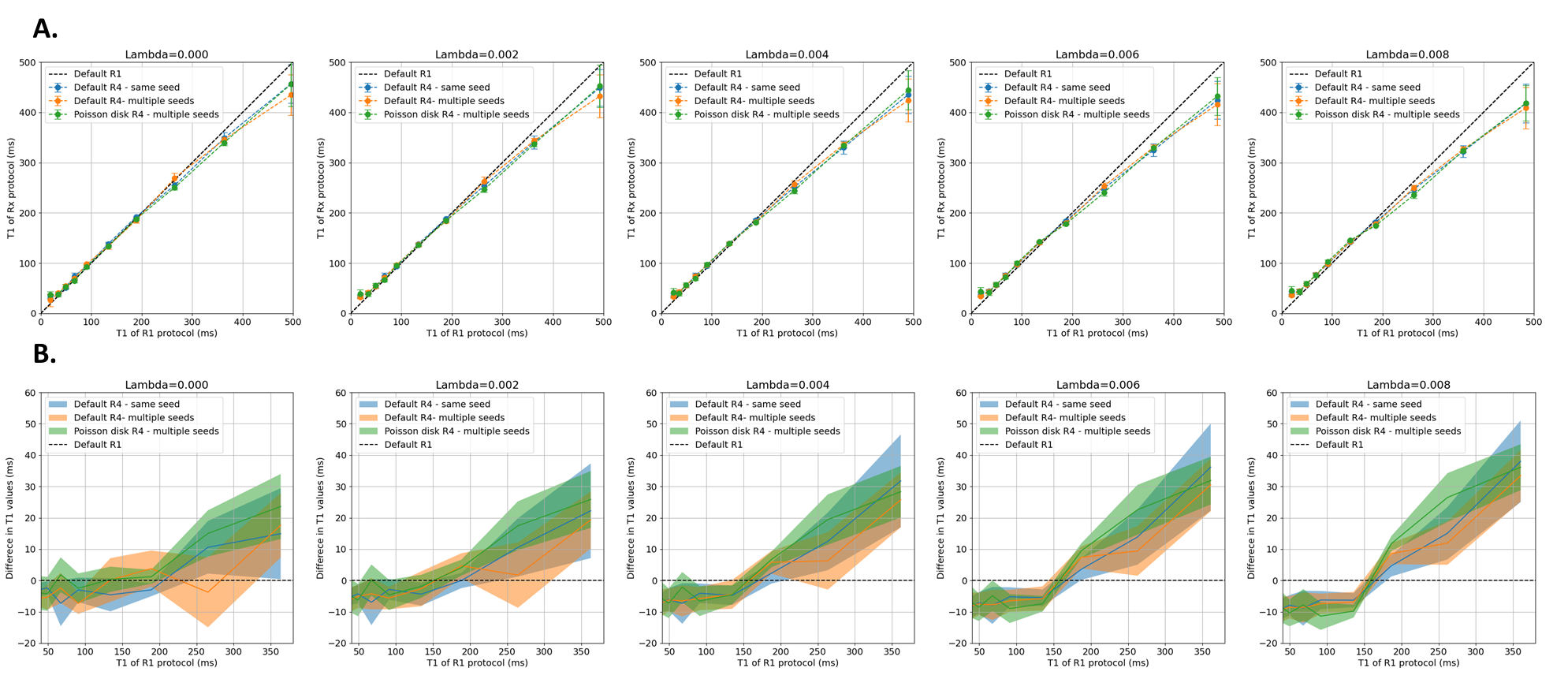
Figure 1. Plots of different regularization factors for A. T1 values (mean std in the ROIs for each tube) measured in the first 10 spheres of the calibrated phantom: accelerated protocol (Rx) vs fully sampled (R1). B. Difference in T1 values of the accelerated protocols respect to the fully sampled one. The shade represents the standard deviation.
Default = customized Hyperfine trajectory, Poisson disk = VD Poisson disk trajectory, R1= fully sampled, R4= undersampling factor 4, same/multiple seed(s) = without/with incoherence in the temporal dimension
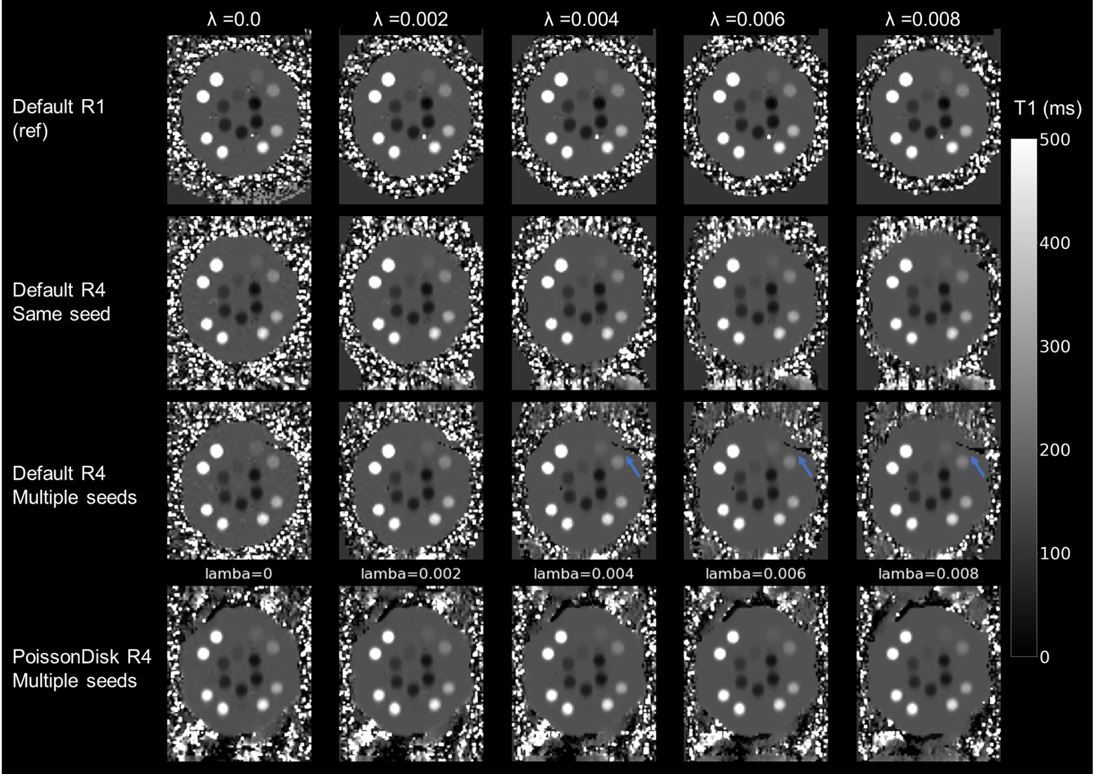
Figure 2. T1 maps of the calibrated phantom for different T1 protocols and regularization factors. Beside an artefact for the Default R4 - multiple seeds probably due to the distortion correction (blue arrows) and minor blurring, the image quality is similar for all accelerated protocols respect to the reference.
Default = customized Hyperfine trajectory, Poisson disk = VD Poisson disk trajectory, R1= fully sampled, R4= undersampling factor 4, same/multiple seed(s) = without/with incoherence in the temporal dimension
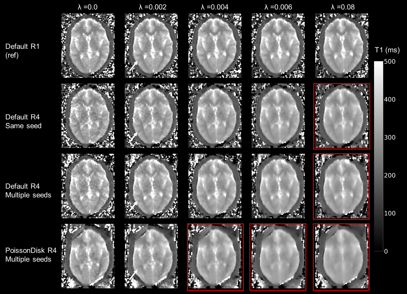
Figure 3. T1 maps of one volunteer for different trajectories and regularization factors. Some maps are clearly overregularized (red squares). Most maps are blurrier than the reference for λ > 0.04. Some fine details are visible for lambda 0.004 (white arrow), especially for Default R4, and get lost at higher lambda.
Default = customized Hyperfine trajectory, Poisson disk = VD Poisson disk trajectory, R1= fully sampled, R4= undersampling factor 4, same/multiple seed(s) = without/with incoherence in the temporal dimension
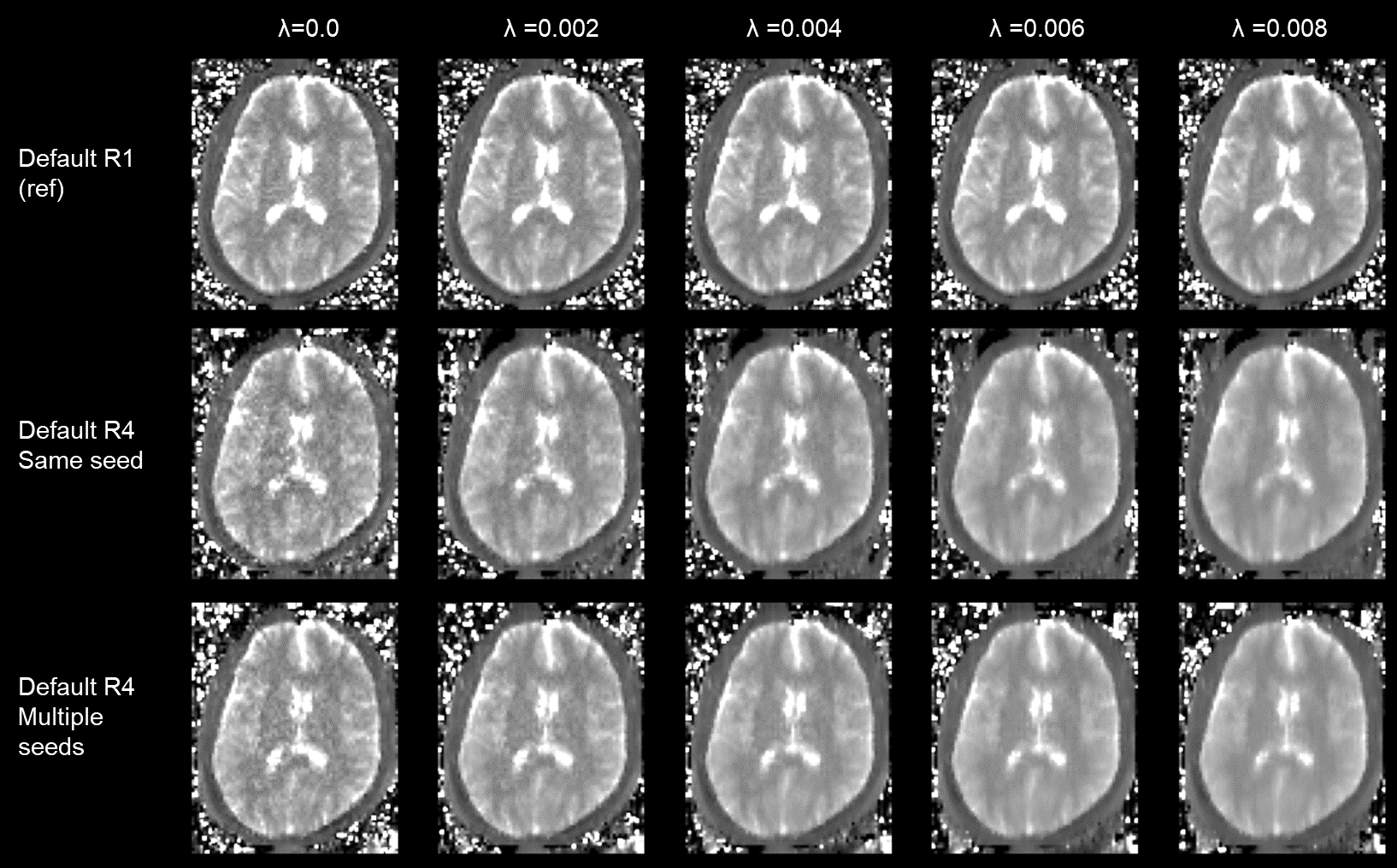
Figure 4. T1 maps of the second volunteer for different trajectories and regularization factors.
Default = customized Hyperfine trajectory, Poisson disk = VD Poisson disk trajectory, R1= fully sampled, R4= undersampling factor 4, same/multiple seed(s) = without/with incoherence in the temporal dimension
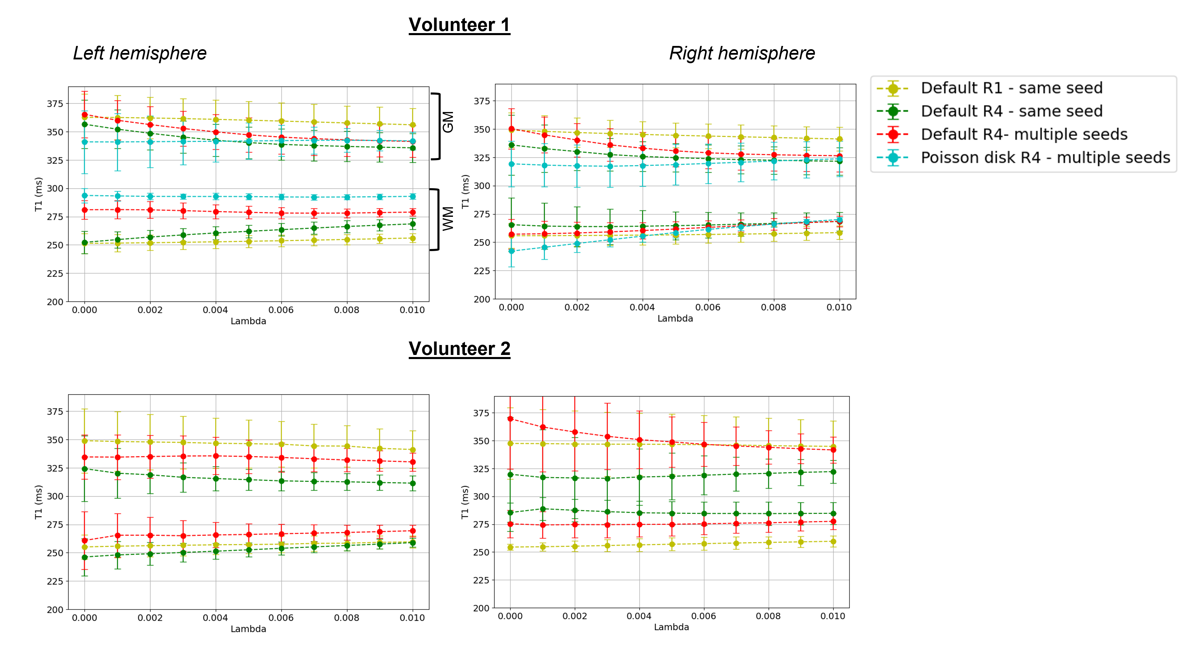
Figure 5. T1 values vs λ for different trajectories: the values were measured in the white and grey matter of 2 volunteers, in two ROIs placed on the right and left hemisphere. The std of the ROIs in the grey matter was larger because of partial volume voxels, between grey matter and CSF. For almost all cases, the Default R4 multiple seeds was closer to the reference fully sampled values. Accuracy and precision of the Default R4 protocol is maximized above λ=0.04.
R1= fully sampled, R4= undersampling factor 4, same/multiple seed(s) = without/with incoherence in the temporal dimension