2690
Radial xSPEN for Non-Fourier Single-Sided MRI1Promaxo, Oakland, CA, United States, 2Biomedical Engineering, Case Western Reserve University, Cleveland, OH, United States
Synopsis
Keywords: Low-Field MRI, Low-Field MRI
Motivation: Developing methods for rapidly imaging in an inhomogeneous magnetic field is necessary for the general adoption of single sided MRI, which would bring MR imaging into the doctor's office.
Goal(s): We aim to develop a fully spatiotemporal pulse sequence for collecting images with a single sided scanner.
Approach: A hyperbolic phase is imparted to the magnetization which results in a time domain signal that is a profile of the phantom. The angle of this profile is rotated to produce an image.
Results: Images with no Fourier encoding were produced and the images could be cropped without aliasing.
Impact: This novel technique allows for rapid imaging in an inhomgeneous magnetic field. Furthermore, this technique allows for improved resolution because the field of view of the images collected with it can be smaller than the object without aliasing.
Introduction
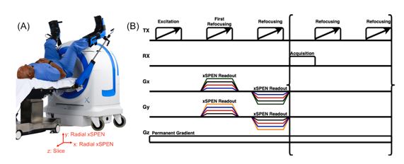
Low-field single-sided MRI scanners enable new point-of-care imaging applications requiring low-cost and open scanner design for physician access to the patient. Our single-sided prostate-dedicated MRI scanner (Promaxo, Oakland, CA, USA) has a permanent magnetic field gradient which requires different imaging approaches (Figure 1A). xSPEN is one such approach which enables rapid image acquisition in the presence of a permanent magnetic field gradient [1-3]. We report a new multislice radial xSPEN method that uses no phase or frequency encoding, and instead rotates the axis of the xSPEN spatiotemporal encoding function to collect a complete MRI sinogram from which a 2D image can be reconstructed.Methods
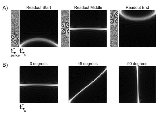
Figure 1B illustrates the radial xSPEN pulse sequence which is based on a CPMG acquisition with WURST excitation and refocusing pulses[4-6]. The images shown in figure 4 were collected with 8 echoes while those in figure 5 had 12 echoes. Echo times varied from 3 to 5.4 ms, depending on the field of view of the xSPEN scan. xSPEN encoding is achieved with rotating x- and y-gradient pulses with opposite polarities during the first two refocusing pulses. Because these pulses are applied simultaneously with the scanner’s permanent z gradient, a bilinear phase is developed across the slice and an angled in-plane dimension. Figure 2A illustrates this bilinear phase which is then shifted during the readout by phase evolution caused by the Gz gradient. Figure 2A illustrates how a system matrix is generated for radial xSPEN image reconstruction. The bilinear phase for a given radial angle and readout time point is calculated using sequence parameters and gradient field maps, and is summed through the slice to generate a point-spread function for that angle and readout time point. Figure 2B shows point-spread functions in the middle of the readout for different angles. These are collected into a matrix which is inverted to reconstruct a two-dimensional image. A simulation was performed to demonstrate the undersampling characteristics of radial xSPEN, and radial xSPEN scans of two phantoms were collected: one of the ACR extremity phantom surrounded by material that mimics tissue, and another of a resolution phantom consisting of a grid of tubes filled with silicon oil. Images were reconstructed using two variations of a CGLS model based reconstruction, for a 120x120 image matrix size with a field of view of 180x180 mm2.Results
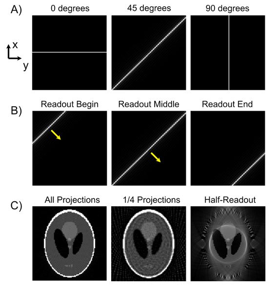
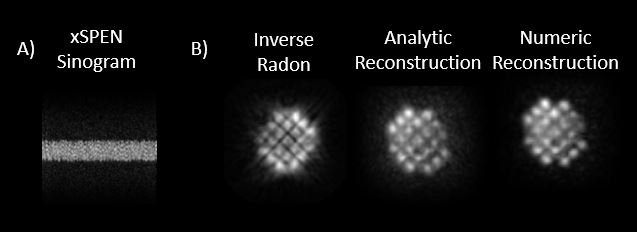
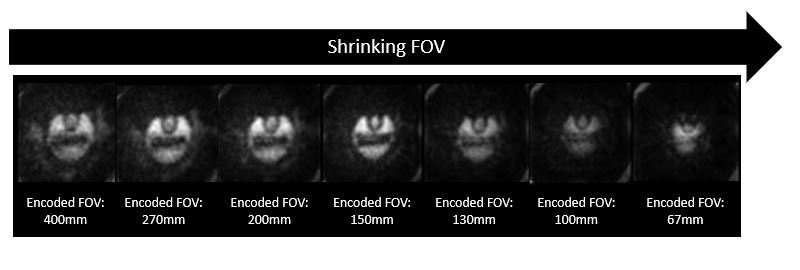
Figure 3 shows the simulation in which an ideal xSPEN point spread function was generated and rotated (Figure 3a) and swept in each direction (Figure 3b). Figure 3c shows that the scan’s angular sampling relationships are the same as Fourier encoding, wherein subsampling the angles leads to streaking artifacts. However, unlike Fourier radial imaging wherein truncating the length of the readout leads to a lower resolution image, truncating an xSPEN readout acquires a reduced FOV. Figure 4A shows a raw radial xSPEN dataset of the resolution phantom, which resembles an x-ray sinogram. Figure 4B shows three reconstructions of this data using an inverse radon transform which ignores the scanner’s gradient non-linearity, an analytic PSF-based reconstruction, and the numerical PSF-based reconstruction described above. The numerical reconstruction has the least blurring. Figure 5 shows ACR phantom reconstructions using the numerical PSF-based method, for different readout durations. As expected from Figure 3b, truncating each radial xSPEN readout leads to a decreasing FOV, without aliasing.Discussion and Conclusion
We have collected fully spatiotemporally encoded images using a novel radial xSPEN acquisition. This approach enables time-efficient acquisitions on a single-sided low-field MRI scanner by flipping the role of the scanner’s built-in z gradient which would normally perform frequency encoding across a small matrix dimension, instead using it for in-plane encoding. A numerical point-spread function-based reconstruction was described which accounts for the scanner’s non-linear gradients, and the acquisition’s reduced-FOV imaging capability was demonstrated. The scan is expected to be well-suited to interventional applications requiring dynamic imaging on this scanner.Acknowledgements
NoneReferences
1. Zhang, Z., Seginer, A. & Frydman, L. Single-scan MRI with exceptional resilience to field heterogeneities. Magn. Reson. Med. 77, 623–634 (2017).2. M Gomes et al. Multiecho xSPEN for Single-Sided Low-Field MRI. ISMRM Low-Field MRI Workshop, (2022).
3. M Gomes et al. In Vivo xSPEN Imaging with a Model-Based Reconstruction for Efficient Spatial Encoding in a Single-Sided Prostate MRI Scanner. ISMRM, (2023).
4. Power, J. E. et al. Increasing the quantitative bandwidth of NMR measurements. Chem. Commun. 52, 2916–2919 (2016).
5. O’Dell, L. A. The WURST kind of pulses in solid-state NMR. Solid State Nucl. Magn. Reson. 55–56, 28–41 (2013).
6. Casabianca, L. B., Mohr, D., Mandal, S., Song, Y. Q. & Frydman, L. Chirped CPMG for well-logging NMR applications. J. Magn. Reson. 242, 197–202 (2014).
Figures