2689
Three-Dimensional xSPEN Imaging on a Single-Sided MRI Scanner1Promaxo, Oakland, CA, United States, 2Biomedical Engineering, Case Western Reserve University, Cleveland, OH, United States
Synopsis
Keywords: Low-Field MRI, Low-Field MRI
Motivation: Imaging with a single sided MRI allows for a smaller scanner footprint, making it possible to bring the scanner into the doctor's office.
Goal(s): An xSPEN pulse sequence capable of producing three dimensional images is presented here, allowing for rapid image collection in an inhomogeneous magnetic field.
Approach: An additional phase encode array is incorporated into the xSPEN pulse sequence, allowing for the excited slab to be resolved along the z axis.
Results: A two dimensional multislice technique was converted into a three dimensional volumetric imaging by adding a phase encode array to the spatiotemporal axis.
Impact: A method for rapidly collecting three dimensional images in an inhomogeneous magnetic field is presented here. This technique will help bring MRI into the doctor's office by allowing for higher quality images to be collected in less time.
Introduction
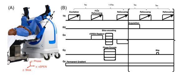
Single-sided low-field MRI scanners provide image guidance during interventions such as prostate biopsies without restricting surgical access. For example, the Promaxo scanner in Figure 1A (Promaxo, Oakland, CA, USA) has a built-in 58-73 mT z-gradient. Imaging is performed using a multi-slab 3D sequence in which the large matrix dimensions are phase-encoded by the pulsed x and y gradients, while the small matrix dimension is frequency encoded by the permanent z gradient. This leads to anatomic scan times less than 10 minutes. However, faster scans are desirable for localizers and intraoperative imaging but are fundamentally limited by the fact that the fastest spatial encoding mechanism is used to encode the smallest matrix dimension. We recently demonstrated how spatiotemporal encoding using xSPEN can be leveraged to swap phase and frequency encoding dimensions [1] on this scanner, however, only thick-slice multislice imaging was achieved[2][3]. Here we report a 3D multi-slab acquisition that uses additional phase encoding in the xSPEN dimension along with a numerical point-spread function-based reconstruction, which together could enable xSPEN to completely replace conventional Fourier imaging on this scanner[4].Methods
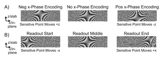
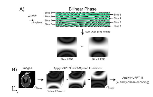
Figure 1B shows the multi-slab 3D xSPEN pulse sequence, in which xSPEN encodes both the x- and z-dimensions[5-7]. Figure 2A shows that phase encoding in x moves the xSPEN bilinear saddle/sensitive point to different locations in the slice, and Figure 2B shows that, conversely, phase evolution due to Gz during the readout moves the sensitive point to different locations in x. Figure 3A illustrates the reconstruction approach which accounts for gradient non-linearity. Given the sequence parameters and measured gradient field maps, for each readout time point the xSPEN bilinear phase pattern is defined over a finely sampled grid covering an imaged slab. The slab is then divided into the sub-slices to be reconstructed, and the phase is summed over each sub-slice to obtain a point spread function for each subslice and readout time point. Figure 3B then shows how this is used in the forward model for reconstruction: an input image is multiplied into each point-spread function, and the result is multiplied into a Type-III NUFFT to apply the phase encoding terms, generating the complete dataset. This forward model was used in a CGLS reconstruction. A three-dimensional image of the extremity ACR phantom was collected using 3D xSPEN. A chirped pulse CPMG style sequence was used to collect 8 echoes per excitation. Eight phase subslices within the slab were collected with a phase encode array of x gradient pulses. The data were reconstructed to a 120x120x40 image matrix with a field of view of 180x180x110 mm3.Results
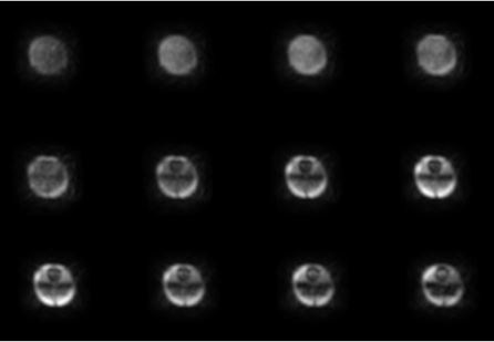
Figure 3 shows 12 contiguous central slices out of the 40 reconstructed slices of the ACR phantom. These slices span two collected slabs. Phantom features smoothly appear and disappear between slices, reflecting the scan’s ability to resolve details across the sub-slices within each slab.Discussion
A method for encoding three dimensional information with xSPEN in a single sided system is presented here, along with a reconstruction technique for it. Previously, 3D xSPEN images were generated from 2D images. The resolution along the z axis depended on the thickness of the slice. Therefore, reducing the pixel size along the slice direction required collecting a larger quantity of thinner slices. Eventually the number of slices needed to cover the whole volume will not fit with a single repetition, necessitating a second one which will slow down scanning. Furthermore, the performance of the chirped pulses tends to degrade as the bandwidth is decreased and switching away from a pulse sequence based on chirp pulses introduces a strong vulnerability to B1 inhomogeneity. Figure 2 shows that we were able to excite a thick slice and then further subdivide it with the phase encode array in the xSPEN axis. As the image moves from slice to slice, features that would otherwise be obscured by projecting the entire slab onto a single 2D image become resolved.Conclusion
With a three-dimensional xSPEN sequence and reconstruction, it is possible to rapidly collect volumetric images in a single sided system. Resolution along the slice selection axis can now be improved without reducing the slice thickness. In the future, this method can be applied to clinical imaging, allowing us to collect high quality images with a single sided MRI scanner.Acknowledgements
NoneReferences
1. Zhang, Z., Seginer, A. & Frydman, L. Single-scan MRI with exceptional resilience to field heterogeneities. Magn. Reson. Med. 77, 623–634 (2017).
2. M Gomes et al. Multiecho xSPEN for Single-Sided Low-Field MRI. ISMRM Low-Field MRI Workshop, (2022).
3. M Gomes et al. In Vivo xSPEN Imaging with a Model-Based Reconstruction for Efficient Spatial Encoding in a Single-Sided Prostate MRI Scanner. ISMRM, (2023).
4. Zhang, Z., Lustig, M. & Frydman, L. Magnetic Resonance in Medicine Phase-encoded xSPEN : A novel high-resolution volumetric alternative to RARE MRI. Magn. Reson. Med. 1–15 (2018).
5. Power, J. E. et al. Increasing the quantitative bandwidth of NMR measurements. Chem. Commun. 52, 2916–2919 (2016).
6. O’Dell, L. A. The WURST kind of pulses in solid-state NMR. Solid State Nucl. Magn. Reson. 55–56, 28–41 (2013).
7. Casabianca, L. B., Mohr, D., Mandal, S., Song, Y. Q. & Frydman, L. Chirped CPMG for well-logging NMR applications. J. Magn. Reson. 242, 197–202 (2014).Figures