2687
3D Balanced Steady-State Free Precession (bSSFP) Imaging at 0.05 T1Department of Electrical and Electronic Engineering, The University of Hong Kong, Hong Kong, China, 2Laboratory of Biomedical Imaging and Signal Processing, The University of Hong Kong, Hong Kong, China
Synopsis
Keywords: Low-Field MRI, Low-Field MRI
Motivation: Ultra-low-field (ULF) MRI technology holds significant promise for advancing medical imaging by offering low-cost and portable solutions for point-of-care applications. These advancements have the potential to improve access to medical imaging in resource-limited settings, thereby benefiting underserved populations and enhancing diagnostic capabilities to ultimately improve patient care.
Goal(s): The implementation of a highly efficient protocol for ULF MRI.
Approach: A 3D bSSFP protocol was implemented and optimized.
Results: The study successfully implemented bSSFP protocol at 0.05 T and demonstrated its utility for imaging the brain, cervical spine, and knee.
Impact: In this study, a bSSFP protocol was successfully implemented at 0.05 T by demonstrating its utility for imaging the brain, cervical spine, and knee. These findings enable the potential of high-quality ULF MRI.
Introduction
In recent years, there has been a growing focus on the development of ultra-low-field (ULF) magnetic resonance imaging (MRI) technologies, which have the potential to provide low-cost and portable imaging solutions in point-of-care scenarios, particularly in low- and middle-income countries1-5. Among the available imaging protocols, the balanced steady-state free precession (bSSFP) protocol offers unique advantages, including high signal-to-noise ratio (SNR) efficiency and distinctive T2/T1 tissue contrast6-7. Furthermore, bSSFP demonstrates high acquisition efficiency owing to its short repetition time (TR), which is particularly important in ULF MRI, where the T1 values of various biological tissues are significantly reduced8. Despite these benefits, ULF MRI using bSSFP has not been extensively investigated. In this study, we successfully implemented and optimized a 3D bSSFP protocol on a 0.05T MRI scanner, and applied it for high-efficiency brain, cervical spine, and knee imaging.Theory and Method
The experiments were performed on a 0.05T MRI scanner that was similar to the system developed in our recent research8. This scanner did not require radiofrequency (RF) shielding, as it employed active electromagnetic interference (EMI) sensing and utilized deep-learning algorithms for EMI prediction and cancellation9-11.Protocol
To improve image quality, a 3D bSSFP protocol was optimized. Specifically, RF excitation phase cycling was employed in all scans to mitigate banding artifacts. The number of phase cycles were selected with respect to practical issues, such as the distribution of field inhomogeneity, main field drift and banding artifact’s location. In order to address the impact of gradient eddy currents, specifically the short-term components, gradient ramp time and phase encoding gradient lobe duration were adjusted.
In vivo experiments
Imaing parameters of the 3D bSSFP protocol were optimized (TR/TE = 8ms/4ms, bandwidth = 33.3kHz, and acquisition resolution = 1.8×1.8×5mm³, and NEX = 15) . The total acquisition time was around 5 minutes. A 6 kHz bandwidth sinc radiofrequency (RF) pulse with durations of 0.6 ms was used. The RF pulse started with an initial zero phase. For subsequent TRs, phase cycling was implemented with an increment of 2π/NEX (equivalent to 24°).
C-spine: TR of 8 ms, TE of 4 ms, bandwidth of 33.3 kHz, and acquisition resolution of 2×2×6 mm³. NEX was set to 10, acquisition time 5 minutes.
Knee: TR of 8 ms, TE of 4 ms, bandwidth of 33.3 kHz, and acquisition resolution of 2×2×6 mm³. The NEX was set to 10, total acquisition time of 5 minutes.
Prior to scanning, linear shimming was performed using typical FID spectral full-width at half maximum (FWHM) and full width at tenth maximum (FWTM) values of approximately 20 Hz and 100 Hz, respectively.
Image denoising was performed after image reconstruction using the standard block matching with 4D filtering (BM4D).
Results
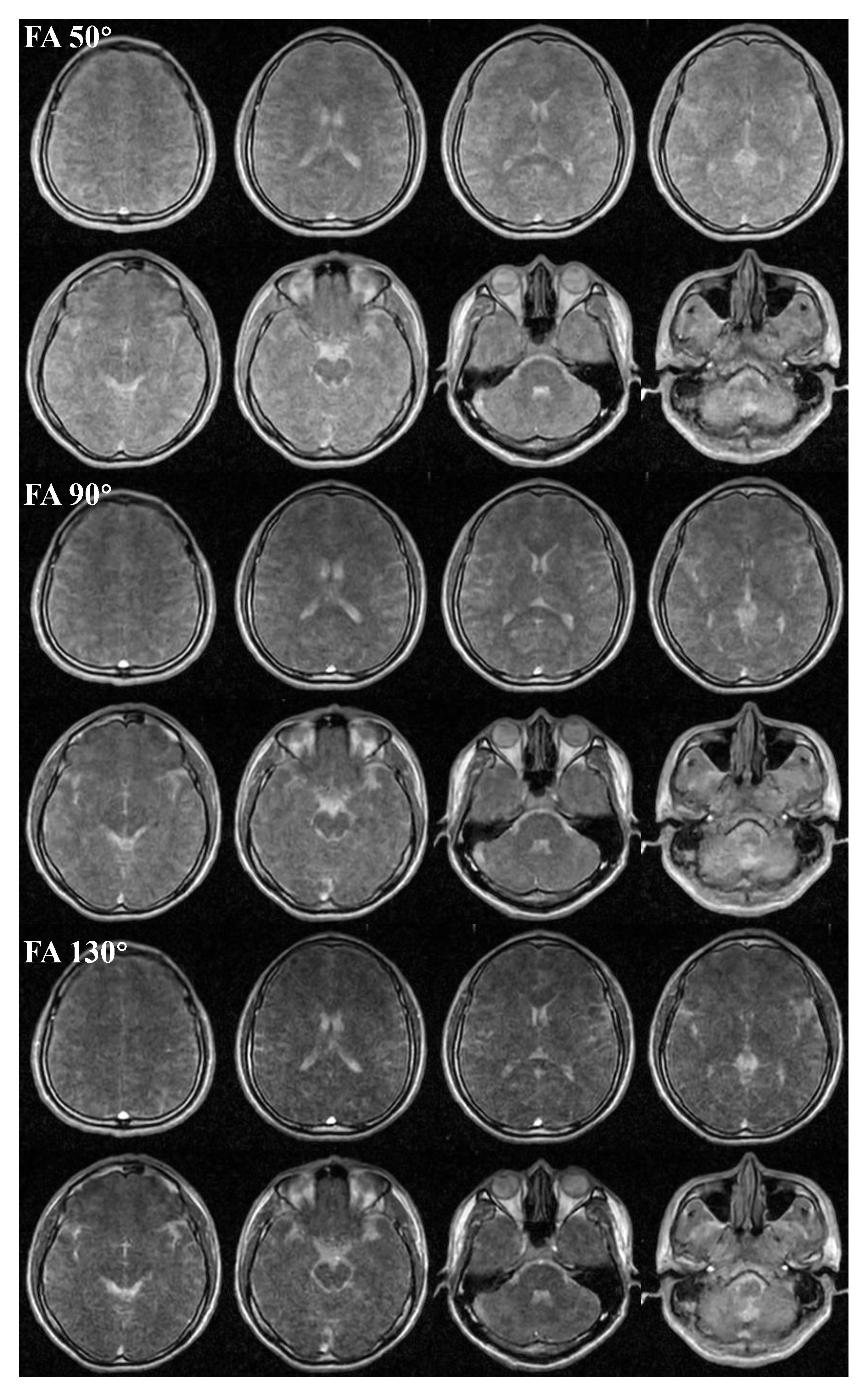
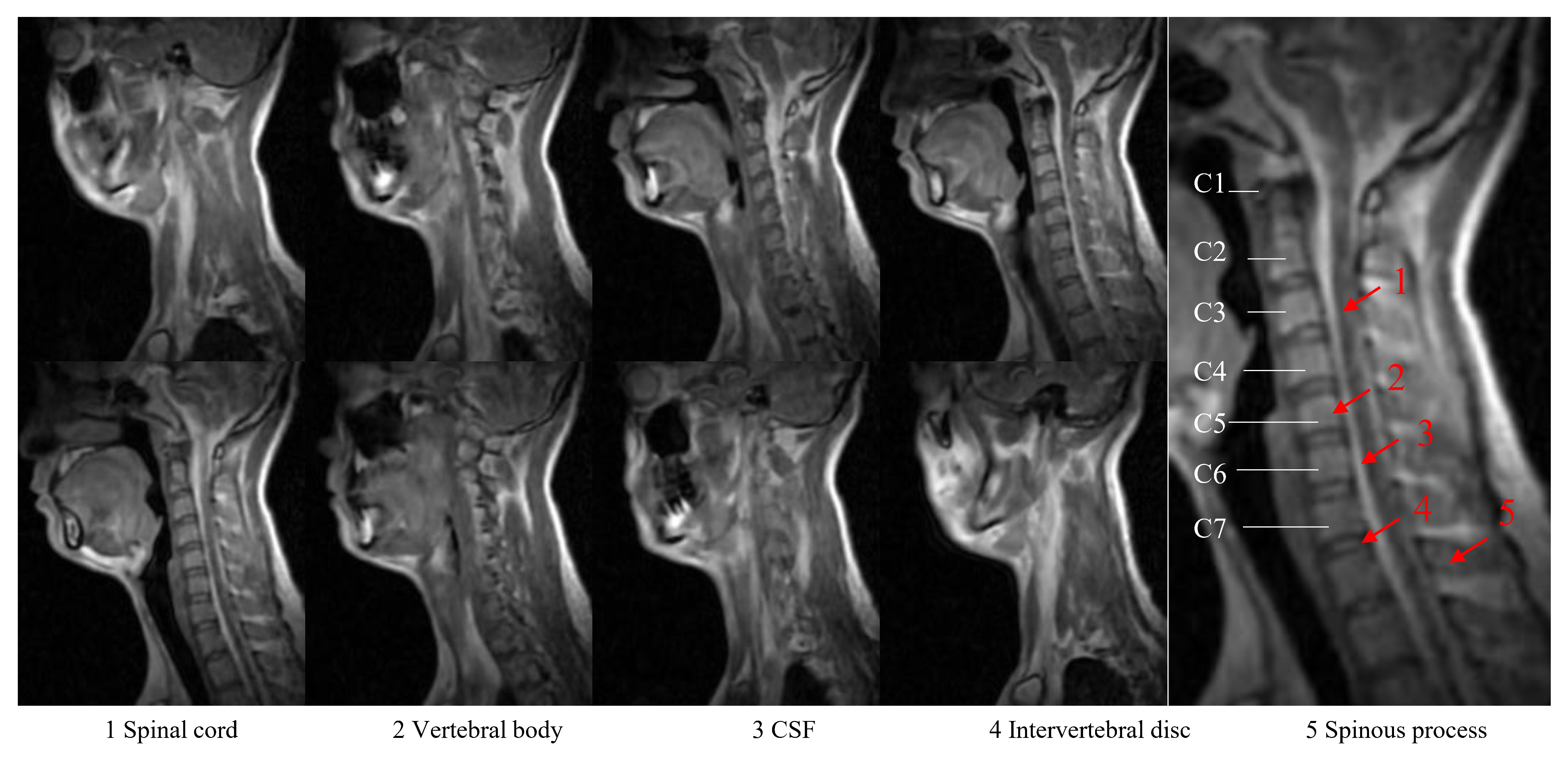
Fig.1 depicts the 3D brain images obtained using the bSSFP sequence with different flip angles. The image contrast was observed to change with the flip angles. This observation suggests the potential for manipulation of bSSFP imaging contrast on ULF systems, although T1 and T2 values have smaller difference at ULF in comparison to the high field.Fig.2 illustrates the results of 3D bSSFP C-spine imaging. This imaging technique enables identification of various anatomical structures, including the intervertebral disk and body, the spinous process, the spinal cord, and cerebrospinal fluid (CSF) within the spinal canal. The zoomed view identifies the C1 to C7 vertebral bodies of the cervical spine.
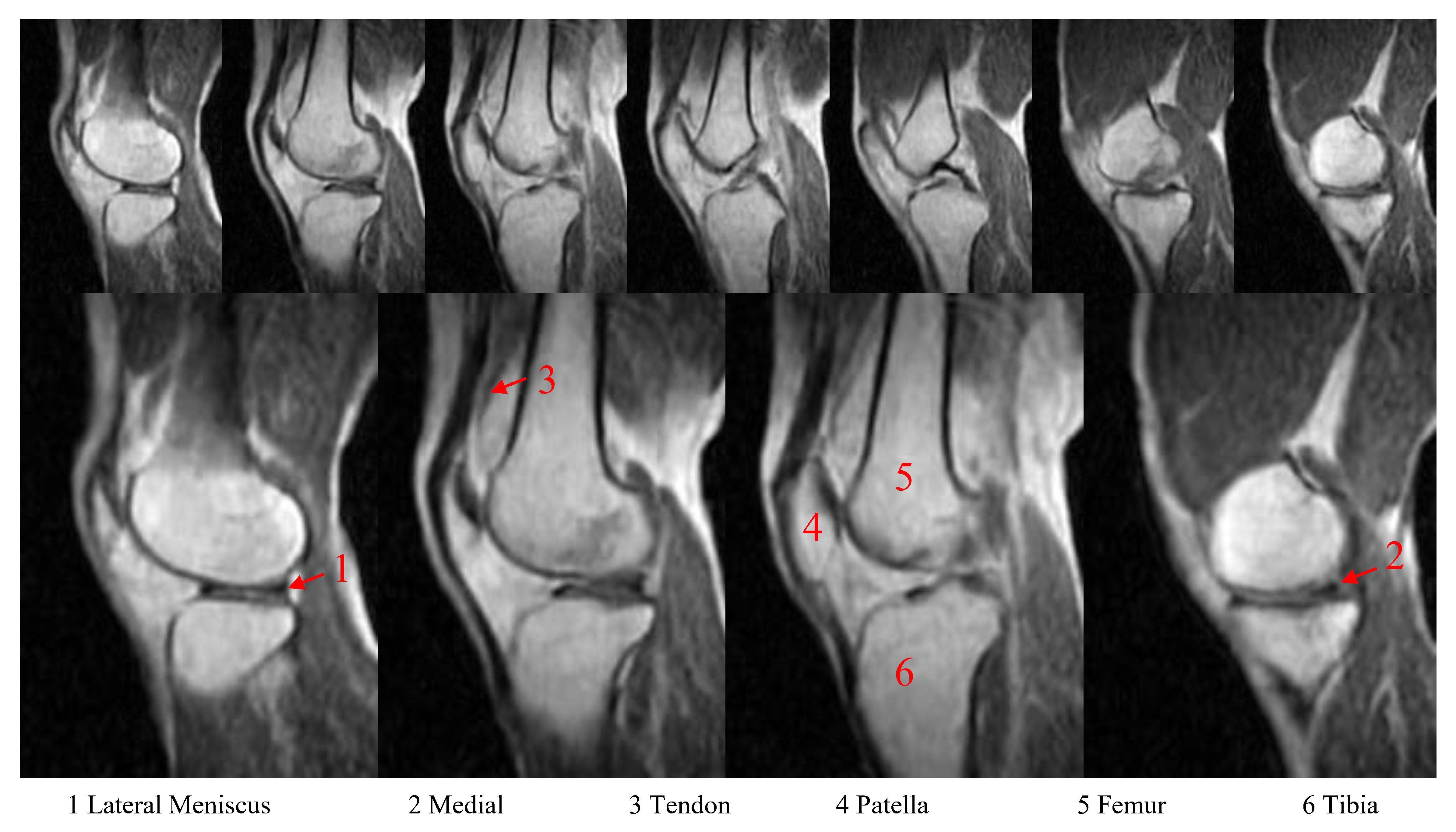
Fig.3 presents the sagittal knee 3D bSSFP images. These images allow for the identification of multiple knee structures, including the tendon, femur, lateral tibia, patella, and the posterior horn of both the lateral and medial meniscus.
Discussion and Conclusions
ULF MRI offers unique imaging opportunities due to the distinct nuclear magnetic resonance (NMR) properties of tissues12-15. At ULF, the T1 and T2 relaxation times of various tissues undergo significant changes12,16. The shorter T1 values at ULF enable rapid relaxation and repetitions, while the lower RF specific absorption rate (SAR) compared to high-field MRI permits swift RF excitations and flexible use of various RF envelopes. The bSSFP protocol is particularly advantageous due to minimal geometric distortion compared to EPI. Despite the relatively poor B0 inhomogeneity (in ppm) observed on ULF MRI scanners, the resulting banding artifacts on bSSFP images remain acceptable. Moreover, due to its fully coherent steady-state magnetization, the bSSFP sequence offers the highest possible SNR per unit time among all known sequences17. This feature is particularly valuable for ULF imaging, where acquisition time is a critical factor. Therefore, bSSFP imaging provides ULF systems with the ability to acquire high-quality images efficiently and within acceptable time.Acknowledgements
This work was supported in part by Hong Kong Research Grant Council (R7003-19F, HKU17112120, HKU17127121, HKU17127022 and HKU17127523 to E.X.W).References
1.Yuen MM, Prabhat AM, Mazurek MH, Chavva IR, Crawford A, Cahn BA, Beekman R, Kim JA, Gobeske KT, Petersen NH, Falcone GJ, Gilmore EJ, Hwang DY, Jasne AS, Amin H, Sharma R, Matouk C, Ward A, Schindler J, Sansing L, de Havenon A, Aydin A, Wira C, Sze G, Rosen MS, Kimberly WT, Sheth KN. Portable, low-field magnetic resonance imaging enables highly accessible and dynamic bedside evaluation of ischemic stroke. Sci Adv 2022;8(16): eabm3952.
2.He Y, He W, Tan L, Chen F, Meng F, Feng H, Xu Z. Use of 2.1 MHz MRI scanner for brain imaging and its preliminary results in stroke. J Magn Reson 2020; 319:106829.
3.O'Reilly T, Teeuwisse WM, de Gans D, Koolstra K, Webb AG. In vivo 3D brain and extremity MRI at 50 mT using a permanent magnet Halbach array. Magn Reson Med 2021;85(1):495-505.
4.Sheth KN, Mazurek MH, Yuen MM, Cahn BA, Shah JT, Ward A, Kim JA, Gilmore EJ, Falcone GJ, Petersen N, Gobeske KT, Kaddouh F, Hwang DY, Schindler J, Sansing L, Matouk C, Rothberg J, Sze G, Siner J, Rosen MS, Spudich S, Kimberly WT. Assessment of Brain Injury Using Portable, Low-Field Magnetic Resonance Imaging at the Bedside of Critically Ill Patients. JAMA Neurol 2020;78(1):41-47.
5.Cooley CZ, McDaniel PC, Stockmann JP, Srinivas SA, Cauley SF, Śliwiak M, Sappo CR, Vaughn CF, Guerin B, Rosen MS, Lev MH, Wald LL. A portable scanner for magnetic resonance imaging of the brain. Nat Biomed Eng 2021;5(3):229-239.
6.Bieri O, Scheffler K. Fundamentals of balanced steady state free precession MRI. J Magn Reson Imaging. 2013 Jul;38(1): 2-11. doi:10.1002/jmri.24163. Epub 2013 Apr 30. PMID: 23633246.
7.Deshpande VS, Chung YC, Zhang Q, Shea SM, Li D. Reduction of transient signal oscillations in true-FISP using a linear flip angle series magnetization preparation. Magn Reson Med 2003; 49:151–157.
8.Liu, Yilong, et al. A low-cost and shielding-free ultra-low-field brain MRI scanner [J]. Nature communications, 2021, 12(1): 1-14.
9.Lau V, Xiao L, Zhao Y, Su S, Ding Y, Man C, Wang X, Tsang A, Cao P, Lau GK. Pushing the limits of low‐cost ultralow‐field MRI by dual‐acquisition deep learning 3D superresolution. Magnetic resonance in medicine 2023.
10.Man C, Lau V, Su S, Zhao Y, Xiao L, Ding Y, Leung GK, Leong AT, Wu EX. Deep learning enabled fast 3D brain MRI at 0.055 tesla. Science advances 2023;9(38):eadi9327.
11.Zhao Y, Xiao L, Liu Y, Leong AT, Wu EX. Electromagnetic Interference (EMI) Elimination via Active Sensing and Deep Learning Prediction for RF Shielding‐free MRI. NMR in Biomedicine 2023:e4956.
12.Bottomley, P. A., Foster, T. H., Argersinger, R. E. & Pfeifer, L. M. A review of normal tissue hydrogen NMR relaxation times and relaxation mechanisms from 1-100 MHz: dependence on tissue type, NMR frequency, temperature, species, excision, and age. Med. Phys. 11, 425–448 (1984).
13.Koenig, S. H., Brown, R. D. 3rd, Adams, D., Emerson, D. & Harrison, C. G. Magnetic field dependence of 1/T1 of protons in tissue. Invest Radio. 19, 76–81 (1984).
14.Fischer, H. W., Rinck, P. A., Van Haverbeke, Y. & Muller, R. N. Nuclear relaxation of human brain gray and white matter: analysis of field dependence and implications for MRI. Magn. Reson. Med. 16, 317–334 (1990).
15.Wansapura, J. P., Holland, S. K., Dunn, R. S. & Ball, W. S. NMR relaxation times in the human brain at 3.0 tesla. J. Magn. Reson. Imaging 9, 531–538 (1999).
16.Stanisz, G.J., Odrobina, E.E., Pun, J., Escaravage, M., Graham, S.J., Bronskill, M.J. and Henkelman, R.M. (2005), T1, T2 relaxation and magnetization transfer in tissue at 3T. Magn. Reson. Med., 54: 507-512. https://doi.org/10.1002/mrm.20605
17.Scheffler K, Lehnhardt S. Principles and applications of balanced SSFP techniques. Eur Radiol. 2003;13(11):2409-2418. doi:10.1007/s00330-003-1957-x
Figures