2681
Deep learning enhanced DWI MUSE at 0.5T1GE HealthCare, Bengaluru, India
Synopsis
Keywords: Low-Field MRI, Diffusion/other diffusion imaging techniques
Motivation: Single-shot diffusion weighted imaging at 0.5T has low SNR, increased blurring and is limited by spatial resolution.
Goal(s): We want to demonstrate image quality improvement in multi-shot DWI at 0.5T with MUSE DL reconstruction.
Approach: Multi-shot DWI is reconstructed using MUSE algorithm followed by ARDL.
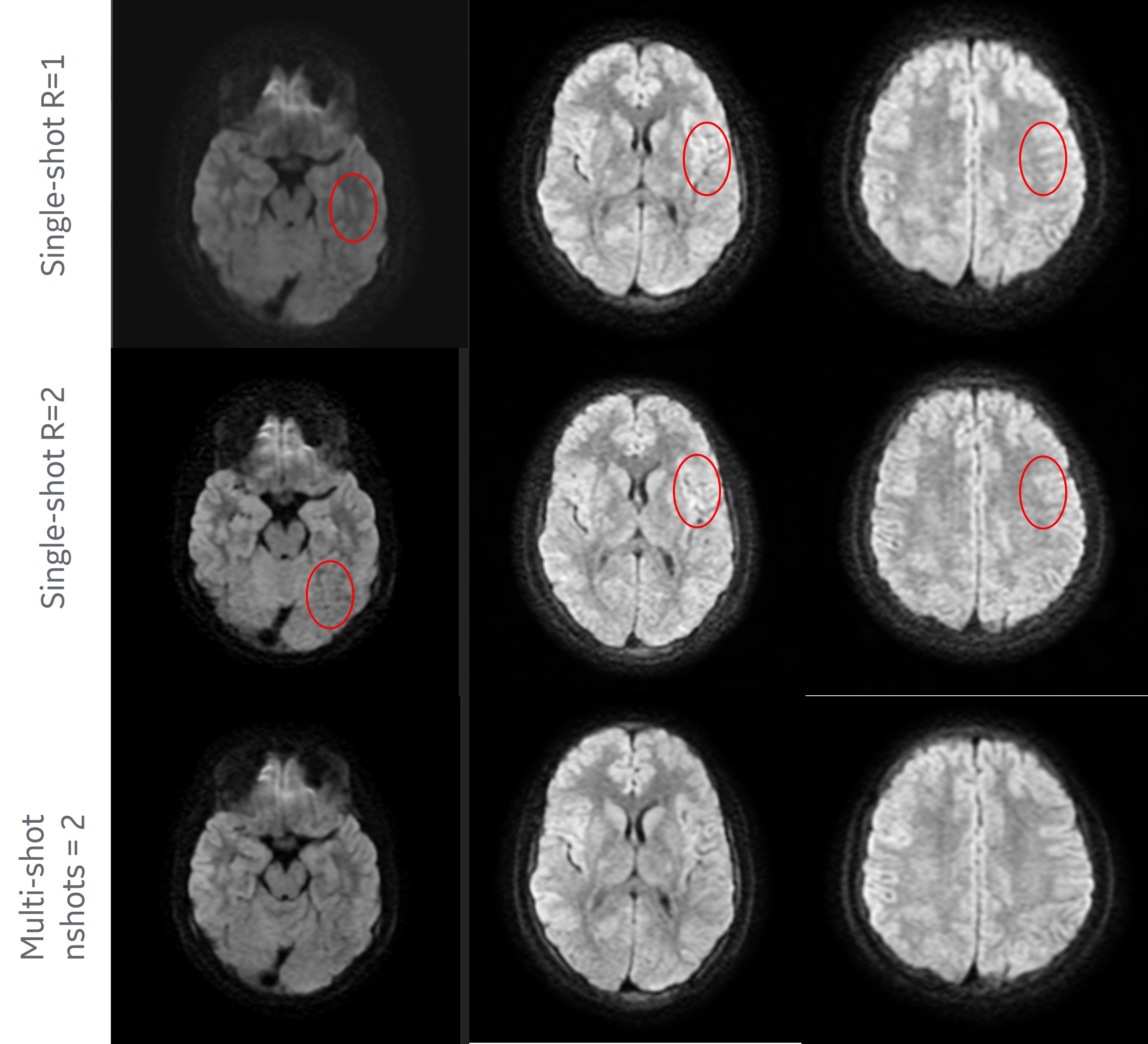
Results: The qualitative DWI results show improved image quality and less blurring with MUSE ARDL reconstruction.
Impact: Using multi-shot DWI and the MUSE ARDL recon increases spatial resolution and image quality at 0.5T and provides reliable imaging for low SNR DWI acquisitions.
Introduction
Diffusion weighted Magnetic resonance imaging (DWI) provides key insights into neuronal microstructures, oncology, and whole-body imaging1. DWI is usually performed using single-shot EPI sequences to avoid motion-induced phase errors. This coupled with lesser distortion at lower field strengths such as 0.5T makes it an attractive technique for diffusion imaging at 0.5T like field strengths. However, some of the main drawbacks associated with single shot DWI are distortion, limited spatial resolution and T2 decay2. Also, at low field strengths such as 0.5T, the SNR noticeably reduces with increased blurring and image quality is affected at high b-values. The spatial resolution issues are addressed using parallel imaging and multi-shot techniques3,4. But the use of parallel imaging with single-shot imaging is limited by g-factor noise amplifications.To overcome these drawbacks, a technique called MUSE4,5 was introduced to enable higher spatial resolution, and addresses phase inconsistencies between shots. This technique made use of an interleaved EPI sequence for acquisition followed by an ASSET3 based reconstruction to estimate motion-induced phase error for each shot and joint reconstruction of the estimated shots. The use of joint formulation improved the matrix conditioning and produced diffusion images with improved SNR. However lower field systems like 0.5T suffer from less SNR than high field systems and require image enhancement algorithms to produce diagnostic quality images. In this study, we acquire an interleaved EPI reconstructed using MUSE followed by the ARDL6 algorithm trained for Diffusion imaging. The MUSE reconstructed images with ARDL based denoising improves the image quality and significantly reduces blurring.
Method
Data Acquisition: In this study, we use vendor provided interleaved EPI sequence with 2 shots which can be extended to higher number of shots. Since this is a reconstruction method, it didn’t require any pulse sequence modifications.Reconstruction: The acquired interleaved EPI is first divided into n shots. Since each of these shots is undersampled, we first use ASSET reconstruction on the shots and determine the motion induced phase map for each shot. This is shown in figure 1.
This is followed by a joint reconstruction which constructs pseudo-sensitivities from all channels associated with the individual shots. These pseudo-sensitivities are used in a virtual channel-based ASSET processing. These images are then passed through the ARDL network to further denoise and remove residual artifacts. The ARDL filter strength was set to 50% so as not to over-smooth the images. The reconstruction procedure is shown in figure 2.
Volunteer scanning: A healthy subject was recruited and scanned under IRB approved protocols using a 1.5T GE creator (GE HealthCare, Milwaukee) modified to operate at 0.5T. A 14-channel HNU coil was used to image the brain. DWI data was acquired with a single shot spin echo EPI sequence with following specifications: TR=5430ms, TE=99ms, FOV= 24cm, matrix size=98x128, slice thickness=5mm with 1.5mm spacing between the slices, b-values acquired = 0 s/mm2 and 800 s/mm2. The number of signal averages was 4 for b0 and 6 for b-800 images. Acceleration factors of 1 and 2 were used. An interleaved EPI data with 2 shots was acquired using the following parameters: TR=5037ms, TE=106.5ms with all the other parameters like the single shot sequence. The number of signal averages was 2 for b0 and 3 for b-800 to match the scan duration of the single shot acquisition.
Results
Reconstruction results are shown in figure 3 comparing single shot EPI with acceleration factors of 1,2 and multi-shot EPI with number of shots as 2. Qualitative comparison highlights the improvement in image quality and less blurring in the ARDL processed MUSE reconstruction.Discussion and conclusion
In this study we have shown the improved image quality enabled using multi-shot DWI with MUSE and ARDL reconstruction at 0.5T field strength. Though we haven’t stretched the spatial resolution in this study, we discuss that the use of MUSE and ARDL recon improves the overall image quality at 0.5T field strength compared to single-shot imaging with/without parallel imaging. Our future work will focus on improving the spatial resolution of DWI at 0.5T.Acknowledgements
No acknowledgement found.References
[1] Taouli, B. and Koh, D.M.,2010. Diffusion-weighted MR imaging of the liver. Radiology, 254(1), pp.47-66.
[2] Le Bihan, D., Poupon, C.,Amadon, A. and Lethimonnier, F., 2006. Artifacts and pitfalls in diffusion MRI.Journal of Magnetic Resonance Imaging: An Official Journal of the International Society for Magnetic Resonance in Medicine, 24(3), pp.478-488
[3] King, K. F. (2004). ASSET--parallel imaging on the GE Scanner. In 2nd international workshop on parallel MRI; October (pp. 15–17)
[4] Chen, N.K., Guidon, A.,Chang, H.C. and Song, A.W., 2013. A robust multi-shot scan strategy for high-resolution diffusion weighted MRI enabled by multiplexed sensitivity-encoding (MUSE). Neuroimage, 72, pp.41-47.
[5] Navigated Multi-shot Diffusion-Weighted Imaging with Multiplexed Sensitivity Encoding, Valentina Taviani, Ann Shimakawa, Lloyd Estkowski, Arnaud Guidon, Ersin Bayram, and Robert Peters, in Proceedings of ISMRM 2018.
[6] Lebel RM. Performance characterization of a novel deep learning-based MR image reconstruction pipeline. 2020. arXiv preprint, arXiv:2008.06559.
Figures
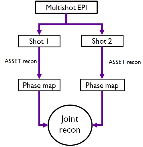
Figure 1 : Flowchart of 2 shot EPI reconstruction where each shot is reconstructed individually with ASSET processing to generate phase maps which is an input to the joint reconstruction algorithm.
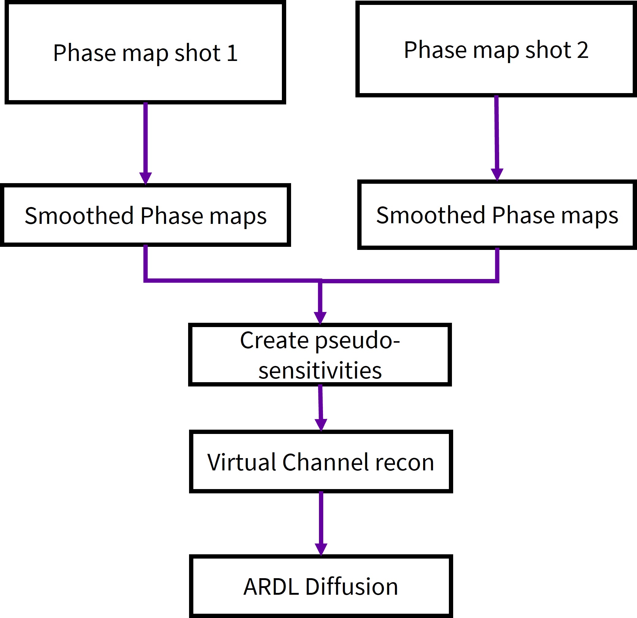
Figure 2 : Flowchart of reconstruction used in the study where the phase maps are smoothed and used to create pseudo-sensitivities which is then used in a virtual channel recon and processed using ARDL Diffusion models.