2679
Initial Experience Using 0.55T MRI for Detection of Liver Metastases in NET Patients1Department of Radiology and Biomedical Imaging, University of California San Francisco, San Francisco, CA, United States, 2Siemens Healthineers, Erlangen, Germany, 3Helen Diller Family Comprehensive Cancer Center, University of California San Francisco, San Francisco, CA, United States
Synopsis
Keywords: Low-Field MRI, Low-Field MRI
Motivation: As assessment of liver metastases requires multiple imaging modalities, it is essential to compare diagnostic capabilities and patient experiences of the low-cost 0.55T MRI to conventional imaging techniques.
Goal(s): We analyzed the sensitivity for neuroendocrine tumor metastases detection of gadoxetate-enhanced 0.55T MRI to 68Ga-DOTATATE PET/CT and 3.0T MRI while evaluating patient experience
Approach: Patients were imaged on 0.55T MRI with 68Ga-DOTATATE PET/CT and/or 3.0T MRI for comparison. Sound pressure levels (SPLmax) were compared between 0.55T and 3.0T.
Results: Hepatobiliary phase imaging at 0.55T showed increased detection rates over 68Ga-DOTATATE PET/CT. SPLmax of 0.55T was significantly lower. Patients found the 0.55T MRI less stressful.
Impact: Routine imaging of patients with neuroendocrine tumor metastases is feasible at 0.55T MRI. Low-field MR imaging has the potential to improve patient experience and accessibility without sacrificing diagnostic capability.
Introduction
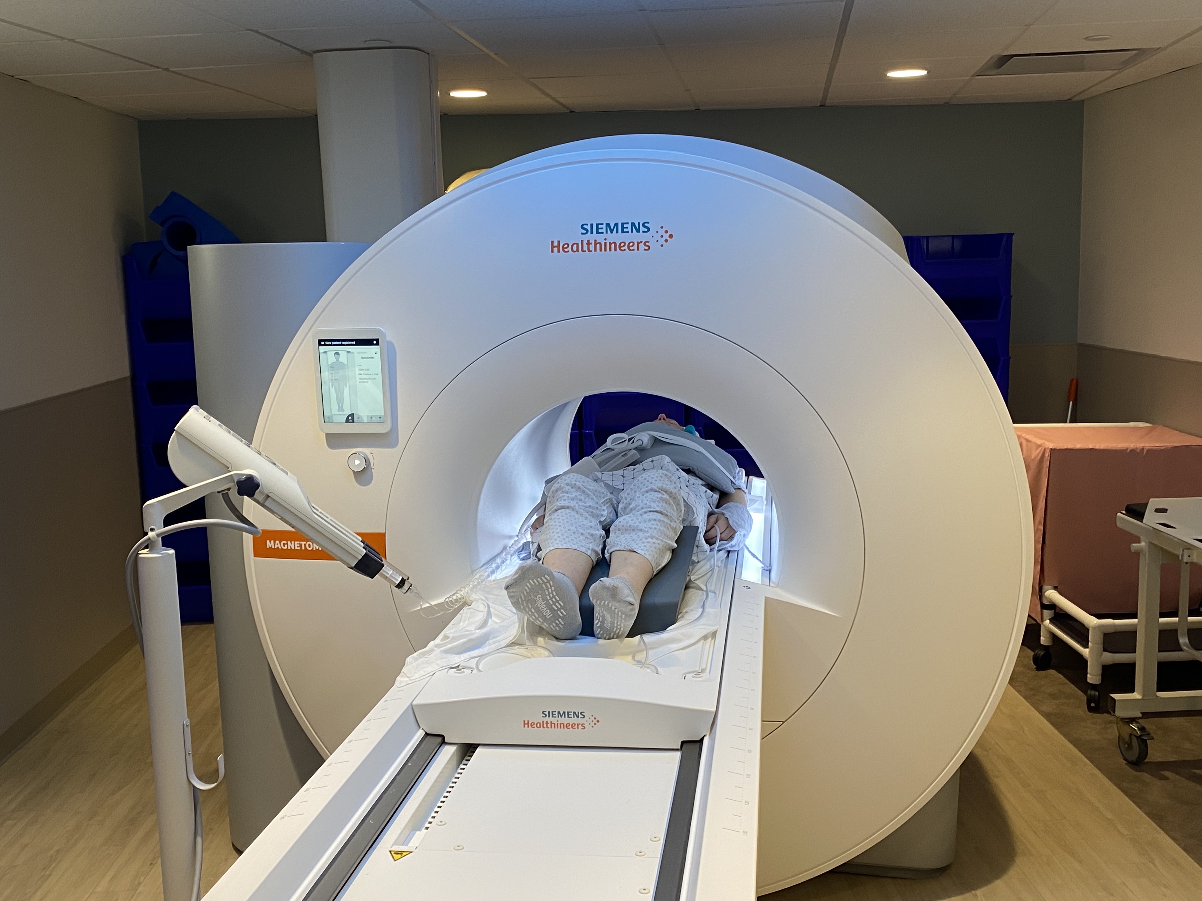
0.55T MRI is a new MRI technology offering increased patient comfort with a wider bore (80cm) and reduced acoustic noise.1 It is lower cost and more accessible than imaging at higher fields, with scanners available for routine clinical use.2 Gadoxetate-enhanced MRI has shown high sensitivity for the detection of metastatic liver disease with diffusion weighted imaging (DWI) showing high sensitivity for the detection of hepatic neuroendocrine metastases.3,4 As the assessment of neuroendocrine tumors necessitates the use of multiple imaging modalities, it is important to compare the diagnostic capability and patient experience of 0.55T MRI to conventional imaging techniques.5We compared the sensitivity for liver metastases detection on gadoxetate-enhanced 0.55T MRI to 68Ga-DOTATATE PET/CT and 3.0T MRI to determine the potential diagnostic capability of 0.55T MRI for patients with neuroendocrine tumors. Furthermore, we evaluated patient-reported experience measures (PREMs) comparing the 0.55T MRI experience to other modalitiesroutinely used to image liver metastases.
Methods
Nine patients with neuroendocrine tumor metastases were imaged using gadoxetate-enhanced 0.55T MRI (MAGNETOM Free.Max, Siemens Healthineers, Erlangen, Germany). Eight patients were imaged with 68Ga-DOTATATE PET/CT and five patients with 3.0T MRIs for comparison. The presence of liver metastases was assessed on CT, PET from PET/CT, diffusion weighted imaging (DWI), and hepatobiliary phase imaging (HBP). Detection rates were compared. Up to five lesions measuring under 1cm and five over 1cm per patient were included for analysis. Maximum standardized uptake values (SUVmax) of DOTATATE PET/CT were determined.Sound pressure level (SPLmax) of DWI and HBP were measured at 0.55T and 3.0T by placing a decibel meter 2m from each magnet’s front panel and SPLmax was calculated over five repetitions. This was compared using an unpaired t-test.
Fifteen patients were prospectively recruited for imaging of various liver metastases and diseases on 0.55T MRI. Following completion of their scans, patients completed PREMs comparing their 0.55T MRI exam to previous imaging scans.
Results
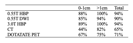
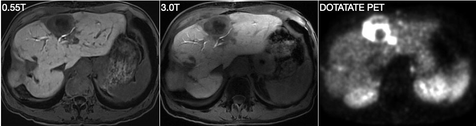
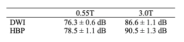
Nine patients (average age 63 [5 female, age range 47-75]) with 69 total liver lesions were successfully imaged at 0.55T MRI. 63 lesions were visualized at 0.55T, 30 lesions at 3.0T, and 42 on DOTATATE PET/CT. Mean SUVmax of hepatic lesions was 18.7 ± 10.5 on DOTATATE PET/CT. HBP and DWI at 0.55T had increased detection of liver metastases over DOTATATE PET/CT. HBP at 3.0T and 0.55T had equivalent detection of metastases. Qualitatively, HBP was superior on 0.55T compared to 3.0T for 4 of 5 patients.SPLmax was significantly lower for DWI and HBP sequences on a 0.55T system (DWI 76.3 ± 0.6 dB, HBP 78.5 ± 1.1 dB) compared to a 3.0T system (DWI 86.6 ± 1.1 dB, HBP 90.5 ± 1.3 dB) (p value < 0.0001).
Fifteen patients who completed exams on the 0.55T MRI for various liver diseases and metastases compared the scanner to previous experiences with MRI (n = 14), PET-MRI (n = 2), CT (n = 8), PET-CT (n = 5), X-Ray (n = 11), and Ultrasound (n = 11). PREMs show that 60% of patients experience at least some anxiety about out-of-pocket expenses for their diagnostic imaging scans. With the 0.55T MRI, 86% of patients felt less or no anxiety and 80% found the 0.55T MRI less stressful than scans on other modalities. 66% reported that the 0.55T MRI was noticeably quieter than other modalities.
Discussion / Conclusion
Routine imaging of patients with neuroendocrine tumor metastases is feasible on 0.55T MRI. Both HBP and DWI at 0.55T showed an increased detection rate for hepatic metastases compared to 68Ga-DOTATATE PET/CT. Quantitative assessment and PREMs show reduced noise levels at 0.55T. Overall, patients experienced less anxiety and stress during their 0.55T MRI compared to imaging scans on other modalities. Low-field MR imaging has the potential to improve the patient experience and reduce routine imaging costs without sacrificing diagnostic capability.Acknowledgements
No acknowledgement found.References
1. Rusche, T. et al. More Space, Less Noise – New-generation Low-Field Magnetic Resonance Imaging Systems Can Improve Patient Comfort: A Prospective 0.55T-1.5T-Scanner Comparison. J. Clin. Med. 2022 Nov 12;11, 6705.
2. Hori, M. et al. Low-Field Magnetic Resonance Imaging: Its History and Renaissance. Investigative Radiology Invest. Radiol. 2021 Jul 21;56:669-679.
3. Hope, TA et al. Simultaneous 68Ga-DOTATATE PET/MRI with gadoxetate disodium in patients with neuroendocrine tumor. Abdom. Imaging. 2015 Mar 28;40:1432-1440.
4. Flechsig, P. et al. Qualitative and quantitative image analysis of CT and MR imaging in patients with neuroendocrine liver metastases in comparison to 68Ga-DOTATOC PET. Euro. J. of Radiol. 2015 Apr 16;84:1593-1600.
5. Maxwell JE, Howe JR. Imaging in neuroendocrine tumors: an update for the clinician. Int J Endocr Oncol. 2015;2:159-168.
Figures