2669
Prospective Motion Correction with Radial MRI for Improved Neuroimaging during Severe Motion1Department of Clinical Neuroscience, Karolinska Institute, Stockholm, Sweden, 2Department of Neuroradiology, Karolinska University Hospital, Stockholm, Sweden, 3MR Applied Science Laboratory Europe, GE Healthcare, Munich, Germany
Synopsis
Keywords: Motion Correction, Data Acquisition
Motivation: Prospective motion correction can correct motion-induced sampling errors in real time but is still sensitive to large motion due to position based non-linear phase differences.
Goal(s): We aim to increase motion robustness of 3D sequences.
Approach: By combining prospective motion correction with radial sampling, non-linear phase errors can be mitigated due to repeated sampling of the k-space center.
Results: We show that prospective motion correction alone is enough for subtle motion but during severe motion (rotations well above ±5°) radial sampling can outperform Cartesian sampling.
Impact: Radial sampling in combination with prospective motion correction allows for imaging during severe motion corruption. This is crucial in imaging e.g. paediatric populations.
Introduction
Motion artifacts are a significant challenge in neuroimaging, compromising the quality of acquired data. Radial MRI sequences have gained attention for their inherent resistance to motion artifacts due to repeated sampling of the center of k-space, which averages out phase errors1. Additionally, many motion correction methods, including prospective motion correction (PMC) techniques2-5 have been developed to counteract motion-induced artifacts. These methods are powerful, but cannot in isolation counteract motion artifacts in 3D sequences because of non-linear spatially and temporally varying image phase when the motion is severe6 (e.g. fast head rotations well above ±5°), as recorded in, for example, paediatric neuroimaging7. This work combines PMC and radial sampling for robust neuroimaging in cases with severe motion.Methods
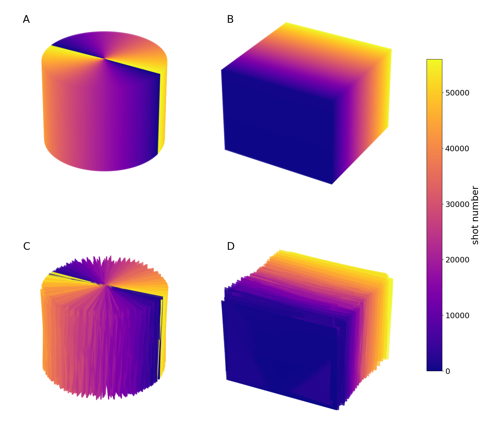
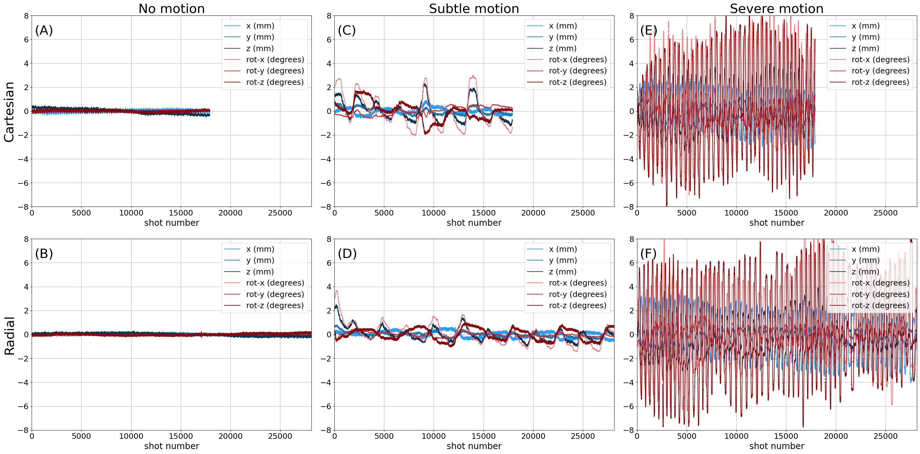
In this project, the prospective motion correction technique Wireless Radio Frequency Triggered Acquisition Device (WRAD)5 was integrated into a radial MRI acquisition protocol. A healthy volunteer was scanned using a GE Healthcare Signa 3T scanner equipped with a 48-channel head coil (discarding the 12 channels with the highest signal in the neck). Both radial 3D stack-of-stars with uniform radial spokes and Cartesian spoiled gradient echo MRI (Figure 1) were acquired (TR/TE/FA/BW = 8 ms, 2 ms, 15 degrees, ±50 kHz), with and without PMC. The resolution was 1x1x2 mm3, and FOV 224x224x160 mm3. In the radial case, 352 spoke angles were acquired for full Nyquist sampling, leading to a standard 57% increase in scan time compared to the Cartesian case1. Retrospective undersampling (R=2 and R=3) was also done to explore the radial sequence’s robustness to undersampling. Motion estimates from the WRAD were acquired and applied every TR and used to adjust the acquisition trajectory in real-time for three different motion conditions: 1. no motion, 2. subtle motion , and 3. severe motion. The volunteer was verbally coached before the scan on how to perform the motion to get similar motion patterns for the Cartesian and radial acquisitions. The recorded motion patterns are shown in Figure 2 and shared on brainmrimotion.org (doi: ISMRM24_PMC_Radial). Reconstruction was performed using custom Python scripts using the SigPy8 package. The radial data was density compensated using the Pipe-Menon method9 and transformed using an adjoint NUFFT in the kx-ky-plane, followed by a 1D iFFT applied in the kz-direction. The Cartesian data was reconstructed using a standard 3D iFFT. The images were then coil combined using root-sum-of-squares and denoised using an ITK-based adaptive denoiser10.Results
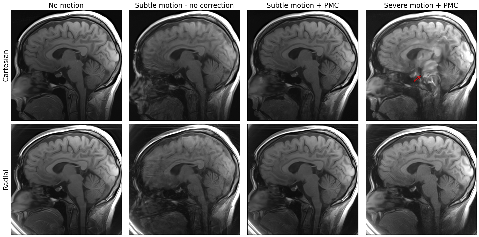
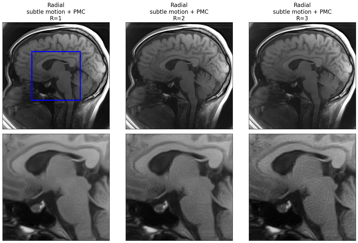
The results show significant benefits of prospective motion correction in combination with radial MRI for neuroimaging (Figure 3). Without PMC, motion artifacts were prominently visible in the reconstructed images even with subtle motion, leading to distortions and blurring in various brain regions, both for radial and Cartesian imaging. Conversely, when PMC was employed, motion-induced artifacts were substantially reduced, resulting in sharp and high-quality images in the subtle motion case. During severe motion, the image fidelity around the midbrain was completely lost in the 3D Cartesian case, whereas for PMC in combination with a 3D stack-of-stars trajectory only minor streaking artifacts were present.Retrospective undersampling of the radial trajectory resulted in increased noiselike artifacts whilst retaining image fidelity (Figure 4), showing that Nyquist sampling of the radial trajectory is unnecessary even using a simple reconstruction method as long as the SNR is sufficient.
Discussion
Without PMC, severe motion causes large gaps in the sampled k-space. With PMC, the designed trajectory that fills k-space evenly can be acquired. But even with perfect PMC, the method cannot mitigate motion-induced non-linear phase errors. This work highlights the effectiveness of PMC whilst also showcasing that it is not sufficient when large non-linear phase errors are present during Cartesian sampling. These errors can be mitigated by radial trajectories.Future work could explore optimizing the radial sequence parameters such as sampling order and angle selection along with PMC update frequency for reduced temporal footprint of the navigator. Kooshball radial sampling should also be explored as the center of k-space is then sampled every spoke for even more motion robustness and potential for self-navigation and retrospective refinement of the motion estimates. Lastly, as it is easier to lie still during a shorter scan, we will explore more sophisticated methods such as parallel imaging11,12, compressed sensing13, and deep learning14 based reconstruction methods to reduce scan time for the various radial trajectories in combination with WRAD PMC.
Conclusion
Prospective motion correction in combination with radial MRI represents an advancement in the pursuit of high-quality, motion-robust neuroimaging.Acknowledgements
The authors acknowledge research support from GE Healthcare and Barncancerfonden.References
- Bernstein MA, King KF, Zhou XJ. Advanced Pulse Sequence Techniques. In: Handbook of MRI Pulse Sequences. Elsevier Academic Press; 2004:802-954.
- Zaitsev M, Dold C, Sakas G, Hennig J, Speck O. Magnetic resonance imaging of freely moving objects: prospective real-time motion correction using an external optical motion tracking system. NeuroImage. 2006;31(3):1038-1050. doi:10.1016/j.neuroimage.2006.01.039
- Wallace TE, Afacan O, Waszak M, Kober T, Warfield SK. Head motion measurement and correction using FIDnavigators. Magnetic Resonance in Med. 2019;81(1):258-274. doi:10.1002/mrm.27381
- Frost R, Wighton P, Karahanoğlu FI, et al. Markerless high‐frequency prospective motion correction for neuroanatomical MRI. Magnetic Resonance in Med. 2019;82(1):126-144. doi:10.1002/mrm.27705
- van Niekerk A, Meintjes E, van der Kouwe A. A Wireless Radio Frequency Triggered Acquisition Device (WRAD) for Self-Synchronised Measurements of the Rate of Change of the MRI Gradient Vector Field for Motion Tracking. IEEE Trans Med Imaging. 2019 Jul;38(7):1610-1621. doi: 10.1109/TMI.2019.2891774. Epub 2019 Jan 10. PMID: 30629498; PMCID: PMC7192240.
- Berglund J, Sprenger T, Van Niekerk A, et al. Motion‐insensitive susceptibility weighted imaging. Magnetic Resonance in Med. 2021;86(4):1970-1982. doi:10.1002/mrm.28850
- Skare S. Brain MRI Motion DB. http://brainmrimotion.org. Accessed November 8, 2023.
- Ong F, Lustig M. SigPy: A Python Package for High Performance Iterative Reconstruction. In: Proc. Intl. Soc. Mag. Reson. Med. 27. Montreal, Canada; :4819.
- Pipe JG, Menon P. Sampling density compensation in MRI: Rationale and an iterative numerical solution. Magn Reson Med. 1999;41(1):179-186. doi:10.1002/(sici)1522-2594(199901)41:1<179::aid-mrm25>3.0.co;2-v
- Manjón JV, Coupé P, Martí‐Bonmatí L, Collins DL, Robles M. Adaptive non‐local means denoising of MR images with spatially varying noise levels. Magnetic Resonance Imaging. 2010;31(1):192-203. doi:10.1002/jmri.22003
- Pruessmann KP, Weiger M, Scheidegger MB, Boesiger P. SENSE: Sensitivity Encoding for Fast MRI. Magn Reson Med. 1999;42(5):952-962. doi:10.1002/(SICI)1522-2594(199911)42:5<952::AID-MRM16>3.0.CO;2-S
- Griswold MA, Jakob PM, Heidemann RM, et al. Generalized autocalibrating partially parallel acquisitions (GRAPPA). Magn Reson Med. 2002;47(6):1202-1210. doi:10.1002/mrm.10171
- Lustig M, Donoho D, Pauly JM. Sparse MRI: The application of compressed sensing for rapid MR imaging. Magn Reson Med. 2007;58(6):1182-1195. doi:10.1002/mrm.21391
- Liang D, Cheng J, Ke Z, Ying L. Deep Magnetic Resonance Image Reconstruction: Inverse Problems Meet Neural Networks. IEEE Signal Process Mag. 2020;37(1):141-151. doi:10.1109/MSP.2019.2950557
Figures