2668
Comparison of Fat Navigators and Optical Camera Based Retrospective Motion Correction1School of Biomedical Engineering, ShanghaiTech University, Shanghai, China, 2Shanghai Clinical Research and Trial Center, Shanghai, China
Synopsis
Keywords: Motion Correction, Motion Correction, Retrospective Motion Correction, Fat Navigator, Markerless Optical Camera, Brain MRI
Motivation: A comparison between efficacies of navigators and optical camera in retrospective motion correction (MC) on brain MR images remains largely unknown.
Goal(s): Our goal was to compare two motion tracking techniques (fat navigators [FatNav] and markerless optical camera [MoCAP]) and their performance in MC.
Approach: Twenty-one healthy subjects were imaged by T2-weighted turbo-spin-echo sequence with their head movements monitored simultaneously by both techniques. Performance was evaluated by image sharpness calculated at lateral ventricle/white matter boundary.
Results: Compared to MoCAP, FatNav had lower motion scores and less fluctuations at small motion. Images after FatNav-based MC showed greater sharpness than MoCAP.
Impact: The better performance of FatNav-based MC despite low temporal resolution suggest that FatNav can be integrated with MoCAP to achieve robust image quality in the presence of both abrupt and slow head motions.
Introduction
The accuracy of motion estimation is a key determinant of motion correction (MC) performance1. Navigators and optical camera are two commonly used methods for obtaining motion parameters. However, their relative efficacies in reducing motion artifacts remain largely unknown, except for some studies using T1-weighted gradient echo sequeces2-3. In this study, we simultaneously measured motion parameters using fat navigators (FatNav)4 and markerless optical camera (MoCAP)5 during a high resolution T2-weighted turbo-spin-echo (TSE) sequence and compared their motion parameters and retrospective motion correction performances.Methods
All images were acquired on a 3T scanner (United Imaging Healthcare, Shanghai, China) equipped with a 64-channel head coil. Twenty (aged 19-45 years, 14 males; named Subject 1-20) and one (aged 5 years, male; named Subject 21) healthy subjects were enrolled in this study after obtaining informed consents. The 3D variable flip angle TSE sequence was performed for Subject 1-20 (Subject 21) using the following parameters: TR/TE = 3000/408 (465) ms; resolution = 0.6×0.6×0.6 (0.8×0.8×0.8) mm3. The 3D FatNav was embedded within each TR using the following parameters: TR/TE = 4.6/2.3 ms; resolution = 3×3×3 mm3. ESPIRiT6 and GRAPPA7 were used to reconstruct TSE and FatNav images, respectively. The six rigid-body motion parameters of FatNav were obtained by registration of imaged volumes. The motion parameters of MoCAP were measured at a sampling rate of 30 Hz by registration of constructed point cloud face data. Due to higher temporal resolution of MoCAP (30 Hz) than FatNav (~0.33 Hz), the motion parameters of MoCAP during echo train acquisition were averaged to reduce noise and subsequently used for MC. MC was performed by non-uniform inverse Fourier transform of ESPIRiT reconstructed images in the k-space after correcting for their phases and positions. Motion parameters for unsampled k-space positions were assigned to be the same as those of the nearest sampled positions. To quantify motion severity, motion score was calculated by combining the root sum square of translational and rotational motion ranges8. To evaluate performance of MC, sharpness was quantified by the full width at half maximum (FWHM) of s-shaped edge function across lateral ventricle/white matter (WM) boundary9. Increased FWHM indicates decreased sharpness.Results
MoCAP produced noiseless constant motion parameters during parts of the scan (≥31%) in two subjects, indicating a failure in head movement measurement (success rate=90%), while FatNav possessed a success rate of 100%. Fig. 1 shows an example where abrupt motion was present, and the sharpnesses on corrected images were improved using both methods (Fig. 1A). The abrupt motion was captured by both FatNav and MoCAP (Fig. 1C and 1E), although a small mismatch (~0.1 mm and deg) was still observed in some periods (Fig. 1G). Fig. 2 shows an example in which FatNav and MoCAP measured motion parameters showed a large difference (≥0.5 mm and deg) and two spikes were observed in MoCAP parameters but was largely absent in FatNav. However, the FatNav-based MC resulted in decreased ringing artifacts compared to MoCAP. The overall motion scores from MoCAP were significantly higher than from FatNav (Fig. 3A), due to larger variances in MoCAP parameters in the case of mild motion (±0.1 mm and deg) as denoted by data points within red ovals in Fig. 3(B). Instead, higher consistencies between FatNav and MoCAP parameters were observed when motion was large. Fig. 4(A) shows significant correlation (corrected p=0.002, Spearman test) between FWHM and motion score without MC. The correlation was reduced and became less significant (p≥0.04) after MC using both FatNav and MoCAP motion parameters. Fig. 4(B) shows that FWHMs were significantly less after FatNav-based MC (mean FWHM=0.55 mm; p=0.04, Wilcoxon test) than MoCAP (mean FWHM=0.60 mm).Discussion
Our results suggest that both FatNav- and MoCAP-based MC can mitigate the negative impacts of motion artifacts and improve image sharpness. FatNav-based MC achieved higher cerebrospinal fluid/WM boundary sharpness than MoCAP-based despite having lower temporal resolution. This may be explained by the higher sensitivity of MoCAP to facial expression changes and eye movement during scan, as reflected by its high motion scores compared to FatNav, consistent with an earlier study3. On the other hand, the ability of MoCAP in more accurately capturing abrupt motion suggest that it may be combined with FatNav to achieve better image quality than using either method alone.Conclusion
Our results suggest that there is systematic difference in estimated motion parameters between FatNav- and optical camera-based methods. FatNav-based MC achieved increased image sharpness despite possibility of missing abrupt motion due to its lower temporal resolution compared to MoCAP.Acknowledgements
No acknowledgement found.References
1. Godenschweger F, Kägebein U, Stucht D, et al. Motion correction in MRI of the brain. Phys Med Biol. 2016;61(5):R32-R56.
2. Gretsch F, Mattern H, Gallichan D, Speck O. Fat navigators and Moiré phase tracking comparison for motion estimation and retrospective correction. Magn Reson Med. 2020;83(1):83-93.
3. Marchetto E, Murphy K, Glimberg SL, Gallichan D. Robust retrospective motion correction of head motion using navigator-based and markerless motion tracking techniques. Magn Reson Med. 2023;90(4):1297-1315.
4. Gallichan D, Marques JP, Gruetter R. Retrospective correction of involuntary microscopic head movement using highly accelerated fat image navigators (3D FatNavs) at 7T. Magn Reson Med. 2016;75(3):1030-1039.
5. Chen H, Dai K, Zhong S, et al. High-resolution multi-shot diffusion-weighted MRI combining markerless prospective motion correction and locally low-rank constrained reconstruction. Magn Reson Med. 2023;89(2):605-619.
6. Uecker M, Lai P, Murphy MJ, et al. ESPIRiT--an eigenvalue approach to autocalibrating parallel MRI: where SENSE meets GRAPPA. Magn Reson Med. 2014;71(3):990-1001.
7. Griswold MA, Jakob PM, Heidemann RM, et al. Generalized autocalibrating partially parallel acquisitions (GRAPPA). Magn Reson Med. 2002;47(6):1202-1210.
8. Zong X, Nanavati S, Hung SC, Li T, Lin W. Effects of motion and retrospective motion correction on the visualization and quantification of perivascular spaces in ultrahigh resolution T2-weighted images at 7T. Magn Reson Med. 2021;86(4):1944-1955.
9. Bazin PL, Nijsse HE, van der Zwaag W, et al. Sharpness in motion corrected quantitative imaging at 7T. Neuroimage. 2020;222:117227.
Figures
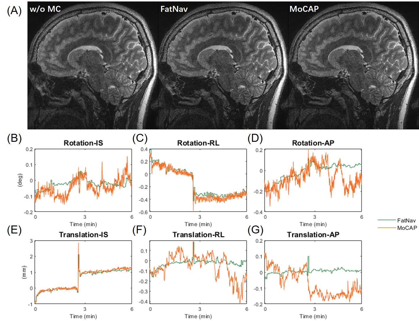
Figure 1: (A) Comparison of uncorrected and corrected images of the subject with abrupt motion. (B-G) are the corresponding motion profiles obtained from FatNav and MoCAP.
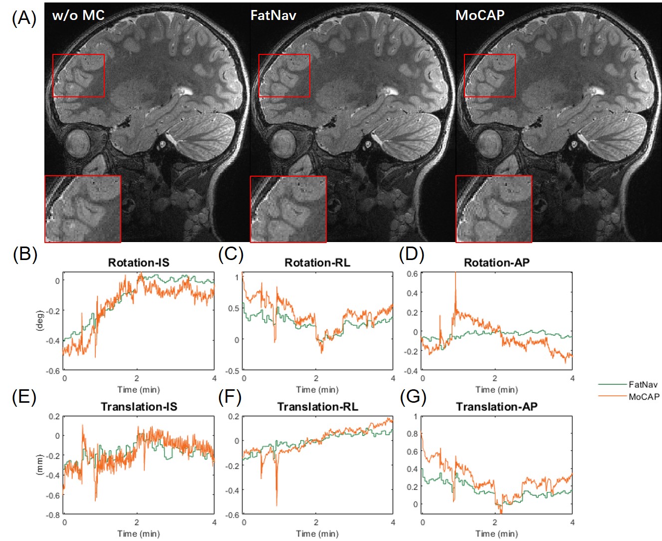
Figure 2: (A) Comparison of uncorrected and corrected images of the 5-year-old subject. (B-G) are the corresponding motion profiles obtained from FatNav and MoCAP.
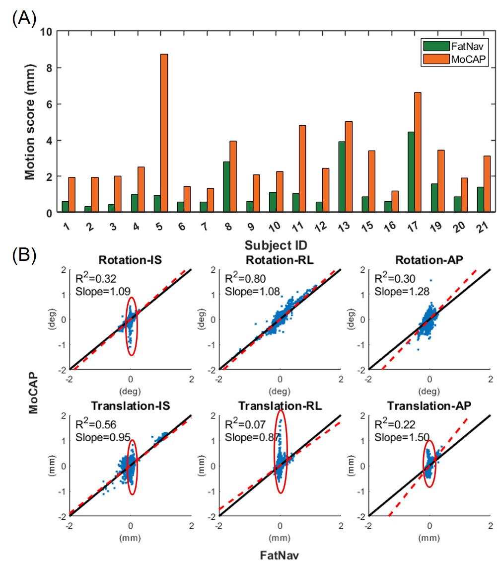
Figure 3: (A) Comparison between motion scores calculated from motion profiles of FatNav and MoCAP. (B) Scatter plots of six motion parameters obtained by MoCAP versus FatNav across all subjects. Slopes and R square of linear regression (y=kx; red dotted line) were shown. The line of identity (y=x) was also shown as the black line.
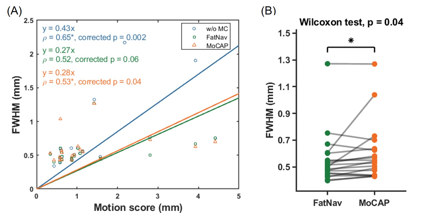
Figure 4: (A)Scatter plot of sharpness versus motion score on images w/o MC, w/ FatNav-based MC and w/ MoCAP-based MC. Linear regression and Spearman correlation coefficients are shown, where asterisk denotes significance after Bonferroni correction. (B) Comparison of sharpnesses between images with FatNav-based and MoCAP-based MC. The asterisk denotes significant difference from a Wilcoxon signed rank test.