2667
Streaming-MoCo: Real-Time Motion-Compensated Image Reconstruction from 3D Non-Cartesian MRI1Computational Radiology Laboratory, Boston Children's Hospital, Boston, MA, United States, 2Department of Radiology, Harvard Medical School, Boston, MA, United States
Synopsis
Keywords: Motion Correction, Brain, streaming, motion correction, real-time, image reconstruction, 3D, radial, non-cartesian
Motivation: A real-time motion compensation monitoring system is vital for structural MRI:
- to assess the effectiveness of motion correction even during k-space data collection,
- to ensure the rapid delivery of corrected images afterwards.
Goal(s): Develop a system that enables real-time:
- motion-compensation,
- image reconstruction,
- and visualization,
Approach: Introduced a framework that includes online:
- coil compression,
- rigid motion-tracking,
- motion-compensated gridding,
- image generation/streaming.
- no motion,
- instructed motion.
Results: Streaming motion-compensated image reconstructions from 3D radial MRI and the progression of image quality improvements were demonstrated in real-time during k-space data collection.
Impact: Our streaming-MoCo framework enables rapid reconstruction of motion-compensated images both during k-space data collection and post scan. This facilitates immediate assessment of image quality during scans, and potentially trigger corrective action well in advance before the scan concludes.
Introduction
Patient motion during MRI scans degrades image quality, especially in high-resolution 3D imaging due to long acquisition times. Non-Cartesian MRI offers more robustness to motion by oversampling the central k-space region. The use of pseudo-random sampling patterns (e.g., the golden-angle radial sequence1) prevents large k-space gaps2 when the patient moves, thereby improving their response to motion-compensation.Even though it is computationally demanding3 to reconstruct 3D non-Cartesian MRI mainly due to the need for k-space interpolation4,5, recent advancements in implicit gridding6 enable near real-time image reconstruction. This formulation does not rely on predetermined k-space trajectories, thus allowing for motion-compensated gridding during k-space data collection7.
Despite the progress, if motion compensation fails, the failure often goes unnoticed until the scan is complete. Our streaming-MoCo framework integrates streaming image reconstruction6 with the motion-compensated online gridding technique7 to provide rapid streaming of motion-compensated images on scanner display for feedback during k-space data collection.
This could allow technologists to monitor motion compensation efficacy, making informed decisions about whether to continue or repeat the scan, well in advance before the scan ends. If continued, the quality of the final feedback image can be refined using the accumulated (already motion-compensated) data once the scan ends.
Methods
The framework includes online coil compression, rigid-motion estimation, motion-compensated gridding, and image reconstruction to efficiently stream images on the scanner display. We customize our implementation to reconstruct from 3D golden-angle radial kooshball acquisitions in real-time on the scanner because it is possible to estimate motion from the data itself8 although any source of motion sensing, preferably with higher temporal resolution9, can be used.Gridding is the problem of finding a Cartesian k-space approximation $$$\mathbf{Z}\in\mathbb{C}^{N\times{D}}$$$ to non-Cartesian k-space measurements $$$\mathbf{Y}\in\mathbb{C}^{M\times{D}}$$$. Implicit gridding mechanism estimates $$$\mathbf{Z}$$$ by means of solving the normal equations $$\mathbf{\Phi}^{\top}\mathbf{\Phi}\mathbf{Z}=\mathbf{\Phi}^{\top}\mathbf{Y}$$ where $$$\mathbf{\Phi}\in\mathbb{R}^{M\times{N}}$$$ is a sparse k-space interpolation operator. Both sides of this system can be cumulatively calculated during k-space data collection and periodically solved to exhibit streaming reconstruction capability6. In addition, the system can be expressed as a sum over mutually exclusive subsets of data, such as $$\sum_s\mathbf{\Phi}_s^{\top}\mathbf{\Phi}_s\mathbf{Z}=\sum_s\mathbf{\Phi}_s^{\top}\mathbf{Y}_s$$ to incorporate motion information7.
There is no restriction on the size of subsets. One subset could be as small as one shot or even a spoke depending on the temporal resolution of motion sensing. Subsets can also be adapted to motion states10,11. We reconstruct each (fixed-width) subset to produce a low-resolution image, which is then aligned to the image from the first subset11 to obtain motion information per subset. An approximate $$$\mathbf{Z}$$$ is estimated at times when a new motion-compensated feedback image needs to be rendered. Once the scan concludes, the final feedback image can be refined using the accumulated (already motion-compensated) data.
To validate the proposed framework, two volunteers were scanned at 3T using a MAGNETOM Prisma (Siemens Healthcare, Erlangen, Germany) with a 64-channel head coil. The framework was deployed on the scanner using the Siemens FIRE prototype12, and motion-compensated feedback images were streamed to the scanner console via inline display. Each volunteer underwent two scans: one while holding still and another with instructed head repositioning. T1-weighted structural images with 1 mm isotropic resolution were acquired from Volunteer #1, and with 0.875 mm isotropic resolution from Volunteer #2. The total acquisition time for each scan was 6.4 minutes, with 45k spokes.
Results
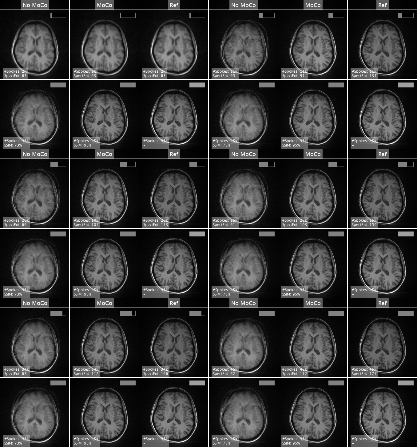
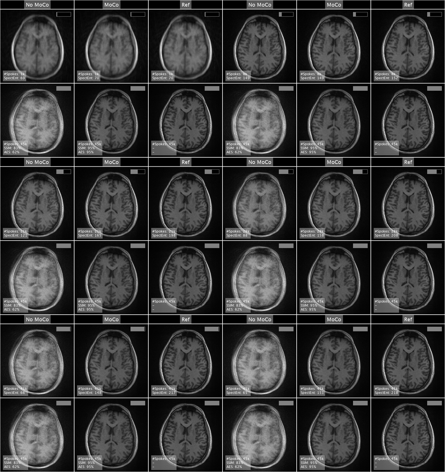
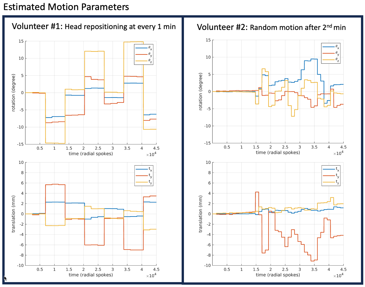
Figures 1 and 2 show the real-time reconstruction of Volunteer #1's scans at increasing progress from 5% to 100%. Figure 3 shows the progression for Volunteer #2's scans. The images illustrate successful compensation for motion during the 3D radial kooshball acquisition. Image quality metrics of structural similarity (SSIM), and average edge strength (AES) demonstrate improvements as compared to not compensating for motion.Figure 4 shows the respective head movements of each volunteer, as estimated during the streaming-MoCo process, where Volunteer #1 repositioned their head at every 60 seconds, and Volunteer #2 moved their head randomly after the second minute.
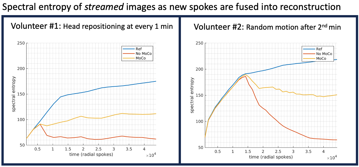
Figure 5 traces spectral entropy13 of streamed images as new spokes are integrated into reconstructions. Ref and MoCo images show increasing trends, while No-MoCo reverts to initial levels, further confirming the effectiveness of motion-compensation.
Discussion
We have proposed streaming-MoCo, an effective framework for real-time motion-compensated image reconstruction from 3D non-Cartesian MRI. The framework efficiently adjusts (implicit) gridding during k-space data collection based on motion (either estimated or provided by a sensor), without relying on predetermined trajectories as they change during patient motion.Conclusion
Streaming-MoCo enables rapid reconstruction of motion-compensated images both during data collection and post-scan, facilitates real-time monitoring of motion compensation effectiveness, and potentially triggers early corrective action before the scan concludes.Acknowledgements
This work was supported in part by the NIH under award numbers S10 OD025111 and R01 EB019483.References
- Chan RW, Ramsay EA, Cunningham CH, Plewes DB. Temporal stability of adaptive 3D radial MRI using multidimensional golden means. Magn Reson Med 2009; 61:354–363.
- Wallace TE, Warfield SK, Afacan O. Robust retrospective correction of 3D golden-ratio radial MRI using electromagnetic tracking. Proceedings 27th Meeting of the ISMRM, Paris, France, 2019
- Smith DS, Sengupta S, Smith SA, Brian Welch E. Trajectory optimized NUFFT: Faster non-Cartesian MRI reconstruction through prior knowledge and parallel architectures. Magn Reson Med 2019; 81:2064–2071.
- Fessler JA, Sutton BP. Nonuniform fast Fourier transforms using min-max interpolation. IEEE Trans Signal Process 2003; 51:560–574.
- Pipe JG, Menon P. Sampling density compensation in MRI: rationale and an iterative numerical solution. Magn Reson Med 1999; 41:179–186.
- Calakli F, Wallace TE, Chow K, Warfield SK. Real Time Streaming Image Reconstruction from Non-Cartesian MRI. ISMRM Workshop on Data Sampling & Image Reconstruction, 2023
- Calakli F, Wallace TE, Warfield SK. Simultaneous Motion Compensation and Image Reconstruction During Acquisition of 3D Radial MRI. In Proceedings of 31st Annual Meeting ISMRM, 2023
- Kecskemeti S, Samsonov A, Velikina J, et al. Robust Motion Correction Strategy for Structural MRI in Unsedated Children Demonstrated with Three-dimensional Radial MPnRAGE. Radiology 2018; 289:509–516.
- Wallace TE, Ariyurek C, Calakli F, Kober T, Warfield SK. High Temporal Resolution Head Motion Tracking using Pilot Tone and 3D Radials. In Proceedings of 31st Annual Meeting ISMRM, 2023
- Anderson AG 3rd, Velikina J, Block W, Wieben O, Samsonov A. Adaptive retrospective correction of motion artifacts in cranial MRI with multicoil three-dimensional radial acquisitions. Magn Reson Med 2013; 69:1094–1103.
- Calakli F, Wallace TE, Warfield SK. Real Time Motion Monitoring and Adaptive Motion Compensation in 3D Radial MRI. ISMRM Workshop on Motion Detection & Correction, 2022
- Chow, K, et al. An ISMRMRD-compatible image reconstruction server written in MATLAB. https://github.com/kspaceKelvin/matlab-ismrmrd-server/ 2019
- Zhang A, Yang B, Huang L. Feature Extraction of EEG Signals Using Power Spectral Entropy. 2008 International Conference on BioMedical Engineering and Informatics, 2008; 435–439
Figures
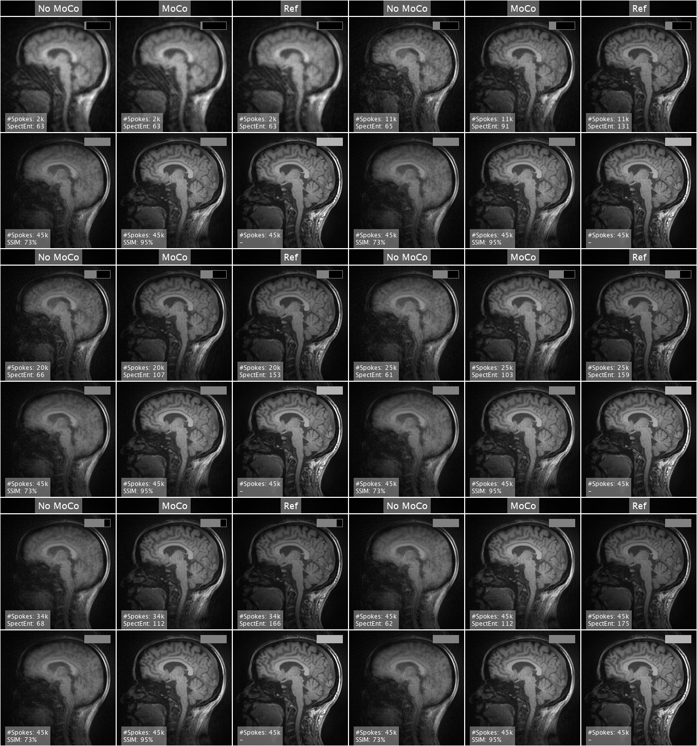
Figure 1: (Vol #1: Sagittal) Real-time streaming reconstructions from Volunteer #1's scans. The first two columns show the progression of motion-data reconstructions with no motion-correction (No-MoCo) and with motion-correction (MoCo) applied. The final column shows reference-data reconstructions (Ref) where the volunteer remained still. The MoCo image quality improves as new spokes are acquired, also confirmed by increase in the spectral entropy metric. The second row assesses the quality of final images against the Ref image, using the structural similarity index (SSIM).