2665
Volumetric self-navigated (vSNav) 3D-EPI for motion-robust functional MRI1University of Cape Town, Cape Town, South Africa, 2Athinoula A. Martinos Center for Biomedical Imaging, Charlestown, MA, United States
Synopsis
Keywords: Motion Correction, fMRI
Motivation: 3D-EPI overcomes spatial resolution, spin-history and acceleration limitations of 2D-EPI but is rarely used for functional MRI (fMRI) due to its greater sensitivity to motion.
Goal(s): To examine the performance of our volumetric self-navigated (vSNav) 3D-EPI sequence for fMRI acquisition.
Approach: We acquired fMRI data using both our vSNav 3D-EPI and the standard 2D-EPI sequence during a block design finger tapping experiment both without and with intentional motion to compare the quality of the BOLD signal and the impact of different pre-processing steps.
Results: Although data quality was similar, 3D data were more robust to spatial smoothing.
Impact: A motion-robust 3D-EPI sequence will permit functional MRI with higher spatial and temporal resolutions. However, since the sequence acquires data and performs motion correction in a new way, it requires suitable preprocessing and analyses pipelines.
Introduction
Since functional MRI (fMRI) measures the temporal evolution over hundreds of seconds of signal changes ≤1% of the average BOLD signal, it is highly susceptible to motion. Although 3D-EPI overcomes the high SAR1,2, spin-history3 and acceleration4,5 limitations of 2D-EPI, its use has been limited by its greater susceptibility to motion artifacts. Previously we developed and implemented a volumetric self-navigated (vSNav) 3D-EPI sequence for prospective motion tracking and correction (MoCo)6. Here we compare the performance of our vSNav 3D-EPI sequence to a standard 2D-EPI sequence with PACE (Prospective Acquisition CorrEction7) MoCo enabled on a block design finger tapping fMRI experiment, both without and with intentional head motion.Methods
Four healthy male volunteers (age 30 – 37 years) were scanned with a 20-channel head/neck coil on a 3T Skyra (Siemens, Erlangen) while performing block design finger tapping tasks using both our vSNav 3D-EPI and PACE MoCo 2D-EPI sequences, both without and with intentional head motion. Participants received visual cues to tap either their left- or right-hand fingers for 20s, interleaved with 20s rest blocks. The order of left- and right-hand tapping blocks were randomized across acquisitions, and the order of acquisitions were randomized across participants. During acquisitions with intentional motion (Mo), participants were instructed to move at specific times during rest blocks. Acquisitions without intentional head motion involved either no motion (NoMo) or leg motion; intentional head motion involved either nodding or rotating the head. Acquisition parameters were TE 30ms, FOV 210x210 mm2, 64x64 matrix, 3.3x3.3x3.1 mm3 resolution, 100 volumes. For 2D-EPI, 52 interleaved slices were acquired per volume, with volume TR 3400ms, flip-angle 90° and bandwidth 1736Hz/px. For 3D-EPI we used our vSNav 3D-EPI sequence with 52 partitions per volume acquired using a center-out acquisition scheme, volume TR 3328ms, flip-angle 16° and bandwidth . Real-time motion correction (MoCo) was active in all acquisitions, and all subjects were instructed to lie still except when instructed to move. De-spiking, motion correction and 2D-EPI slice scan time correction were performed using AFNI8,9, while non-brain tissue removal, intensity normalization, high-pass temporal filtering with cut-off 0.025 Hz, spatial filtering, and 3D-EPI slice scan time correction were performed with FSL10-12. For each data set, pre-processing was repeated with 6 different Gaussian spatial filters (FWHM 0 mm, 3.3 mm, 6.6 mm, 8 mm, 9.9 mm and 12 mm). Each volunteer’s fMRI data were co-registered to his .0 mm3 structural T1 weighted images and normalized to the MNI152_T1_2mm_brain standard space using a linear transform calculated on the anatomical images. First-level statistical analyses were performed in FEAT using a general linear model with predictors for left- and right-hand finger tapping convolved with a double-gamma hemodynamic response function, and motion parameters added as predictors of no interest. We compare the quality of the BOLD data between acquisitions and for different amounts of spatial smoothing using voxel-wise temporal signal-to-noise ratios (tSNR) defined as the mean of the signal time course divided by the variance of the residuals after model fitting. We also examine the effect of different amounts of spatial smoothing on the mean and standard deviation of the percent BOLD signal change, and the number of activated voxels, in the motor cortices during finger tapping. The activations in the motor cortices were extracted from the intersection of the primary motor cortices and the Z-statistic maps. Finally, we compare activation maps for 2D- and 3D-EPI acquisitions with 6.6 mm smoothing without and with intentional motion.Results
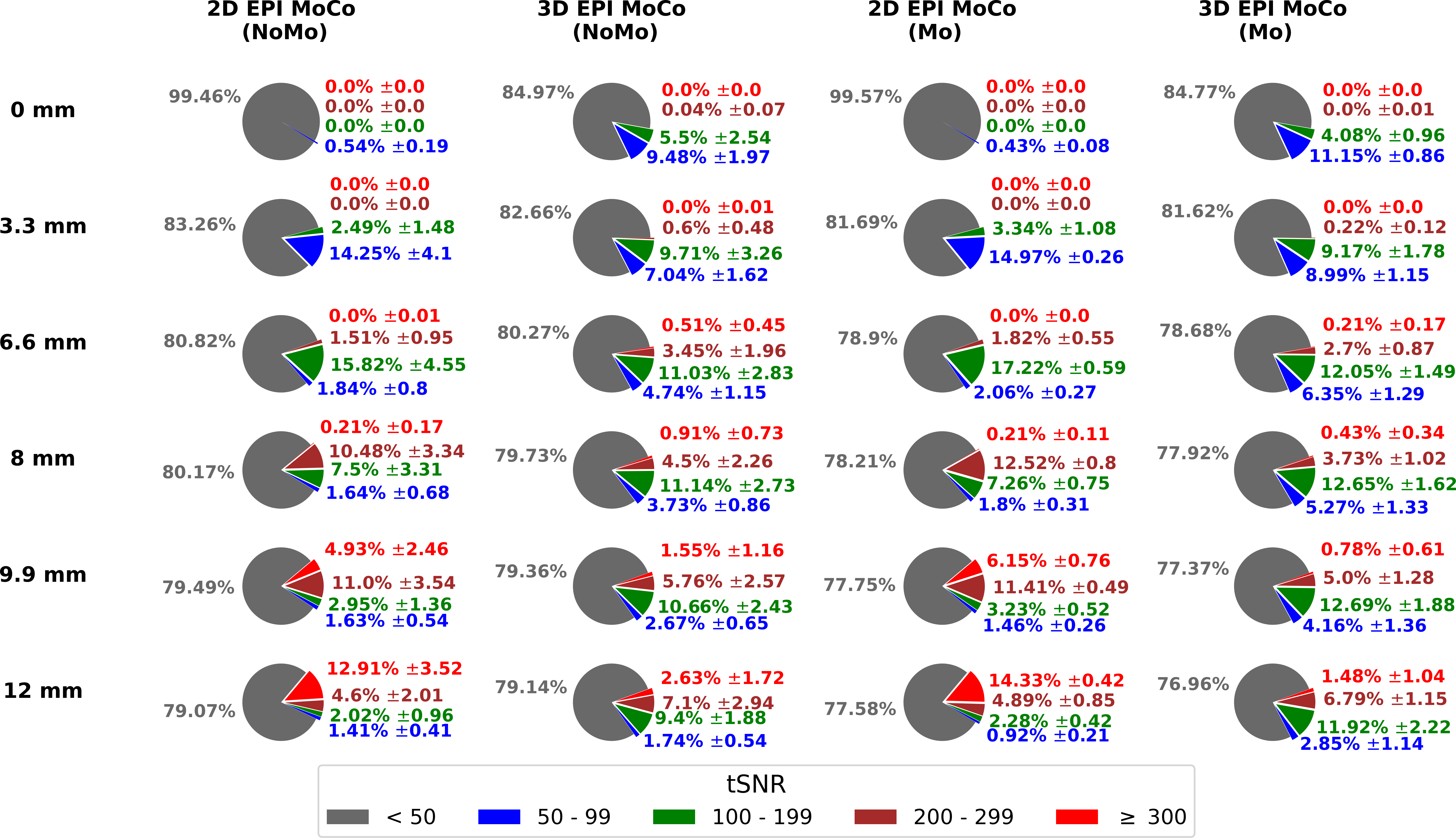
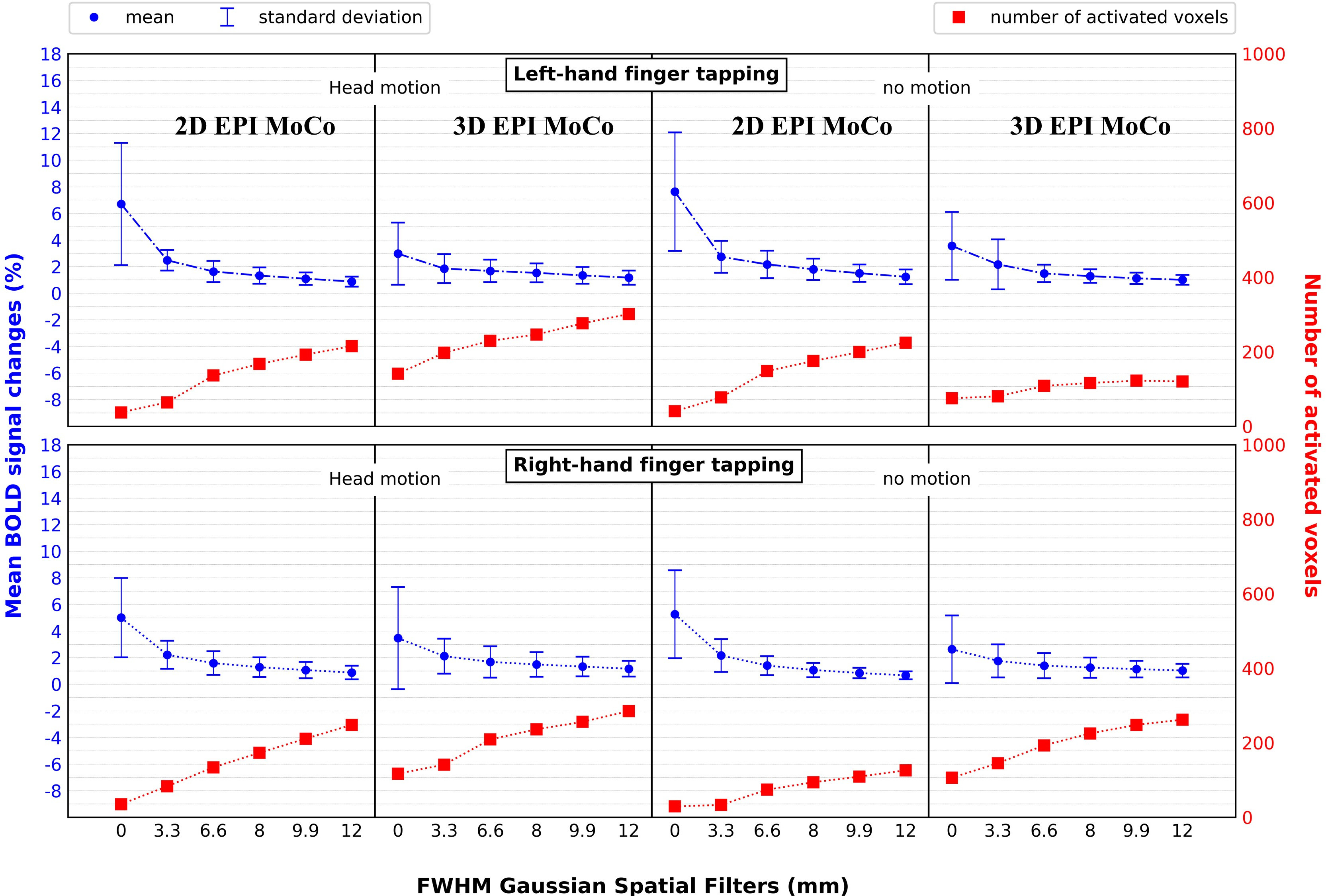
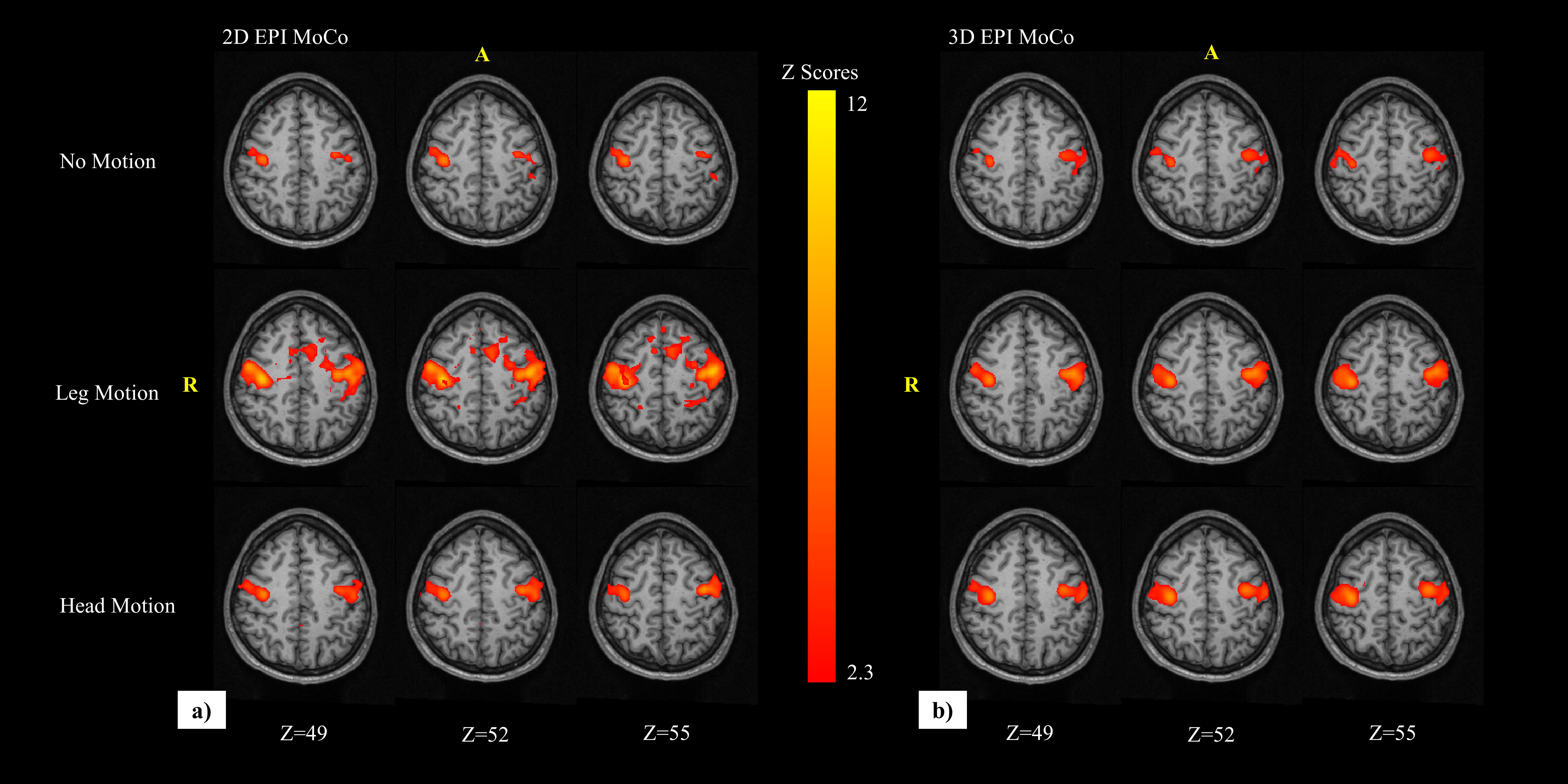
Figure 1 shows that the distribution of tSNR values was similar for acquisitions without and with intentional motion. However, compared to 3D data for which there are limited increases in tSNR following smoothing with filters >6.6 mm, 2D data show a substantial impact of increased smoothing. After smoothing of 2D data with filters of 9.9 and 12 mm, >15% of voxels demonstrate tSNR>200 and <5% have tSNR in the range 50 to 199. Figure 2 shows that reductions in mean % BOLD signal change, and increases in the number of activated voxels, with increasing smoothing in 2D and 3D acquisitions both with and without intentional head motion are similar. Although activation maps in 2D- and 3D-EPI data are similar, leg motion introduces more noise in 2D data (Figure 3).Discussion and Conclusion
These results demonstrate comparable quality of 3D-EPI fMRI data acquired with our vSNav 3D-EPI sequence to 2D-EPI even in the presence of motion. Moreover, 3D data are affected less by pre-processing steps and excess smoothing. Future work should implement acceleration to improve the temporal resolution.Acknowledgements
South African National Research Foundation grant 48337; National Institutes of Health (NIH) grants R01HD085813, R01HD099846 and R01HD093578.References
1.Collins et.al., Magn. Res. in Med., 2011.
2. Bernstein et al., 2004.
3. Karl J. Friston et al., Magn. Res. in Med., 1996.
4. Hu & Glover et.al., Magn. Res. in Med., 2007.
5. Poser et al., NeuroImage, 2010.
6. Bayih et.al., Magn. Res. in Med., 2022.
7. Thesen et al., Magn. Res. in Med., 2000.
8. Cox J.S., Comp. & Biomed. Research., 1996.
9. Cox & Hyde et. al., NMR Biomed., 1997.
10. Jenkinson et al., NeuroImage, 2012;
11. Smith et al., NeuroImage, 2004.
12. Woolrich et al., NeuroImage, 2009.
Figures